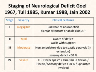
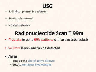
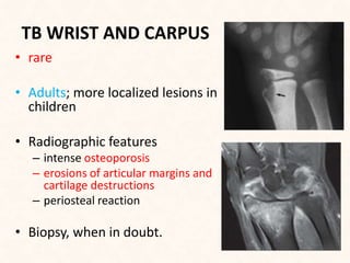
DR. BIPUL BORTHAKUR
PROFESSOR & HOD
DEPARTMENT OF ORTHOPAEDICS
SILCHAR MEDICAL COLLEGE
BONE TUBERCULOSIS
• Spine – Pott’s spine (50% of all cases of skeletal
Tuberculosis)
• Joints - Tubercular arthritis:
 Hip Joint
 Knee joint and Triple deformity
 Shoulder joint and Caries Sicca
 Elbow joint
 Wrist and Carpus
 Sacroiliac joints
• Long and flat bones - Tubercular osteomyelitis
• Short bones – Tubercular dactylitis (spina ventosa)
PRINCIPLES OF MANAGEMENT
• General and systemic treatment is like that of
TB in general
• Any concomitant disease must be treated
• Hospitalization required only for-
 Complications
 Deformity correction under supervision
SURGERY IN BONE AND JOINT
TUBERCULOSIS
• Exploration and appropriate operation
mandatory when-
 Lesion not responding favorably to drugs
 Doubt in diagnosis
 Refractory recrudescence of infection
 Juxta-articular osseous focus threatening the joint
TUBERCULOSIS OF SPINE
• first described by Sir Percival Pott in 1779, hence the
name Potts disease
• Usually two continuous vertebrae involved but
several vertebrae maybe affected, skip lesions and
solitary vertebral involvement may occur
• skip lesions -- 4 -10 % of cases.
• 12% cases - has associated other osteo-articular
tuberculous involvement
CLINICAL FEATURES
• any age group; majority <30 years
• Male=female
• Rare in the 1st year of life but when it occurs, tends
to be more severe
• Constitutional symptoms:
 Malaise
 Loss of weight/appetite
 Night sweats
 Evening rise of temperature
CLINICAL FEATURES
• Specific Symptoms:
 Pain/Night cries
 Stiffness/spasm of vertebral muscles
 Localized/Persistent backache
 Deformity
 Restricted ROM
 Enlarged lymph nodes
 Abscess and sinuses
 Neurological deficit
NEUROLOGICAL COMPLICATIONS
• 10-30% cases – Neurological deficit
• Age: 1st 3 decades
• Disease below L1 vertebrae rarely causes Paraplegia
• Highest incidence of paraplegia: TB of lower thoracic
vertebrae
• Commonest pathology for non traumatic paraplegia in
developing countries still remains Tuberculosis
Staging of Neurological Deficit Goel
1967, Tuli 1985, Kumar 1988, Jain 2002
Stage Severity Clinical Features
I Negligible unaware of neurodeficit
plantar extensors or ankle clonus +
II Mild aware of deficit
walks with support
III Moderate Non ambulatory due to spastic paralysis (in
extension)
sensory deficit <50 %
IV Severe III + Flexor spasm / Paralysis in flexion /
Flaccid/ Sensory deficit >50 % / Sphincter
Involved
CLINICAL FEATURES OF POTT’S
PARAPLEGIA
• Paraplegia itself – Rare
• Spontaneous muscle twitching in lower limbs
• Clumsiness while walking
• Extensor plantar response
• Exagerrated reflexes – Sustained patellar and ankle clonus
• Motor affected first – then Sensory
• Sense of position and vibration – last to disappear
Regional distribution of Spine TB
(in pre-MRI era)
• Cervical – 12%
• Cervicodorsal – 5%
• Dorsal – 42%
• Dorsolumbar – 12%
• Lumbar – 26%
• Lumbosacral (including sacrum) – 3%
Types of vertebral lesions
5 types:
1. Paradiscal- Arterial spread
2. Central – Venous spread
3. Anterior- Subperiosteal spread
4. Appendiceal/ neural arch
5. Synovial
LABORATORY INVESTIGATIONS
• CBC:
– Hb% ↓
– ESR:
• Raised:active stage of disease
• Normal ESR over period of 3 months : stage of repair
• CRP
• Biopsy
– In case of doubt, mandatory to prove the diagnosis by
obtaining the diseased tissues
• Smear and culture
– Culture: gold standard
– Staining: Zeill- Neilson stain - sensitivity 25 to 75
%
– Culture: Lowenstein jensen- median incubation
period: 4 to 6weeks
• Bactec For faster culture of Mycobacterium
tuberculosis(Bactec radiometric culture)
– takes < 2 weeks (7-14 days)
PCR
• Ideal for detection of paucibacillary TB case
• ADVANTAGES
– Highly efficient, sensetive & rapid method for Dx –
3days
– Can differentiate typical mycobacteria from atypical
mycobacteria
• DISADVANTAGES
– Not able to differentiate live from dead organism
– Does not tell about the activity of the disease
• PCR – not a substitute for culture
IMAGING MODALITIES
• Conventional Radiographs –
 often negative in early disease
 >30 to 50 % of mineral must be lost before a
radiolucent lesion becomes conspicuous; takes
about 2 to 5 months
Computed tomography
 early detection of bone and soft tissue changes
 better anatomic localization and evaluation of difficult
areas such as cranio-vertebral junction, cervico-dorsal
junction, sacrum
 guidance for biopsy, surgical approach
• Disadvantages-
– Early signs (inflammatory marrow changes) in
vertebral body not well depicted
– Effect on the thecal sac and spinal cord and neural
elements: difficult to evaluate properly
Magnetic resonance imaging
• modality of choice
• MRI Scores over CT in
 Detection of early disease (marrow edema)
 Skip lesions more easily and more often detected
 Detection of epidural, meningeal and cord involvement
 Planning the surgical approach
• Diffusion weighted MR imaging: distinguish between
tubercular and neoplastic vertebral disease (metastasis ,
myeloma)
PET CT
• high sensitivity: chronic osteomyelitis
• determine multiple occult foci of involvement in
single scan
• Baseline for monitoring response to treatment and
information on disease spread
• Guide the site of biopsy or other interventional
procedures
• Limitations–
 uptake patterns are indistinguishable from malignant
processes
USG
• to find out primary in abdomen
• Detect cold abscess
• Guided aspiration
Radionucleotide Scan T 99m
• ↑uptake in up to 60% patients with active tuberculosis
• >= 5mm lesion size can be detected
• Aid to
– localize the site of active disease
– detect multilevel involvement
BASIC PRINCIPLES OF MANAGEMENT
• Early Diagnosis
• Expeditious medical treatment with ATT DOTS
daily regimen
• Aggressive surgical approach
• Prevent Deformity
Present management
• Conservative treatment with ATT
• Radical surgery
• Monitoring
– Radiographs and ESR at 3-6 months interval
– MRI at 6 months interval for 2 years
• Gradual mobilization
– Encouraged in absence of neurological deficit with
support of spinal braces
– As soon as the diseased part permits
• Absolute Indications of surgery
1. No progressive recovery after fair trial of conservative
treatment
2. Neurological complications develops during
conservative treatment
3. Worsening of neurological deficit during t/t
4. Recurrence of neurological complications
5. Pressure effects (deglutition/respiration)
6. Advanced cases of neurological involvement(Sphincter
disturbances, flaccid paralysis or severe flexor spasm)
FOLLOW UP
• evaluated at 3 months interval upto 2 years
• Evaluation
• Clinical:
o Weight gain
o Pain relief
o Free ROM
o Resolution of abscesses
o Neurological recovery
• Radiological:
o Decreased soft tissue
shadow
o Disappearance of
erosions
o Return of mineralization
o Graft incorporation
o Bony ankylosis
TB HIP
• 2nd only to spine
• Spine: Hip ratio – 10:7
• Hematogenous dissemination
• Articular cartilage destruction begins peripherally
• TB Arthritis- does not form proteolytic enzymes in joint
space.
• Hence central areas of articular cartilage preserved for long
time.
COMMON SITES
• Initial focus may start in
1. Acetabular roof –most
common
2. Epiphysis/Femur Head-joint
involved rapidly
3. Metaphyseal region/ Femur
neck
4. Greater trochanter- least
common; may involve the
overlying trochanteric bursa
CLINICAL FEATURES
• Commonest age : 1st three decades
• Limping – earliest, commonest symptom
• Antalgic gait
• Pain – referred to medial aspect of knee - max
towards end of the day
• Deformity
DIAGNOSIS
• Clinico-radiological- X-Rays, CT Scan, MRI and
USG
• Synovial fluid aspiration
– AFB positive in 10 – 20% of cases
– Cultures positive in 50% of cases
• Aspiration of cold abscess
• Synovial Biopsy
– Cultures positive in 80% cases
• HPE & PCR –diagnostic
MANAGEMENT
• Early diagnosis , effective chemotherapy – vital to
save the joint
• Depends upon the stage of clinical presentation
• Rx includes :
– ATT
– Absolute bed rest
– Traction
– Arthroplasty
– Arthrodesis
– THA
• After 4-6 months of Rx – Ambulation with
crutches / orthosis
• Ambulation :
– 1st 12 weeks :non weight bearing
– 2nd 12 weeks :partial weight bearing
– Unprotected weight bearing :18 -24 months after
onset of Rx
TB KNEE
• 10 % of osteo-articular tuberculosis
• Any age group
• Symptoms
– Pain
– palpable synovial thickening
– Tenderness in the medial or lateral joint line and
patello-femoral segment of the joint
• Initial focus: synovium or subchondral bone of
distal femur, proximal tibia or patella
TB ANKLE AND FOOT
ANKLE
• Swelling - front of joint, around the
malleoli and tendoachilles insertion
• Marked osteoporosis with/ without
erosion, unsharpness of articular
surfaces with reduction joint space
FOOT
• Common: calcaneum, subtalar and
midtarsal joints
• Radiograph: osteolytic lesion with or
without coke-like sequestrum
TB SHOULDER
• Rare; more frequent in adults
• The classical sites could be –
 head of humerus
 Glenoid
 spine of the scapula
• Classical dry type: more
common- adults
• fulminating variety with cold
abscess/sinus formation: more
common- children
TB ELBOW• 2-5 % cases
• Most frequent sites
 medial and lateral condyles of humerus
 articular surface of olecranon
 head of radius
• Rarely synovial in origin, Infants and children: sequestra
may be present
• Radiographic features
 Osteoporosis
 blurring of articular cortex and early diminution of joint space
 Periostitis
TB WRIST AND CARPUS
• rare
• Adults; more localized lesions in
children
• Radiographic features
– intense osteoporosis
– erosions of articular margins and
cartilage destructions
– periosteal reaction
• Biopsy, when in doubt.
TB SACROILIAC JOINTS
• frequently missed
• Young adults > children
• usually unilateral
• Clinically: Tenderness over sacroiliac joint
• MRI ideal
• Radiographic features –
– Irregularity, fuzziness of articular surfaces
– Both sclerosis and erosions predominate on
the iliac side while punched out lesions may
be seen in ilium or sacrum
Long and flat bones- TB osteomyelitis
• TB osteomyelitis: 3 % of MSK tuberculosis
• In 7 % of them, multiple skeletal site of lesions
• Earliest lesion: eccentric osteolytic lesion in the
shaft near the epiphysis or metaphysis
• Solitary involvement predominant
• Multiple sites of involvement are seen in children,
while in adults, involvement is more often
confined to a single bone
Tuberculosis of short bones(TB Dactylitis)
• primarily childhood; hands > feet
• affects short tubular bones distal to wrist;
Monostotic and diaphyseal involvement
• Clinically
– marked swelling on the dorsum of the hand
– soft tissue abscess
– Often follows a benign course, as opposed to
acute osteomyelitis
• Radiography
– Cystic expansion of short tubular bones have
led to the name of "spina ventosa”
Atypical Mycobacterial Infection
• immunocompromised patients ,renal transplants
or those receiving cortico-steroids
• Infection can lead to osteomyelitis, septic
arthritis, tenosynovitis and bursitis
• Radiologically –
– multiple lesions may be seen
– metaphysis and diaphysis of long bones usually
affected
– osteoporosis is NOT marked
Skeletal tuberculosis

Skeletal tuberculosis

  • 1.
    DR. BIPUL BORTHAKUR PROFESSOR& HOD DEPARTMENT OF ORTHOPAEDICS SILCHAR MEDICAL COLLEGE
  • 2.
    BONE TUBERCULOSIS • Spine– Pott’s spine (50% of all cases of skeletal Tuberculosis) • Joints - Tubercular arthritis:  Hip Joint  Knee joint and Triple deformity  Shoulder joint and Caries Sicca  Elbow joint  Wrist and Carpus  Sacroiliac joints • Long and flat bones - Tubercular osteomyelitis • Short bones – Tubercular dactylitis (spina ventosa)
  • 3.
    PRINCIPLES OF MANAGEMENT •General and systemic treatment is like that of TB in general • Any concomitant disease must be treated • Hospitalization required only for-  Complications  Deformity correction under supervision
  • 4.
    SURGERY IN BONEAND JOINT TUBERCULOSIS • Exploration and appropriate operation mandatory when-  Lesion not responding favorably to drugs  Doubt in diagnosis  Refractory recrudescence of infection  Juxta-articular osseous focus threatening the joint
  • 5.
    TUBERCULOSIS OF SPINE •first described by Sir Percival Pott in 1779, hence the name Potts disease • Usually two continuous vertebrae involved but several vertebrae maybe affected, skip lesions and solitary vertebral involvement may occur • skip lesions -- 4 -10 % of cases. • 12% cases - has associated other osteo-articular tuberculous involvement
  • 6.
    CLINICAL FEATURES • anyage group; majority <30 years • Male=female • Rare in the 1st year of life but when it occurs, tends to be more severe • Constitutional symptoms:  Malaise  Loss of weight/appetite  Night sweats  Evening rise of temperature
  • 7.
    CLINICAL FEATURES • SpecificSymptoms:  Pain/Night cries  Stiffness/spasm of vertebral muscles  Localized/Persistent backache  Deformity  Restricted ROM  Enlarged lymph nodes  Abscess and sinuses  Neurological deficit
  • 8.
    NEUROLOGICAL COMPLICATIONS • 10-30%cases – Neurological deficit • Age: 1st 3 decades • Disease below L1 vertebrae rarely causes Paraplegia • Highest incidence of paraplegia: TB of lower thoracic vertebrae • Commonest pathology for non traumatic paraplegia in developing countries still remains Tuberculosis
  • 9.
    Staging of NeurologicalDeficit Goel 1967, Tuli 1985, Kumar 1988, Jain 2002 Stage Severity Clinical Features I Negligible unaware of neurodeficit plantar extensors or ankle clonus + II Mild aware of deficit walks with support III Moderate Non ambulatory due to spastic paralysis (in extension) sensory deficit <50 % IV Severe III + Flexor spasm / Paralysis in flexion / Flaccid/ Sensory deficit >50 % / Sphincter Involved
  • 10.
    CLINICAL FEATURES OFPOTT’S PARAPLEGIA • Paraplegia itself – Rare • Spontaneous muscle twitching in lower limbs • Clumsiness while walking • Extensor plantar response • Exagerrated reflexes – Sustained patellar and ankle clonus • Motor affected first – then Sensory • Sense of position and vibration – last to disappear
  • 11.
    Regional distribution ofSpine TB (in pre-MRI era) • Cervical – 12% • Cervicodorsal – 5% • Dorsal – 42% • Dorsolumbar – 12% • Lumbar – 26% • Lumbosacral (including sacrum) – 3%
  • 12.
    Types of vertebrallesions 5 types: 1. Paradiscal- Arterial spread 2. Central – Venous spread 3. Anterior- Subperiosteal spread 4. Appendiceal/ neural arch 5. Synovial
  • 13.
    LABORATORY INVESTIGATIONS • CBC: –Hb% ↓ – ESR: • Raised:active stage of disease • Normal ESR over period of 3 months : stage of repair • CRP • Biopsy – In case of doubt, mandatory to prove the diagnosis by obtaining the diseased tissues
  • 14.
    • Smear andculture – Culture: gold standard – Staining: Zeill- Neilson stain - sensitivity 25 to 75 % – Culture: Lowenstein jensen- median incubation period: 4 to 6weeks • Bactec For faster culture of Mycobacterium tuberculosis(Bactec radiometric culture) – takes < 2 weeks (7-14 days)
  • 15.
    PCR • Ideal fordetection of paucibacillary TB case • ADVANTAGES – Highly efficient, sensetive & rapid method for Dx – 3days – Can differentiate typical mycobacteria from atypical mycobacteria • DISADVANTAGES – Not able to differentiate live from dead organism – Does not tell about the activity of the disease • PCR – not a substitute for culture
  • 16.
    IMAGING MODALITIES • ConventionalRadiographs –  often negative in early disease  >30 to 50 % of mineral must be lost before a radiolucent lesion becomes conspicuous; takes about 2 to 5 months
  • 17.
    Computed tomography  earlydetection of bone and soft tissue changes  better anatomic localization and evaluation of difficult areas such as cranio-vertebral junction, cervico-dorsal junction, sacrum  guidance for biopsy, surgical approach • Disadvantages- – Early signs (inflammatory marrow changes) in vertebral body not well depicted – Effect on the thecal sac and spinal cord and neural elements: difficult to evaluate properly
  • 18.
    Magnetic resonance imaging •modality of choice • MRI Scores over CT in  Detection of early disease (marrow edema)  Skip lesions more easily and more often detected  Detection of epidural, meningeal and cord involvement  Planning the surgical approach • Diffusion weighted MR imaging: distinguish between tubercular and neoplastic vertebral disease (metastasis , myeloma)
  • 19.
    PET CT • highsensitivity: chronic osteomyelitis • determine multiple occult foci of involvement in single scan • Baseline for monitoring response to treatment and information on disease spread • Guide the site of biopsy or other interventional procedures • Limitations–  uptake patterns are indistinguishable from malignant processes
  • 20.
    USG • to findout primary in abdomen • Detect cold abscess • Guided aspiration Radionucleotide Scan T 99m • ↑uptake in up to 60% patients with active tuberculosis • >= 5mm lesion size can be detected • Aid to – localize the site of active disease – detect multilevel involvement
  • 21.
    BASIC PRINCIPLES OFMANAGEMENT • Early Diagnosis • Expeditious medical treatment with ATT DOTS daily regimen • Aggressive surgical approach • Prevent Deformity
  • 22.
    Present management • Conservativetreatment with ATT • Radical surgery • Monitoring – Radiographs and ESR at 3-6 months interval – MRI at 6 months interval for 2 years • Gradual mobilization – Encouraged in absence of neurological deficit with support of spinal braces – As soon as the diseased part permits
  • 23.
    • Absolute Indicationsof surgery 1. No progressive recovery after fair trial of conservative treatment 2. Neurological complications develops during conservative treatment 3. Worsening of neurological deficit during t/t 4. Recurrence of neurological complications 5. Pressure effects (deglutition/respiration) 6. Advanced cases of neurological involvement(Sphincter disturbances, flaccid paralysis or severe flexor spasm)
  • 24.
    FOLLOW UP • evaluatedat 3 months interval upto 2 years • Evaluation • Clinical: o Weight gain o Pain relief o Free ROM o Resolution of abscesses o Neurological recovery • Radiological: o Decreased soft tissue shadow o Disappearance of erosions o Return of mineralization o Graft incorporation o Bony ankylosis
  • 25.
    TB HIP • 2ndonly to spine • Spine: Hip ratio – 10:7 • Hematogenous dissemination • Articular cartilage destruction begins peripherally • TB Arthritis- does not form proteolytic enzymes in joint space. • Hence central areas of articular cartilage preserved for long time.
  • 26.
    COMMON SITES • Initialfocus may start in 1. Acetabular roof –most common 2. Epiphysis/Femur Head-joint involved rapidly 3. Metaphyseal region/ Femur neck 4. Greater trochanter- least common; may involve the overlying trochanteric bursa
  • 27.
    CLINICAL FEATURES • Commonestage : 1st three decades • Limping – earliest, commonest symptom • Antalgic gait • Pain – referred to medial aspect of knee - max towards end of the day • Deformity
  • 28.
    DIAGNOSIS • Clinico-radiological- X-Rays,CT Scan, MRI and USG • Synovial fluid aspiration – AFB positive in 10 – 20% of cases – Cultures positive in 50% of cases • Aspiration of cold abscess • Synovial Biopsy – Cultures positive in 80% cases • HPE & PCR –diagnostic
  • 29.
    MANAGEMENT • Early diagnosis, effective chemotherapy – vital to save the joint • Depends upon the stage of clinical presentation • Rx includes : – ATT – Absolute bed rest – Traction – Arthroplasty – Arthrodesis – THA
  • 30.
    • After 4-6months of Rx – Ambulation with crutches / orthosis • Ambulation : – 1st 12 weeks :non weight bearing – 2nd 12 weeks :partial weight bearing – Unprotected weight bearing :18 -24 months after onset of Rx
  • 31.
    TB KNEE • 10% of osteo-articular tuberculosis • Any age group • Symptoms – Pain – palpable synovial thickening – Tenderness in the medial or lateral joint line and patello-femoral segment of the joint • Initial focus: synovium or subchondral bone of distal femur, proximal tibia or patella
  • 32.
    TB ANKLE ANDFOOT ANKLE • Swelling - front of joint, around the malleoli and tendoachilles insertion • Marked osteoporosis with/ without erosion, unsharpness of articular surfaces with reduction joint space FOOT • Common: calcaneum, subtalar and midtarsal joints • Radiograph: osteolytic lesion with or without coke-like sequestrum
  • 33.
    TB SHOULDER • Rare;more frequent in adults • The classical sites could be –  head of humerus  Glenoid  spine of the scapula • Classical dry type: more common- adults • fulminating variety with cold abscess/sinus formation: more common- children
  • 34.
    TB ELBOW• 2-5% cases • Most frequent sites  medial and lateral condyles of humerus  articular surface of olecranon  head of radius • Rarely synovial in origin, Infants and children: sequestra may be present • Radiographic features  Osteoporosis  blurring of articular cortex and early diminution of joint space  Periostitis
  • 35.
    TB WRIST ANDCARPUS • rare • Adults; more localized lesions in children • Radiographic features – intense osteoporosis – erosions of articular margins and cartilage destructions – periosteal reaction • Biopsy, when in doubt.
  • 36.
    TB SACROILIAC JOINTS •frequently missed • Young adults > children • usually unilateral • Clinically: Tenderness over sacroiliac joint • MRI ideal • Radiographic features – – Irregularity, fuzziness of articular surfaces – Both sclerosis and erosions predominate on the iliac side while punched out lesions may be seen in ilium or sacrum
  • 37.
    Long and flatbones- TB osteomyelitis • TB osteomyelitis: 3 % of MSK tuberculosis • In 7 % of them, multiple skeletal site of lesions • Earliest lesion: eccentric osteolytic lesion in the shaft near the epiphysis or metaphysis • Solitary involvement predominant • Multiple sites of involvement are seen in children, while in adults, involvement is more often confined to a single bone
  • 38.
    Tuberculosis of shortbones(TB Dactylitis) • primarily childhood; hands > feet • affects short tubular bones distal to wrist; Monostotic and diaphyseal involvement • Clinically – marked swelling on the dorsum of the hand – soft tissue abscess – Often follows a benign course, as opposed to acute osteomyelitis • Radiography – Cystic expansion of short tubular bones have led to the name of "spina ventosa”
  • 39.
    Atypical Mycobacterial Infection •immunocompromised patients ,renal transplants or those receiving cortico-steroids • Infection can lead to osteomyelitis, septic arthritis, tenosynovitis and bursitis • Radiologically – – multiple lesions may be seen – metaphysis and diaphysis of long bones usually affected – osteoporosis is NOT marked