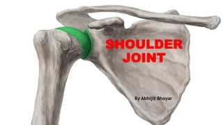
Shoulder joint_nursing.pptx
•Download as PPTX, PDF•
0 likes•149 views

This topic is related to the joints. it is a type of synovial joint. it is a ball and socket type. This is very sensative joint and easy to have fracture to this part.

Recommended
More Related Content
What's hot
What's hot (20)
Similar to Shoulder joint_nursing.pptx
Similar to Shoulder joint_nursing.pptx (20)
More from ABHIJIT BHOYAR
More from ABHIJIT BHOYAR (20)
Recently uploaded
Recently uploaded (20)
Shoulder joint_nursing.pptx
- 2. INTRODUCTION • The shoulder joint is a synovial joint of ball and socket variety. • The shoulder joint (glenohumeral joint) is an articulation between the scapula and the humerus
- 3. Articular Surface • The joint is formed by articulation of the glenoid cavity of scapula and the head of the humerus. Therefore, it is also known as the glenohumeral articulation. • Structurally, it is a weak joint because the glenoid cavity is too small and shallow to hold the head of the humerus in place (the head is four times the size of the glenoid cavity).
- 4. Stability of the joint is maintained by the following factors. The coracoacromial arch or secondary socket for the head of the humerus. The musculotendinous cuff of the shoulder The glenoidal labrum (Latin lip) helps in deepening the glenoid fossa. Stability is also provided by the muscles attaching the humerus to the pectoral girdle, Atmospheric pressure also stabilizes the joint. 1 2 3 4 5
- 5. Ligaments • 1 The capsular ligament: It is very loose and permits free movements. It is least supported inferiorly where dislocations are common. Such a dislocation may damage the closely related axillary nerve. • Medially, the capsule is attached to the scapula beyond the supraglenoid tubercle and the margins of the labrum.
- 6. • Laterally, it is attached to the anatomical neck of the humerus with the following exceptions. – Inferiorly, the attachment extends down to the surgical neck – Superiorly, it is deficient for passage of the tendon of the long head of the biceps brachii
- 7. • Anteriorly, the capsule is reinforced by supplemental bands called the superior, middle and inferior glenohumeral ligaments.
- 8. • The area between the superior and middle glenohumeral ligament is a point of weakness in the capsule (foramen of Weitbrecht) which is a common site of anterior dislocation of humeral head. • The capsule is lined with synovial membrane. • An extension of this membrane forms a tubular sheath for the tendon of the long head of the biceps brachii
- 9. • 2 The coracohumeral ligament: It extends from the root of the coracoid process to the neck of the humerus opposite the greater tubercle. It gives strength to the capsule. • 3 Transverse humeral ligament: It bridges the upper part of the bicipital groove of the humerus (between the greater and lesser tubercles). The tendon of the long head of the biceps brachii passes deep to the ligament. • 4 The glenoidal labrum: It is a fibrocartilaginous rim which covers the margins of the glenoid cavity, thus increasing the depth of the cavity.
- 10. Relations SUPERIORLY: • Coracoacromi al arch, subacromial bursa, supraspinatus and deltoid INFERIORLY: • Long head of the triceps brachii, axillary nerves and posterior circumflex humeral artery. ANTERIORLY: • Subscapularis, coracobrachial is, short head of biceps brachii and deltoid. POSTERIORLY: • Infraspinatus, teres minor and deltoid. WITHIN THE JOINT: • Tendon of the long head of the biceps brachii.
- 12. • Blood Supply • 1 Anterior circumflex humeral vessels • 2 Posterior circumflex humeral vessels • 3 Suprascapular vessels • 4 Subscapular vessels • Nerve Supply • 1 Axillary nerve • 2 Musculocutaneous nerve • 3 Suprascapular nerve
- 13. Movements of Shoulder Joint • Movements of the shoulder joint are considered in relation to the scapula rather than in relation to the sagittal and coronal planes. When the arm is by the side (in the resting position) the glenoid cavity faces almost equally forwards and laterally; and the head of the humerus faces medially and backwards. • Keeping these directions in mind, the movements are analysed as follows.
- 14. 1. Flexion and extension: During flexion, the arm moves forwards and medially, and during extension, the arm moves backwards and laterally. Thus flexion and extension take place in a plane parallel to the surface of the glenoid cavity.
- 15. 2. Abduction and adduction 2. Abduction and adduction take place at right angles to the plane of flexion and extension, i.e. approximately midway between the sagittal and coronal planes. In abduction, the arm moves anterolaterally away from the trunk. This movement is in the same plane as that of the body of the scapula.
- 16. 3 Medial and lateral rotations • 3 Medial and lateral rotations are best demonstrated with a midflexed elbow. In this position, the hand is moved medially across the chest either in front or behind the chest in medial rotation, and laterally in lateral rotation of the shoulder joint
- 17. 4 Circumduction • 4 Circumduction is a combination of different movements as a result of which the hand moves along a circle. The range of any movement depends on the availability of an area of free articular surface on the head of the humerus.
- 19. CLINICAL ANATOMY • Dislocation of joint • Shoulder tip pain • Fracture • Sprain • Frozen shoulder
- 20. • The clavicle may be dislocated at either of its ends. • At the medial end, it is usually dislocated forwards. Backward dislocation is rare as it is prevented by the costoclavicular ligament. • • The main bond of union between the clavicle and the manubrium is the articular disc. Apart from its attachment to the joint capsule, the disc is also attached above to the medial end of the clavicle, and below to the manubrium. This prevents the sternal end of the clavicle from tilting upwards when the weight of the arm depresses the acromial end.
- 21. • The clavicle dislocates upwards at the acromio-clavicular joint, because the clavicle overrides the acromion process. • The weight of the limb is transmitted from the scapula to the clavicle through the coraco-clavicular ligament, and from the clavicle to the sternum through the sternoclavicular joint. Some of the weight also passes to the first rib by the costoclavicular ligament. The clavicle usually fractures between these two ligaments.
- 22. • Dislocation: The shoulder joint is more prone to dislocation than any other joint. This is due to laxity of the capsule and the disproportionate area of the articular surfaces. Dislocation usually occurs when the arm is abducted. In this position, the head of the humerus presses against the lower unsupported part of the capsular ligament. Thus almost always the dislocation is primarily subglenoid. Dislocation endangers the axillary nerve which is closely related to the lower part of the joint capsule. • Optimum attitude: In order to avoid ankylosis, many diseases of the shoulder joint are treated in an optimum position of the joint. In this position, the arm is abducted by 45°–90°.
- 23. Questions ask • Explain shoulder joint under following heads a) Define shoulder joint b) Movements of shoulder joint c) Ligaments of shoulder joints d) Applied anatomy of shoulder joint • Discuss about movements of shoulder joint