
Recommended
More Related Content
What's hot
What's hot (20)
Similar to ANATOMY OF ELBOW JOINT.pptx
Similar to ANATOMY OF ELBOW JOINT.pptx (20)
Recently uploaded
Recently uploaded (20)
ANATOMY OF ELBOW JOINT.pptx
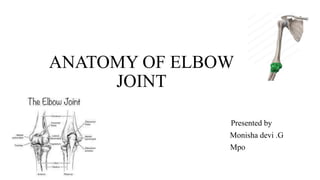
- 1. ANATOMY OF ELBOW JOINT Presented by Monisha devi .G Mpo
- 2. Introduction • The elbow joint is a synovial joint ( HINGE TYPE) found in the upper limb between the arm and the forearm. • It is the point of articulation of three bones: the humerus of the arm and the radius and the ulna of the forearm. • Synovial joints, also called diarthroses, are free movable joints. The articular surfaces of the bones at these joints are separated from each other by a layer of hyaline cartilage. • Smooth movement at these joints is provided by a highly viscous synovial fluid, which acts as a lubricant
- 3. • Synovial joints can be further categorized based on function. The elbow joint is functionally a hinge joint, allowing movement in only one plane (uniaxial).
- 4. Osteology There are three bones that comprise the elbow joint: • the humerus • the radius • the ulna These bones give rise to two joints: • Humeroulnar joint • Humeroradial joint
- 5. Joints • Humeroulnar joint is the joint between the trochlea on the medial aspect of the distal end of the humerus and the trochlear notch on the proximal ulna. • Humeroradial joint is the joint between the capitulum on the lateral aspect of the distal end of the humerus with the head of the radius.
- 6. • The humeroulnar and the humeroradial joints are the joints that give the elbow its characteristic hinge like properties. • The rounded surfaces of the trochlea and capitulum of the humerus rotate against the concave surfaces of the trochlear notch of the ulna and head of the radius. • At the elbow joint, the proximal ends of the radius and ulna articulate with each other at the proximal radioulnar joint
- 7. • This joint, however, is considered to be a separate articulation than those forming the elbow joint itself. • The proximal radioulnar joint is the articulation between the circumferential head of the radius and a fibro-osseous ring formed by the radial groove of the ulna and the annular ligament that hold the head of the radius in this groove. • The proximal radioulnar joint is functionally a pivot joint, allowing a rotational movement of the radius on the ulna.
- 8. Articulating Surfaces The elbow joint consists of two separate articulations: • Trochlear notch of the ulna and the trochlea of the humerus • Head of the radius and the capitulum of the humerus
- 9. • Here are is a mnemonic that can help you remember the articulations involved in the elbow joint. • CRAzy TULips • Capitulum = RAdius (capitulum of the humerus articulates with the head of radius) • Trochlea = ULnar (the trochlea of the humerus articulates with the trochlear notch of the ulna)
- 10. Joint Capsule and Bursae • Like all synovial joints, the elbow joint has a capsule enclosing the joint. • This in itself is strong and fibrous, strengthening the joint. • The joint capsule is thickened medially and laterally to form collateral ligaments, which stabilise the flexing and extending motion of the arm.
- 11. A bursa is a membranous sac filled with synovial fluid. It acts as a cushion to reduce friction between the moving parts of a joint, limiting degenerative damage. There are many bursae in the elbow, but only a few have clinical importance: • Intratendinous – located within the tendon of the triceps brachii. • Subtendinous – between the olecranon and the tendon of the triceps brachii, reducing friction between the two structures during extension and flexion of the arm. • Subcutaneous (olecranon) bursa – between the olecranon and the overlying connective tissue (implicated in olecranon bursitis).
- 12. Ligaments of the elbow joint • There are a collection of ligaments that connect the bones forming the elbow joint to each other, contributing to the stability of the joint. • The humeroulnar and the humeroradial joints each have a ligament connecting the two bones involved at the articulation: • the ulnar collateral ligaments • the radial collateral ligaments
- 13. ulnar collateral ligament • The ulnar collateral ligament extends from the medial epicondyle of the humerus to the coronoid process of the ulna. • It is triangular in shape, and is composed of three parts: an anterior, a posterior and an inferior band.
- 14. The radial collateral ligament • The radial collateral ligament has a low attachment to the lateral epicondyle of the humerus. • The distal fibers blend with the annular ligament that encloses the head of the radius, as well as with the fibers of the supinator and the extensor carpi radialis brevis muscles
- 15. • The annular ligament also reinforces the joint by holding the radius and ulna together at their proximal articulation. • The quadrate ligament is also present at this joint, and maintains constant tension during pronation and supination movements of the forearm.
- 16. Blood supply and innervation • Proximal to elbow joint - Ulnar collateral artery, radial collateral artery, middle collateral artery • Distal to elbow joint - Radial recurrent artery, ulnar recurrent artery
- 17. Nerve supply • Radial nerve • Ulnar nerve • Medial nerve
- 18. Movements • As the elbow joint is a hinge joint, movement is in only one plane. The movements at the elbow joint involve movement of the forearm at the elbow joint. • Flexion of the forearm at the elbow joint involves decreasing the angle between the forearm and the arm at the elbow joint. • Extension involves increasing the angle between the arm and forearm. These movements are performed by two groups of muscles in the arm: the anterior compartment and the posterior compartment of the arm.
- 19. Flexion • Most of the muscles producing flexion are found in the anterior compartment of the arm. There are two muscles in this compartment that produce flexion at the elbow joint: Biceps Brachii Brachialis
- 20. Biceps Brachii originates as two heads. • The tendon of the long head originates from the supraglenoid tubercle of the scapula. It passes through the joint capsule of the shoulder joint and through the bicipital groove on the anterior surface of the humerus. • The short head of the biceps brachii muscle originates from the coracoid process of the scapula. These heads join together to form the biceps brachii muscle belly. • The muscle inserts via a single tendon onto the radial tuberosity distal to the elbow joint. In the forearm, there is a continuation of this tendon as a flattened connective tissue sheath, the bicipital aponeurosis. • This aponeurosis blends with the deep fascia in the anterior forearm.
- 22. • Brachialis originates from the distal half of the anterior surface of the humerus, as well as from the intermuscular septa on either side of the anterior compartment. • It is located deep to the biceps brachii muscle. It forms a singular tendon that inserts onto the tuberosity of the ulna.
- 23. • Both the biceps brachii and brachialis muscles are innervated by the Musculocutaneous nerve. • While the biceps brachii and the brachialis muscles are the main flexors of the elbow joint, the brachioradialis muscle is also involved in the flexion of the forearm at this joint. • Brachioradialis originates from the lateral aspect of the distal humerus above the lateral epicondyle. • It inserts onto the lateral aspect of the distal radius. Although this muscle is primarily in the forearm, it crosses the elbow joint so therefore it acts on the elbow joint. It is innervated by the radial nerve.
- 24. • Mnemonic • Learning the muscles that bend the elbow becomes child's play if you anchor them to a mnemonic like the one below. 3 B's bend the elbow Biceps Brachialis Brachioradialis
- 25. Extension • Extension of the forearm at the elbow joint is the increase of the angle at the elbow to bring the forearm back to the anatomical position from a flexed position. • There is one muscle involved in extension, the triceps brachii muscle. It is the only muscle in the posterior compartment of the arm.
- 26. • Triceps Brachii originates as three heads. • The long head originates from the infra glenoid tubercle of the scapula, the lateral head originates from the lateral aspect of the humerus above the radial groove, and the medial head originates from the medial aspect of the humerus below the level of the radial groove. • The three heads converge on a single tendon that inserts onto the olecranon of the ulna. It is supplied by the radial nerve, which passes down through the arm in the radial groove between the lateral and medial heads of the muscle
- 27. • While flexion and extension are the only movements that can occur at the elbow joint itself, movement is also afforded at the proximal radioulnar joint, which contributes to the elbow joint. • Movements at this joint are called pronation and supination. • These are rotational movements that occur when the distal end of the radius moves over the distal end of the ulna by rotating the radius in the pivot joint formed by the circular head of the radius, the radial groove of the ulna and the annular ligament.
- 28. • Pronation and supination are easily visualized when the elbow is flexed at 90°. • Supination is where the palm of the hand is facing upwards; pronation is the rotation of the forearm so that the palm is facing downwards. • In the anatomical position, the forearm is in the supine position. Pronation in the anatomical position is movement of the forearm so that the palm is facing posteriorly.
- 29. Carrying angle • Normal alignment of elbow is determined by measuring the normal carrying angle Normal carrying angle 5-15degree More in female than in males
- 30. Thank you