Shoulder Joint by Thirumurugan professor.docx

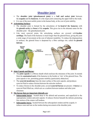
Shoulder joint • The shoulder joint (glenohumeral joint) is a ball and socket joint between the scapula and the humerus. It is the major joint connecting the upper limb to the trunk. • It is one of the most mobile joints in the human body, at the cost of joint stability. Articulating Surfaces • The shoulder joint is formed by the articulation of the head of the humerus with the glenoid cavity (or fossa) of the scapula. This gives rise to the alternate name for the shoulder joint – the glenohumeral joint. • Like most synovial joints, the articulating surfaces are covered with hyaline cartilage. The head of the humerus is much larger than the glenoid fossa, giving the joint a wide range of movement at the cost of inherent instability. To reduce the disproportion in surfaces, the glenoid fossa is deepened by a fibro cartilage rim, called the glenoid labrum. Joint Capsule and Bursae: • The joint capsule is a fibrous sheath which encloses the structures of the joint. It extends from the anatomical neck of the humerus to the border or ‘rim’ of the glenoid fossa. The joint capsule is lax, permitting greater mobility (particularly abduction). • The synovial membrane lines the inner surface of the joint capsule, and produces synovial fluid to reduce friction between the articular surfaces. • To reduce friction in the shoulder joint, several synovial bursae are present. A bursa is a synovial fluid filled sac, which acts as a cushion between tendons and other joint structures. The bursae that are important clinically are: • Subacromial bursa: located deep to the deltoid and acromion, and superficial to the supraspinatus tendon and joint capsule. It reduces friction beneath the deltoid, promoting free motion of the rotator cuff tendons. • Subscapular bursa: located between the subscapularis tendon and the scapula. It reduces wear and tear on the tendon during movement at the shoulder joint. • Subcoracoid bursa: It is located anterior to the subscapularis muscle and inferior to the coracoid process. Its function is to reduce friction & facilitating internal and external rotation of the shoulder Ligaments: In the shoulder joint, the ligaments play a key role in stabilizing the bony structures. • Glenohumeral ligaments (superior, middle and inferior): the joint capsule is formed by this group of ligaments connecting the humerus to the glenoid fossa. They are the main source of stability for the shoulder, holding it in place and preventing it from dislocating anteriorly. They act to stabilize the anterior aspect of the joint. • Coracohumeral ligament: attaches the base of the coracoid process to the greater tubercle of the humerus. It supports the superior part of the joint capsule. • Transverse humeral ligament: spans the distance between the two tubercles of the humerus. It holds the tendon of the long head of the biceps in the intertubercular groove. • Coraco–clavicular ligament: composed of the trapezoid and conoid ligaments

Recommended
More Related Content
Similar to Shoulder Joint by Thirumurugan professor.docx
Similar to Shoulder Joint by Thirumurugan professor.docx (20)
More from thiru murugan
More from thiru murugan (20)
Recently uploaded
Recently uploaded (20)
Shoulder Joint by Thirumurugan professor.docx
- 1. Page 1 of 3 Shoulder joint • The shoulder joint (glenohumeral joint) is a ball and socket joint between the scapula and the humerus. It is the major joint connecting the upper limb to the trunk. • It is one of the most mobile joints in the human body, at the cost of joint stability. Articulating Surfaces • The shoulder joint is formed by the articulation of the head of the humerus with the glenoid cavity (or fossa) of the scapula. This gives rise to the alternate name for the shoulder joint – the glenohumeral joint. • Like most synovial joints, the articulating surfaces are covered with hyaline cartilage. The head of the humerus is much larger than the glenoid fossa, giving the joint a wide range of movement at the cost of inherent instability. To reduce the disproportion in surfaces, the glenoid fossa is deepened by a fibro cartilage rim, called the glenoid labrum. Joint Capsule and Bursae: • The joint capsule is a fibrous sheath which encloses the structures of the joint. It extends from the anatomical neck of the humerus to the border or ‘rim’ of the glenoid fossa. The joint capsule is lax, permitting greater mobility (particularly abduction). • The synovial membrane lines the inner surface of the joint capsule, and produces synovial fluid to reduce friction between the articular surfaces. • To reduce friction in the shoulder joint, several synovial bursae are present. A bursa is a synovial fluid filled sac, which acts as a cushion between tendons and other joint structures. The bursae that are important clinically are: • Subacromial bursa: located deep to the deltoid and acromion, and superficial to the supraspinatus tendon and joint capsule. It reduces friction beneath the deltoid, promoting free motion of the rotator cuff tendons. • Subscapular bursa: located between the subscapularis tendon and the scapula. It reduces wear and tear on the tendon during movement at the shoulder joint.
- 2. Page 2 of 3 • Subcoracoid bursa: It is located anterior to the subscapularis muscle and inferior to the coracoid process. Its function is to reduce friction & facilitating internal and external rotation of the shoulder Ligaments: In the shoulder joint, the ligaments play a key role in stabilizing the bony structures. • Glenohumeral ligaments (superior, middle and inferior): the joint capsule is formed by this group of ligaments connecting the humerus to the glenoid fossa. They are the main source of stability for the shoulder, holding it in place and preventing it from dislocating anteriorly. They act to stabilize the anterior aspect of the joint. • Coracohumeral ligament: attaches the base of the coracoid process to the greater tubercle of the humerus. It supports the superior part of the joint capsule. • Transverse humeral ligament: spans the distance between the two tubercles of the humerus. It holds the tendon of the long head of the biceps in the intertubercular groove. • Coraco–clavicular ligament: composed of the trapezoid and conoid ligaments and runs from the clavicle to the coracoid process of the scapula. They work is to maintain the alignment of the clavicle in relation to the scapula. • The other major ligament is the coracoacromial ligament. Running between the acromion and coracoid process of the scapula it forms the coraco-acromial arch.
- 3. Page 3 of 3 Muscles Acting on Shoulder Joint: • Above - Supraspinatus • Below - Long head of Triceps • Front - Subscapularis • Behind - Infraspinatus and Teres Minor • Deltoid is placed most externally and covers the articulation from its outer side, as well as in front and behind. Movements: As a ball and socket synovial joint - wide range of movement permitted: • Extension • Flexion • Abduction • Adduction Blood Supply: Anterior circumflex humeral vessels, Posterior circumflex humeral vessels, Suprascapular vessels Nerve supply: Axillary nerve, Musculocutaneous nerve, Suprascapular Nerve & Lateral pectoral nerve • Internal rotation. • External rotation • Circumduction