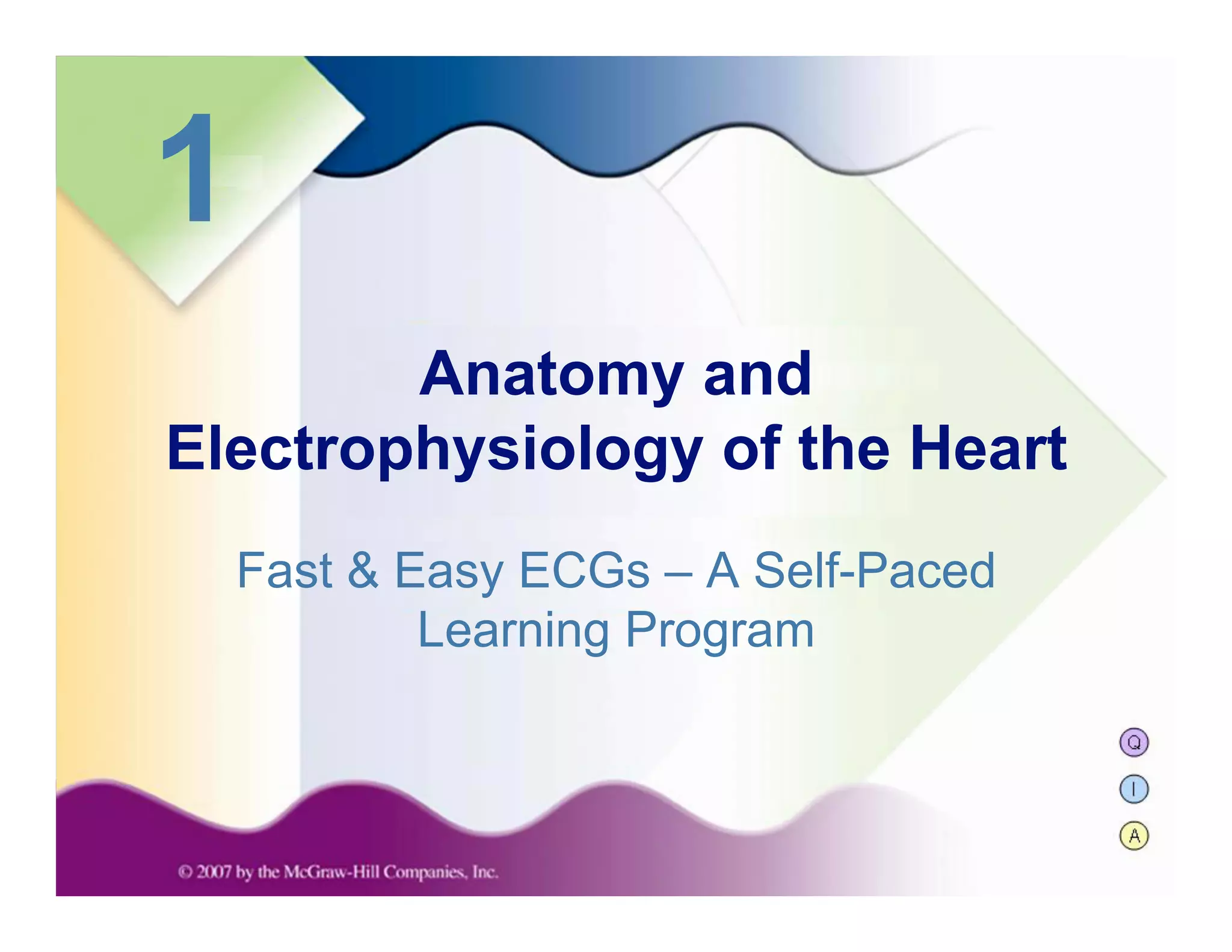
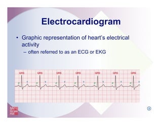
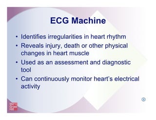
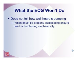
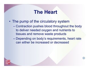
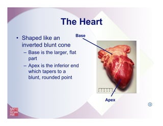
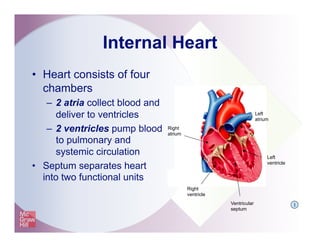
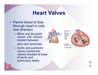
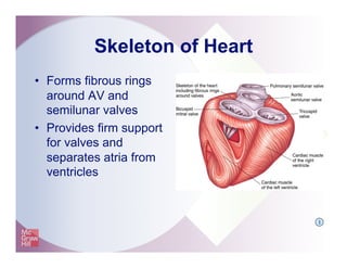
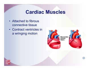
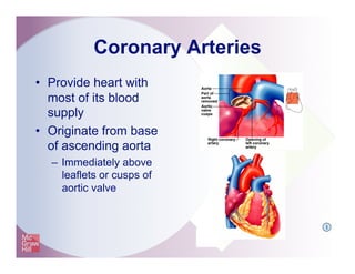
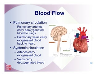
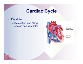
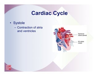
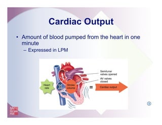
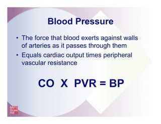
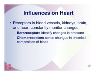
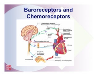
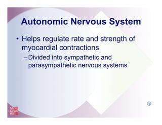
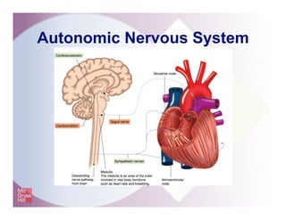
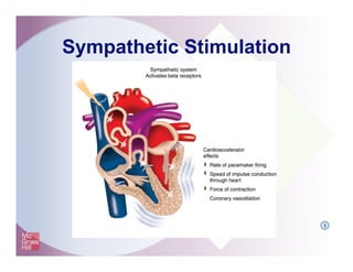
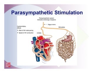
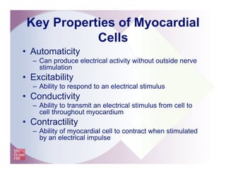
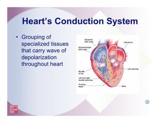
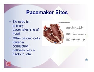
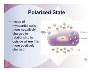
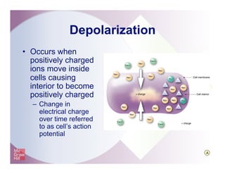
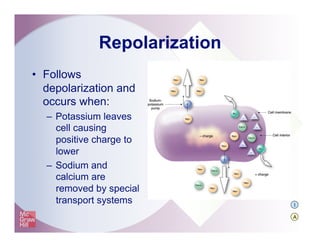
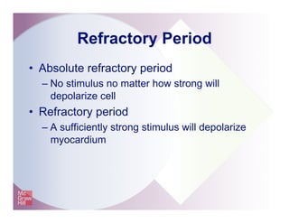
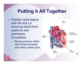
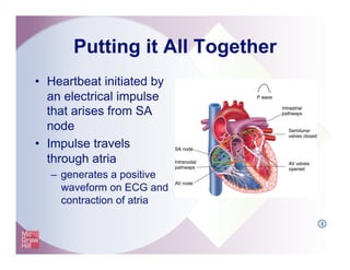
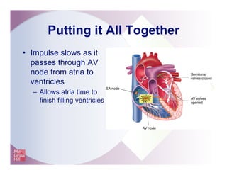
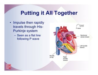
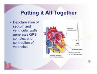
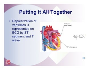
Q
I
A
1
Fast & Easy ECGs – A Self-Paced
Learning Program
Anatomy and
Electrophysiology of the Heart
Electrocardiogram
•  Graphic representation of heart’s electrical
activity
–  often referred to as an ECG or EKG
I
ECG Machine
•  Detects heart’s
electrical current
activity
–  Displays it on a screen
or prints it onto graph
paper
I
ECG Machine
•  Identifies irregularities in heart rhythm
•  Reveals injury, death or other physical
changes in heart muscle
•  Used as an assessment and diagnostic
tool
•  Can continuously monitor heart’s electrical
activity
I
What the ECG Won’t Do
•  Does not tell how well heart is pumping
– Patient must be properly assessed to ensure
heart is functioning mechanically
I
The Heart
•  The pump of the circulatory system
– Contraction pushes blood throughout the body
to deliver needed oxygen and nutrients to
tissues and remove waste products
– Depending on body’s requirements, heart rate
can either be increased or decreased
I
The Heart
•  Shaped like an
inverted blunt cone
–  Base is the larger, flat
part
–  Apex is the inferior end
which tapers to a
blunt, rounded point
Apex
Base
I
The Heart
•  Located between
the two lungs in
mediastinum
behind the
sternum
The Heart
•  Anterior-posterior
orientation in the
chest
–  RV closer to
front of left chest
–  LV closer to
side of left chest
The Heart
•  Surrounded by
pericardial sac (a
double-walled closed
sac)
–  fibrous pericardium
–  serous pericardium
I
Heart Wall
•  Made up of three layers
–  Epicardium (outermost)
–  Myocardium (middle)
–  Endocardium (innermost)
I
Heart Cells
•  Myocardial cells (working cells)
– Contract to propel blood out of heart’s
chambers
•  Electrical conduction system cells
– Initiate and carry impulses throughout heart
I
Myocardial Cells
•  Cylindrical and
branching at their ends
–  Intercalated disks and
gap junctions allow rapid
movement of electrical
impulses from one cell to
another
–  Desmosomes hold cells
together when heart
muscle contracts
I
Working Cells
•  Myocytes
–  Enclosed in
sarcolemma
–  Composed of two
protein filaments
•  Actin (thin)
•  Myosin (thick)
I
A
Internal Heart
•  Heart consists of four
chambers
–  2 atria collect blood and
deliver to ventricles
–  2 ventricles pump blood
to pulmonary and
systemic circulation
•  Septum separates heart
into two functional units
Left
ventricle
Right
ventricle
Ventricular
septum
Right
atrium
Left
atrium
I
Heart Valves
•  Permit blood to flow
through heart in only
one direction
–  Mitral and bicuspid
valves (AV valves)
located between
atria and ventricles
–  Aortic and pulmonic
valves (semilunar
valves) located at base
of aorta and
pulmonary artery I
A
Skeleton of Heart
•  Forms fibrous rings
around AV and
semilunar valves
•  Provides firm support
for valves and
separates atria from
ventricles
I
Cardiac Muscles
•  Attached to fibrous
connective tissue
•  Contract ventricles in
a wringing motion
I
Coronary Arteries
•  Provide heart with
most of its blood
supply
•  Originate from base
of ascending aorta
–  Immediately above
leaflets or cusps of
aortic valve
I
Blood Flow
•  Pulmonary circulation
–  Pulmonary arteries
carry deoxygenated
blood to lungs
–  Pulmonary veins carry
oxygenated blood
back to heart
•  Systemic circulation
–  Arteries carry
oxygenated blood
–  Veins carry
deoxygenated blood I
Cardiac Cycle
•  Diastole
–  Relaxation and filling
of atria and ventricles
Cardiac Cycle
•  Systole
–  Contraction of atria
and ventricles
Cardiac Output
•  Amount of blood pumped from the heart in one
minute
–  Expressed in LPM
I
Blood Pressure
•  The force that blood exerts against walls
of arteries as it passes through them
•  Equals cardiac output times peripheral
vascular resistance
CO X PVR = BP
Influences on Heart
•  Receptors in blood vessels, kidneys, brain,
and heart constantly monitor changes
– Baroreceptors identify changes in pressure
– Chemoreceptors sense changes in chemical
composition of blood
A
Baroreceptors and
Chemoreceptors
Autonomic Nervous System
•  Helps regulate rate and strength of
myocardial contractions
– Divided into sympathetic and
parasympathetic nervous systems
I
Autonomic Nervous System
Sympathetic Stimulation
I
Parasympathetic Stimulation
Key Properties of Myocardial
Cells
•  Automaticity
–  Can produce electrical activity without outside nerve
stimulation
•  Excitability
–  Ability to respond to an electrical stimulus
•  Conductivity
–  Ability to transmit an electrical stimulus from cell to
cell throughout myocardium
•  Contractility
–  Ability of myocardial cell to contract when stimulated
by an electrical impulse
Heart’s Conduction System
•  Grouping of
specialized tissues
that carry wave of
depolarization
throughout heart
I
Pacemaker Sites
•  SA node is
primary
pacemaker site of
heart
•  Other cardiac cells
lower in
conduction
pathway play a
back-up role
I
Polarized State
•  Inside of
myocardial cells
more negatively
charged in
relationship to
outside where it is
more positively
charged
I
Depolarization
•  Occurs when
positively charged
ions move inside
cells causing
interior to become
positively charged
–  Change in
electrical charge
over time referred
to as cell’s action
potential
A
Repolarization
•  Follows
depolarization and
occurs when:
–  Potassium leaves
cell causing
positive charge to
lower
–  Sodium and
calcium are
removed by special
transport systems I
A
Refractory Period
•  Absolute refractory period
– No stimulus no matter how strong will
depolarize cell
•  Refractory period
– A sufficiently strong stimulus will depolarize
myocardium
Putting it All Together
•  Cardiac cycle begins
with RA and LA
receiving blood from
systemic and
pulmonary
circulations
–  Rising pressure within
atria forces tricuspid
and mitral valves open
I
Putting it All Together
•  Heartbeat initiated by
an electrical impulse
that arises from SA
node
•  Impulse travels
through atria
–  generates a positive
waveform on ECG and
contraction of atria
I
Putting it All Together
•  Impulse slows as it
passes through AV
node from atria to
ventricles
–  Allows atria time to
finish filling ventricles
Putting it All Together
•  Impulse then rapidly
travels through His-
Purkinje system
–  Seen as a flat line
following P wave
Putting it All Together
•  Depolarization of
septum and
ventricular walls
generates QRS
complex and
contraction of
ventricles
I
Putting it All Together
•  Repolarization of
ventricles is
represented on
ECG by ST
segment and T
wave
I
Summary
•  Electrocardiogram detects electrical activity occurring in
heart.
•  Nerve impulses stimulate cardiac muscles to contract.
•  Heart consists of two upper chambers, the atria and two
lower chambers, the ventricles.
•  Heart is separated into right and left sides by the septum.
•  Coronary arteries perfuse myocardium during diastole.
Summary
•  Cardiac output is amount of blood pumped through
circulatory system in one minute.
•  Rate and strength of myocardial contractions can be
influenced by autonomic nervous system.
–  Two divisions are the sympathetic and parasympathetic nervous
systems.
Summary
•  Sodium, calcium and potassium are key electrolytes
responsible for initiating electrical charges.
•  Depolarization of cells occurs when positive electrolytes
move from outside to inside cell causing it to become
more positively charged.
•  Depolarization of myocardial cells causes calcium to be
released and come into close proximity with actin and
myosin filaments of muscle fibers leading to myocardial
contraction.
Summary
•  Myocardial depolarization progresses from atria to
ventricles in an orderly fashion.
–  Electrical stimulus causes heart muscle to contract.
•  Electrical impulse that initiates heartbeat arises from SA
node.
•  From there it travels through atria generating a positive
waveform on ECG and contraction of atria.
•  Impulse is slowed as it passes from atria to ventricles
through AV node.
Summary
•  On ECG impulse traveling through His-Purkinje system
is seen as a flat line following the P wave.
•  QRS complex is generated and ventricles contract as a
result of electrical impulse stimulating ventricles.
•  ST segment and T wave represents repolarization of
ventricles.
–  Atrial repolarization occurs but is hidden by QRS complex.
•  Other sites in heart can assume control by discharging
impulses faster than SA node or stepping in when SA
node fails.
I

Shadechapter01.ppt [read only]

  • 1.
    Q I A 1 Fast & EasyECGs – A Self-Paced Learning Program Anatomy and Electrophysiology of the Heart
  • 2.
    Electrocardiogram •  Graphic representationof heart’s electrical activity –  often referred to as an ECG or EKG I
  • 3.
    ECG Machine •  Detectsheart’s electrical current activity –  Displays it on a screen or prints it onto graph paper I
  • 4.
    ECG Machine •  Identifiesirregularities in heart rhythm •  Reveals injury, death or other physical changes in heart muscle •  Used as an assessment and diagnostic tool •  Can continuously monitor heart’s electrical activity I
  • 5.
    What the ECGWon’t Do •  Does not tell how well heart is pumping – Patient must be properly assessed to ensure heart is functioning mechanically I
  • 6.
    The Heart •  Thepump of the circulatory system – Contraction pushes blood throughout the body to deliver needed oxygen and nutrients to tissues and remove waste products – Depending on body’s requirements, heart rate can either be increased or decreased I
  • 7.
    The Heart •  Shapedlike an inverted blunt cone –  Base is the larger, flat part –  Apex is the inferior end which tapers to a blunt, rounded point Apex Base I
  • 8.
    The Heart •  Locatedbetween the two lungs in mediastinum behind the sternum
  • 9.
    The Heart •  Anterior-posterior orientationin the chest –  RV closer to front of left chest –  LV closer to side of left chest
  • 10.
    The Heart •  Surroundedby pericardial sac (a double-walled closed sac) –  fibrous pericardium –  serous pericardium I
  • 11.
    Heart Wall •  Madeup of three layers –  Epicardium (outermost) –  Myocardium (middle) –  Endocardium (innermost) I
  • 12.
    Heart Cells •  Myocardialcells (working cells) – Contract to propel blood out of heart’s chambers •  Electrical conduction system cells – Initiate and carry impulses throughout heart I
  • 13.
    Myocardial Cells •  Cylindricaland branching at their ends –  Intercalated disks and gap junctions allow rapid movement of electrical impulses from one cell to another –  Desmosomes hold cells together when heart muscle contracts I
  • 14.
    Working Cells •  Myocytes – Enclosed in sarcolemma –  Composed of two protein filaments •  Actin (thin) •  Myosin (thick) I A
  • 15.
    Internal Heart •  Heartconsists of four chambers –  2 atria collect blood and deliver to ventricles –  2 ventricles pump blood to pulmonary and systemic circulation •  Septum separates heart into two functional units Left ventricle Right ventricle Ventricular septum Right atrium Left atrium I
  • 16.
    Heart Valves •  Permitblood to flow through heart in only one direction –  Mitral and bicuspid valves (AV valves) located between atria and ventricles –  Aortic and pulmonic valves (semilunar valves) located at base of aorta and pulmonary artery I A
  • 17.
    Skeleton of Heart • Forms fibrous rings around AV and semilunar valves •  Provides firm support for valves and separates atria from ventricles I
  • 18.
    Cardiac Muscles •  Attachedto fibrous connective tissue •  Contract ventricles in a wringing motion I
  • 19.
    Coronary Arteries •  Provideheart with most of its blood supply •  Originate from base of ascending aorta –  Immediately above leaflets or cusps of aortic valve I
  • 20.
    Blood Flow •  Pulmonarycirculation –  Pulmonary arteries carry deoxygenated blood to lungs –  Pulmonary veins carry oxygenated blood back to heart •  Systemic circulation –  Arteries carry oxygenated blood –  Veins carry deoxygenated blood I
  • 21.
    Cardiac Cycle •  Diastole – Relaxation and filling of atria and ventricles
  • 22.
    Cardiac Cycle •  Systole – Contraction of atria and ventricles
  • 23.
    Cardiac Output •  Amountof blood pumped from the heart in one minute –  Expressed in LPM I
  • 24.
    Blood Pressure •  Theforce that blood exerts against walls of arteries as it passes through them •  Equals cardiac output times peripheral vascular resistance CO X PVR = BP
  • 25.
    Influences on Heart • Receptors in blood vessels, kidneys, brain, and heart constantly monitor changes – Baroreceptors identify changes in pressure – Chemoreceptors sense changes in chemical composition of blood A
  • 26.
  • 27.
    Autonomic Nervous System • Helps regulate rate and strength of myocardial contractions – Divided into sympathetic and parasympathetic nervous systems I
  • 28.
  • 29.
  • 30.
  • 31.
    Key Properties ofMyocardial Cells •  Automaticity –  Can produce electrical activity without outside nerve stimulation •  Excitability –  Ability to respond to an electrical stimulus •  Conductivity –  Ability to transmit an electrical stimulus from cell to cell throughout myocardium •  Contractility –  Ability of myocardial cell to contract when stimulated by an electrical impulse
  • 32.
    Heart’s Conduction System • Grouping of specialized tissues that carry wave of depolarization throughout heart I
  • 33.
    Pacemaker Sites •  SAnode is primary pacemaker site of heart •  Other cardiac cells lower in conduction pathway play a back-up role I
  • 34.
    Polarized State •  Insideof myocardial cells more negatively charged in relationship to outside where it is more positively charged I
  • 35.
    Depolarization •  Occurs when positivelycharged ions move inside cells causing interior to become positively charged –  Change in electrical charge over time referred to as cell’s action potential A
  • 36.
    Repolarization •  Follows depolarization and occurswhen: –  Potassium leaves cell causing positive charge to lower –  Sodium and calcium are removed by special transport systems I A
  • 37.
    Refractory Period •  Absoluterefractory period – No stimulus no matter how strong will depolarize cell •  Refractory period – A sufficiently strong stimulus will depolarize myocardium
  • 38.
    Putting it AllTogether •  Cardiac cycle begins with RA and LA receiving blood from systemic and pulmonary circulations –  Rising pressure within atria forces tricuspid and mitral valves open I
  • 39.
    Putting it AllTogether •  Heartbeat initiated by an electrical impulse that arises from SA node •  Impulse travels through atria –  generates a positive waveform on ECG and contraction of atria I
  • 40.
    Putting it AllTogether •  Impulse slows as it passes through AV node from atria to ventricles –  Allows atria time to finish filling ventricles
  • 41.
    Putting it AllTogether •  Impulse then rapidly travels through His- Purkinje system –  Seen as a flat line following P wave
  • 42.
    Putting it AllTogether •  Depolarization of septum and ventricular walls generates QRS complex and contraction of ventricles I
  • 43.
    Putting it AllTogether •  Repolarization of ventricles is represented on ECG by ST segment and T wave I
  • 44.
    Summary •  Electrocardiogram detectselectrical activity occurring in heart. •  Nerve impulses stimulate cardiac muscles to contract. •  Heart consists of two upper chambers, the atria and two lower chambers, the ventricles. •  Heart is separated into right and left sides by the septum. •  Coronary arteries perfuse myocardium during diastole.
  • 45.
    Summary •  Cardiac outputis amount of blood pumped through circulatory system in one minute. •  Rate and strength of myocardial contractions can be influenced by autonomic nervous system. –  Two divisions are the sympathetic and parasympathetic nervous systems.
  • 46.
    Summary •  Sodium, calciumand potassium are key electrolytes responsible for initiating electrical charges. •  Depolarization of cells occurs when positive electrolytes move from outside to inside cell causing it to become more positively charged. •  Depolarization of myocardial cells causes calcium to be released and come into close proximity with actin and myosin filaments of muscle fibers leading to myocardial contraction.
  • 47.
    Summary •  Myocardial depolarizationprogresses from atria to ventricles in an orderly fashion. –  Electrical stimulus causes heart muscle to contract. •  Electrical impulse that initiates heartbeat arises from SA node. •  From there it travels through atria generating a positive waveform on ECG and contraction of atria. •  Impulse is slowed as it passes from atria to ventricles through AV node.
  • 48.
    Summary •  On ECGimpulse traveling through His-Purkinje system is seen as a flat line following the P wave. •  QRS complex is generated and ventricles contract as a result of electrical impulse stimulating ventricles. •  ST segment and T wave represents repolarization of ventricles. –  Atrial repolarization occurs but is hidden by QRS complex. •  Other sites in heart can assume control by discharging impulses faster than SA node or stepping in when SA node fails. I