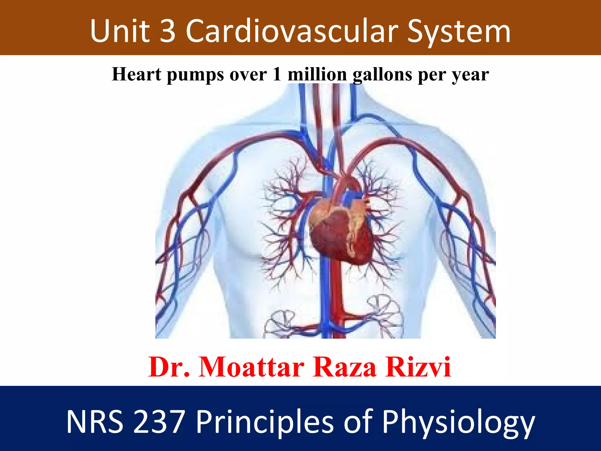
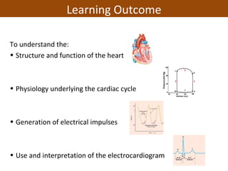
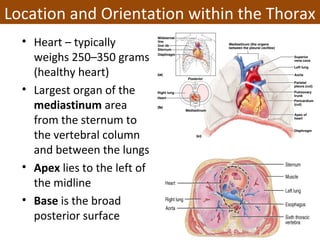
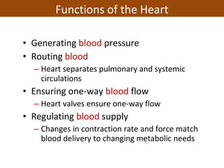
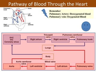
NRS 237 Principles of Physiology
Dr. Moattar Raza Rizvi
Unit 3 Cardiovascular System
Heart pumps over 1 million gallons per year
To understand the:
• Structure and function of the heart
• Physiology underlying the cardiac cycle
• Generation of electrical impulses
• Use and interpretation of the electrocardiogram
Learning Outcome
• Heart – typically
weighs 250–350 grams
(healthy heart)
• Largest organ of the
mediastinum area
from the sternum to
the vertebral column
and between the lungs
• Apex lies to the left of
the midline
• Base is the broad
posterior surface
Location and Orientation within the Thorax
• Generating blood pressure
• Routing blood
– Heart separates pulmonary and systemic
circulations
• Ensuring one-way blood flow
– Heart valves ensure one-way flow
• Regulating blood supply
– Changes in contraction rate and force match
blood delivery to changing metabolic needs
Functions of the Heart
The Double Pump
Lungs
Body cells
THE RIGHT SIDE
OF THE SYSTEM
DEALS WITH
DEOXYGENATED
BLOOD.
THE LEFT SIDE
OF THE SYSTEM
DEALS WITH
OXYGENATED
BLOOD.
The Double Pump
• Protects and anchors the heart
• Prevents overfilling of the heart with blood
• Allows the heart to work in a relatively friction-
free environment
Membrane of Heart: Pericardium
1. Endocardium:
The innermost layer of the heart (endothelial cells)
2. Myocardium:
The thickest main layer, consists of cardiac muscle
Heart Walls 3 Distinct Layers
3. Epicardium:
• The thin, outer covering
around the heart
• Made up of simple
squamous epithelium
Epicardium
Atria- (2) upper chambers
– Thin walled
– Receive blood from
veins
– Send blood to ventricles
Ventricles- (2) lower chambers
– Thick walled
– Receive blood from atria
– Pump blood out through
arteries
Heart Chambers
Heart Chambers
• Both ventricles (Right & left) – thick walled
• Left ventricle – three
times thicker than right
– Left ventricle has to
push the blood to all
the body parts while RV
has to push the blood
to closely lying lungs
only
– Flattens right ventricle
into a crescent shape
Ventricles
• Chordae tendinease
– “Heart strings”
– Cord-like tendons
– Connect papillary
muscles to tricuspid
and mitral valves
– Prevent inversion
of valve
• Papillary muscles
– Small muscles that
anchor the cords
Papillary
muscle
Structures of the Heart
Valves of the Heart
• Semilunar Valves
– Prevent backflow into
ventricles
– At origin of pulmonary
artery & aorta.
– Pulmonary (Right) &
Aortic (Left).
• Atrioventricular Valves
• Allow blood to flow from atria into ventricles.
• Tricuspid (Right) & Mitral (Left).
– Prevent backflow to the atria
– Prolapse is prevented by the chordae tendinae
– Tensioned by the papillary muscles
Function is to prevent backflow of blood
Atrioventricular and Semilunar Valves
• Oxygen-poor blood draining
from the body through
veins into the superior and
inferior vena cava flows to
the right atrium, through
the tricuspid valve, and into
the right ventricle.
• As the right ventricle
contracts, oxygen-poor
blood passes through the
pulmonary valve into the
pulmonary arteries and on
to the lungs to receive
oxygen.
Pathway of Blood Through the Heart
• Oxygen-rich blood from the
lungs enters the heart
through the pulmonary
veins, passing into the left
atrium.
• Then through the mitral
valve to the left ventricle.
Contraction of the left
ventricle forces blood
through the aortic valve into
the aorta.
• Various arteries branch off
from the aorta to supply
blood to all parts of the body.
Pathway of Blood Through the Heart
Remember
Pulmonary Artery: Deoxygenated blood
Pulmonary vein: Oxygenated Blood
• Coronary circulation is blood supply to the heart
• Heart as a very active muscle needs lots of O2
• When the heart relaxes high pressure of blood in
aorta pushes blood into coronary vessels
• Many anastomoses
– connections between arteries supplying blood to the
same region, provide alternate routes if one artery
becomes occluded
Coronary Circulation
Coronary Artery: 2 main coronary arteries, the left and right
coronary arteries, and these branch further to form several
major branches.
Coronary Veins: Collects wastes from cardiac muscle and
drains into a large sinus on posterior surface of heart called
the coronary sinus and coronary sinus empties into right
atrium
Coronary Circulation
• Pulmonary circulation:
– Path of blood from right ventricle through the lungs and back to the
heart.
• Systemic circulation:
– Oxygen-rich blood pumped to all organ systems to supply nutrients.
• Rate of blood flow through
systemic circulation = flow
rate through pulmonary
circulation.
Pulmonary and Systemic Circulations
The Pulmonary and Systemic Circuits ()
Systemic circuit
–Longer than pulmonary
circuit
–Offers greater
resistance to blood flow
1. Pulmonary circuit carries CO2-rich blood from the heart to
the gas-exchange surfaces of the lungs and returns O2-rich
blood back to the heart
2. Systemic circuit transports O2-
rich blood from the heart to the
rest of the body’s cells, returning
CO2-rich blood back to the heart
• To pump effectively, large portions of cardiac muscle
must receive an action potential nearly
simultaneously.
• Cardiac muscle tissue has intrinsic ability to
– Generate and conduct impulses ()
– Signal these cells to contract rhythmically
• Inherent and rhythmical beat is due to autorhythmic
fibers of the cardiac muscle.
• These fibers have 2 important function
- Act as pace maker (Nodal cells)
- Form the conduction system
Conducting System ()
• Specialized muscle cells (autorhythmic cells) conduct APs to time and
synchronize the action of the chambers
• SA node -PACEMAKER, spontaneously depolarizes most rapidly and
initiate heart beat, positioned on back wall of right atrium , transmits
action potential to AV node via intermodal pathway
• AV node - (where the four chambers meet).
– Allows atria to communicate with ventricle
• AV bundle (bundle of His) transmits down top of interventricular
septum where it divides into two
• Bundle branches, one of which supplies each ventricle where they
branch into
• Purkinje fibers reflect up external walls of ventricles and stimulate
contraction of cardiac muscle cells as a unit.
• Purkinje fibers extend into papillary muscles as well
Conducting System of the Heart
– Initiated by the Sino-Atrial node (SA node) which is
myogenic at 70-80 action potentials/minute
• First conducting tissue to be depolarized
– Depolarization is spread through the atria via gap
junctions and internodal pathways to the Atrio-
Ventricular node (AV node)
– A slight delay at the AV node occurs
– Action potentials travel down the Atrioventricular
bundle (Bundle of His) which splits into left and right
atrioventricular bundles (bundle branches) and then
into the conduction myofibers (Purkinje cells)
– Stimulation of Purkinje fibers cause both ventricles
to contract simultaneously .
Electrical Conduction Pathway
1. SA Node
2. Internodal pathways
3. AV Node
4. AV Bundle
5. Bundle Branches
6. Purkinjee fibers
Sino-Atrial node (SA node)
First conducting
tissue to be
depolarized
Cardiac Cycle
• Sequence of events as blood enters the atria, leaves the
ventricles and then starts over
• Synchronizing this is the Intrinsic Electrical Conduction
System
• In each cycle, atria and ventricles alternately contract
(systole) and relax (diastole)
– During atrial systole, ventricles are relaxed
– During ventricle systole, atria are relaxed
• Forces blood from higher pressure to lower pressure
• During relaxation period, both atria and ventricles are
relaxed
– The faster the heart beats, the shorter the relaxation period
– Systole and diastole lengths shorten slightly
• All events associated with one heartbeat
– At 75 beats/min, one cycle requires 0.8 sec.
• Phases of the cardiac cycle
1. Late diastole
• Both atria and ventricles in diastole
• Blood is filling both atria and ventricles due to low
pressure conditions
2. Atrial Systole
• Completes ventricular filling
3. Isovolumetric Ventricular Contraction
• Increased pressure in the ventricles causes the AV valves
to close… why?
– Creates the first heart sound (lub)
• Atria go back to diastole
• No blood flow as semilunar valves are closed as well
Mechanical events of the cardiac cycle:
Heart Chambers and the Beat Sequence
• Phases of the cardiac cycle
4. Ventricular Ejection
• Intraventricular pressure overcomes aortic pressure
– Semilunar valves open
– Blood is ejected
5. Isovolumetric Ventricular Relaxation
• Intraventricular pressure drops below aortic pressure
– Semilunar valves close = second heart sound (dup)
• Pressure still hasn’t dropped enough to open AV valves so
volume remains same (isovolumetric)
Back to Atrial & Ventricular Diastole
Mechanical events of the cardiac cycle:
Heart Chambers and the Beat Sequence
Cardiac Cycle: Mechanical events
Figure 14-25: Mechanical events of the cardiac cycle
Cardiac Cycle: Blood Volumes & Pressure
Cardiac Cycle
• Refers to the repeating pattern of contraction
and relaxation of the heart.
– Systole:
• Phase of contraction.
– Diastole:
• Phase of relaxation.
– End-diastolic volume (EDV):
• Total volume of blood in the ventricles at the end of
diastole.
– Stroke volume (SV):
• Amount of blood ejected from ventricles during systole.
– End-systolic volume (ESV):
• Amount of blood left in the ventricles at the end of
systole.
Heart Sounds and Cardiac Cycle
• “Lub-dup” –
Closing of the AV
and semilunar
valves.
• Lub (first sound):
– Produced by closing
of the AV valves
• Dub (second sound):
– Produced by closing
of the semilunar
valves when
pressure in the
ventricles falls below
pressure in the
arteries.
– Composite record of action
potentials produced by all the
heart muscle fibers
– Compare tracings from
different leads with one
another and with normal
records
– 3 recognizable waves
• P, QRS, and T
Electrocardiogram (ECG/EKG)
ECG• P wave:
– Atrial
depolarization.
• QRS complex:
– Ventricular
depolarization.
– Atrial
repolarization.
• T wave:
– Ventricular
repolarization.
Electrocardiogram (ECG/EKG)
• First heart sound:
– Produced immediately
after QRS wave.
– Rise of intraventricular
pressure causes AV
valves to close.
• Second heart sound:
– Produced after T wave
begins.
– Fall in intraventricular
pressure causes
semilunar valves to close.
Correlation of ECG with Heart Sounds
• Arterial blood pressure is of 2 types:
• Systolic blood pressure- defined as the pressure
which the blood exerts on the wall of the blood
vessels at the end of systolic contraction of ventricles.
It is normally 120 mmHg
• Diastolic blood pressure – defined as the pressure
which the blood exerts on the wall of arteries when
the ventricles are maximally relaxed. It is normally
80mmHg.
Arterial Blood Pressure
• Is via auscultation (to examine by listening)
• No sound is heard during laminar flow (normal,
quiet, smooth blood flow)
• Korotkoff sounds can be heard when
sphygmomanometer cuff pressure is greater than
diastolic but lower than systolic pressure
Measurement of Blood Pressure (BP)
• Blood pressure cuff is
inflated above
systolic pressure,
occluding artery
• As cuff pressure is
lowered, blood flows
only when systolic
pressure is above cuff
pressure, producing
Korotkoff sounds
• Sounds are heard
until cuff pressure
equals diastolic
pressure, causing
sounds to disappear
Measurement of Blood Pressure (BP)
Cardiac Output (CO)
• Cardiac output is amount of blood pumped out of the ventricle
– CO = HR (beats/minute) X SV (liters/beat)
– Normal adult: 4-8 liters/minute
• Stroke volume (SV) = blood pumped/beat by each
ventricle
– The average resting stroke volume is about 70 mL, and
Cardiac output can be affected by changes in either stroke
volume or heart rate.
• Heart rate (HR) = the number of beats/minute
– the heart rate is 60 to 80 beats per minute (bpm)
– CO = SV x HR
– (72 beats/m × 70 ml/beat = 5040 ml)
– Total blood volume = about 5.5L
• The percentage of the end-diastolic volume
that is ejected with each stroke is called the
ejection fraction (EF)
• Ejection fraction = Stroke Volume/ EDV
• (EF) = 50-70%
• Useful clinical diagnostic tool
Ejection Fraction
• Mean arterial pressure (MAP) represents
average arterial pressure during cardiac cycle
– Has to be approximated because period of
diastole is longer than period of systole
– MAP = diastolic pressure + 1/3 pulse pressure
– Pulse pressure = (systolic pressure) – (diastolic
pressure)
– MAP=CO x PR (Peripheral Resistance)
– PR is total resistance against which blood must
be pumped
Mean arterial pressure (MAP)
• Baroreceptor reflexes is
activated by changes in
blood pressure
• Which is detected by
baroreceptors (stretch
receptors) located in aortic
arch & carotid sinuses
– Increase in BP causes walls
of these regions to stretch,
increasing frequency of
Action Potentials
– Baroreceptors send APs to
vasomotor & cardiac
control centers in medulla
• Is most sensitive to decrease
& sudden changes in BP
Baroreceptor and Chemoreceptor Reflexes
Autonomic regulation of the Heart
• Extrinsic regulation: Involves neural and hormonal control
• Sympathetic stimulation (Supplied by cardiac nerves) –
releases NE: Has 2 separate effects
• In SA and AV node speeds rate of spontaneous
depolarization (increase in HR and force of contractions)
through stimulation of beta receptors on nodal and
contractile cells
• In contractile fibers enhances Ca2+
entry increasing
contractility
– Parasympathetic stimulation (Supplied by vagus nerves
release acetylcholine
– Decreases heart rate by slowing rate of spontaneous
depolarization stimulation of muscarinic receptors of nodal and
contractile cells
Autonomic regulation of the Heart
• Blood is carried in a closed system of vessels that begins and
ends at the heart
• 5 Classes of Blood Vessels
1. Arteries:
– carry blood away from heart
1. Arterioles:
– Are smallest branches of arteries
1. Capillaries:
– are smallest blood vessels
– location of exchange between blood and interstitial
fluid
1. Venules:
– collect blood from capillaries
1. Veins:
– return blood to heart
BLOOD VESSELS
Generalized Structure of Blood Vessels
Structure: 3 layers or tunics
• Tunica interna (intima)
– Inner lining in direct contact with blood
– Endothelial layer that lines the lumen of all vessels
– Active role in vessel-related activities
• Tunica media
– Muscular and connective tissue layer
– Greatest variation among vessel types, maintains BP
– Smooth muscle regulates diameter of lumen
• Tunica externa
– Elastic and collagen fibers
– Vasa vasorum
– Helps anchor vessel to surrounding tissue
A Comparison of a Typical Artery and a Typical Vein
• Blood flows through the blood vessels from the
heart and back to the heart in the following
order:
– Elastic Arteries e.g. Aorta, pulmonary artery
– Muscular Arteries
– Arterioles
– Capillaries – the only vessels that allow exchange
– Venules
– Medium Veins
– Large Veins e.g. vena cava, pulmonary vein
Blood Flow Through the Blood Vessels
ARTERIES
• Blood flow through the capillary is regulated by pre-
capillary sphincter.
• Very thin walled; large total cross-sectional area
– Walls consisting of a thin tunica interna, one cell
thick
– Allow only a single RBC to pass at a time
– Pericytes on the outer surface stabilize their walls
• There are three structural types of capillaries:
continuous, fenestrated, and sinusoids
Capillaries
Figure 21-5
Capillary bed or capillary plexus connect 1 arteriole and 1 venule
Capillary Networks
Types of Capillaries
– Thin walled compared to arteries; highly distensible;
large radii
– Primary resistance vessels; determine distribution of
cardiac output
– Veins are formed when venules converge
– Composed of three tunics, with a thin tunica media
and a thick tunica externa consisting of collagen
fibers and elastic networks
– Capacitance vessels (blood reservoirs) that contain
65% of the blood supply
Venous System: Veins
1. Venules:
• very small veins
• collect blood from capillaries
2. Medium-sized veins:
• Thin tunica media and few smooth muscle cells
• Tunica externa with longitudinal bundles of elastic
fibers
3. Large veins:
• Have all 3 tunica layers
• Thick tunica externa
• Thin tunica media
• Tunica intima
Three vein categories
S. No. ARTERIES VEINS
1 Most arteries are located deep in the
body
Veins are situated superficially
2. They appear pink () in color They appear dark red in color
3. They contain oxygenated blood
except pulmonary artery
They contain deoxygenated blood except
pulmonary vein
4. They carry blood away () from heart
into various organs and tissue
They bring blood from various organs and
tissues into or towards () heart
5. Their wall is thick, strong, and less
distensible ()
Their wall is thin, weak and more
distensible
6. They are non collapsible They are collapsible ()
7. Lumen of arteries is small Lumen () of veins is large
8. The flow () of blood is fast, jerky and
with great pressure
The flow of blood is slow, steady and with
less pressure
9. Valve absent Valve present and prevents back flow of
blood
10. They become empty after death They contain blood even after death

Unit 3 cardiovascular system nrs237

  • 1.
    NRS 237 Principlesof Physiology Dr. Moattar Raza Rizvi Unit 3 Cardiovascular System Heart pumps over 1 million gallons per year
  • 2.
    To understand the: •Structure and function of the heart • Physiology underlying the cardiac cycle • Generation of electrical impulses • Use and interpretation of the electrocardiogram Learning Outcome
  • 3.
    • Heart –typically weighs 250–350 grams (healthy heart) • Largest organ of the mediastinum area from the sternum to the vertebral column and between the lungs • Apex lies to the left of the midline • Base is the broad posterior surface Location and Orientation within the Thorax
  • 4.
    • Generating bloodpressure • Routing blood – Heart separates pulmonary and systemic circulations • Ensuring one-way blood flow – Heart valves ensure one-way flow • Regulating blood supply – Changes in contraction rate and force match blood delivery to changing metabolic needs Functions of the Heart
  • 5.
  • 6.
    Lungs Body cells THE RIGHTSIDE OF THE SYSTEM DEALS WITH DEOXYGENATED BLOOD. THE LEFT SIDE OF THE SYSTEM DEALS WITH OXYGENATED BLOOD. The Double Pump
  • 7.
    • Protects andanchors the heart • Prevents overfilling of the heart with blood • Allows the heart to work in a relatively friction- free environment Membrane of Heart: Pericardium
  • 8.
    1. Endocardium: The innermostlayer of the heart (endothelial cells) 2. Myocardium: The thickest main layer, consists of cardiac muscle Heart Walls 3 Distinct Layers 3. Epicardium: • The thin, outer covering around the heart • Made up of simple squamous epithelium Epicardium
  • 9.
    Atria- (2) upperchambers – Thin walled – Receive blood from veins – Send blood to ventricles Ventricles- (2) lower chambers – Thick walled – Receive blood from atria – Pump blood out through arteries Heart Chambers
  • 10.
  • 11.
    • Both ventricles(Right & left) – thick walled • Left ventricle – three times thicker than right – Left ventricle has to push the blood to all the body parts while RV has to push the blood to closely lying lungs only – Flattens right ventricle into a crescent shape Ventricles
  • 12.
    • Chordae tendinease –“Heart strings” – Cord-like tendons – Connect papillary muscles to tricuspid and mitral valves – Prevent inversion of valve • Papillary muscles – Small muscles that anchor the cords Papillary muscle Structures of the Heart
  • 13.
    Valves of theHeart • Semilunar Valves – Prevent backflow into ventricles – At origin of pulmonary artery & aorta. – Pulmonary (Right) & Aortic (Left). • Atrioventricular Valves • Allow blood to flow from atria into ventricles. • Tricuspid (Right) & Mitral (Left). – Prevent backflow to the atria – Prolapse is prevented by the chordae tendinae – Tensioned by the papillary muscles Function is to prevent backflow of blood
  • 14.
  • 15.
    • Oxygen-poor blooddraining from the body through veins into the superior and inferior vena cava flows to the right atrium, through the tricuspid valve, and into the right ventricle. • As the right ventricle contracts, oxygen-poor blood passes through the pulmonary valve into the pulmonary arteries and on to the lungs to receive oxygen. Pathway of Blood Through the Heart
  • 16.
    • Oxygen-rich bloodfrom the lungs enters the heart through the pulmonary veins, passing into the left atrium. • Then through the mitral valve to the left ventricle. Contraction of the left ventricle forces blood through the aortic valve into the aorta. • Various arteries branch off from the aorta to supply blood to all parts of the body. Pathway of Blood Through the Heart
  • 17.
    Pathway of BloodThrough the Heart Remember Pulmonary Artery: Deoxygenated blood Pulmonary vein: Oxygenated Blood
  • 18.
    • Coronary circulationis blood supply to the heart • Heart as a very active muscle needs lots of O2 • When the heart relaxes high pressure of blood in aorta pushes blood into coronary vessels • Many anastomoses – connections between arteries supplying blood to the same region, provide alternate routes if one artery becomes occluded Coronary Circulation
  • 19.
    Coronary Artery: 2main coronary arteries, the left and right coronary arteries, and these branch further to form several major branches. Coronary Veins: Collects wastes from cardiac muscle and drains into a large sinus on posterior surface of heart called the coronary sinus and coronary sinus empties into right atrium Coronary Circulation
  • 20.
    • Pulmonary circulation: –Path of blood from right ventricle through the lungs and back to the heart. • Systemic circulation: – Oxygen-rich blood pumped to all organ systems to supply nutrients. • Rate of blood flow through systemic circulation = flow rate through pulmonary circulation. Pulmonary and Systemic Circulations
  • 21.
    The Pulmonary andSystemic Circuits () Systemic circuit –Longer than pulmonary circuit –Offers greater resistance to blood flow 1. Pulmonary circuit carries CO2-rich blood from the heart to the gas-exchange surfaces of the lungs and returns O2-rich blood back to the heart 2. Systemic circuit transports O2- rich blood from the heart to the rest of the body’s cells, returning CO2-rich blood back to the heart
  • 22.
    • To pumpeffectively, large portions of cardiac muscle must receive an action potential nearly simultaneously. • Cardiac muscle tissue has intrinsic ability to – Generate and conduct impulses () – Signal these cells to contract rhythmically • Inherent and rhythmical beat is due to autorhythmic fibers of the cardiac muscle. • These fibers have 2 important function - Act as pace maker (Nodal cells) - Form the conduction system Conducting System ()
  • 23.
    • Specialized musclecells (autorhythmic cells) conduct APs to time and synchronize the action of the chambers • SA node -PACEMAKER, spontaneously depolarizes most rapidly and initiate heart beat, positioned on back wall of right atrium , transmits action potential to AV node via intermodal pathway • AV node - (where the four chambers meet). – Allows atria to communicate with ventricle • AV bundle (bundle of His) transmits down top of interventricular septum where it divides into two • Bundle branches, one of which supplies each ventricle where they branch into • Purkinje fibers reflect up external walls of ventricles and stimulate contraction of cardiac muscle cells as a unit. • Purkinje fibers extend into papillary muscles as well Conducting System of the Heart
  • 24.
    – Initiated bythe Sino-Atrial node (SA node) which is myogenic at 70-80 action potentials/minute • First conducting tissue to be depolarized – Depolarization is spread through the atria via gap junctions and internodal pathways to the Atrio- Ventricular node (AV node) – A slight delay at the AV node occurs – Action potentials travel down the Atrioventricular bundle (Bundle of His) which splits into left and right atrioventricular bundles (bundle branches) and then into the conduction myofibers (Purkinje cells) – Stimulation of Purkinje fibers cause both ventricles to contract simultaneously . Electrical Conduction Pathway
  • 25.
    Electrical Conduction Pathway 1.SA Node 2. Internodal pathways 3. AV Node 4. AV Bundle 5. Bundle Branches 6. Purkinjee fibers Sino-Atrial node (SA node) First conducting tissue to be depolarized
  • 26.
    Cardiac Cycle • Sequenceof events as blood enters the atria, leaves the ventricles and then starts over • Synchronizing this is the Intrinsic Electrical Conduction System • In each cycle, atria and ventricles alternately contract (systole) and relax (diastole) – During atrial systole, ventricles are relaxed – During ventricle systole, atria are relaxed • Forces blood from higher pressure to lower pressure • During relaxation period, both atria and ventricles are relaxed – The faster the heart beats, the shorter the relaxation period – Systole and diastole lengths shorten slightly • All events associated with one heartbeat – At 75 beats/min, one cycle requires 0.8 sec.
  • 27.
    • Phases ofthe cardiac cycle 1. Late diastole • Both atria and ventricles in diastole • Blood is filling both atria and ventricles due to low pressure conditions 2. Atrial Systole • Completes ventricular filling 3. Isovolumetric Ventricular Contraction • Increased pressure in the ventricles causes the AV valves to close… why? – Creates the first heart sound (lub) • Atria go back to diastole • No blood flow as semilunar valves are closed as well Mechanical events of the cardiac cycle: Heart Chambers and the Beat Sequence
  • 28.
    • Phases ofthe cardiac cycle 4. Ventricular Ejection • Intraventricular pressure overcomes aortic pressure – Semilunar valves open – Blood is ejected 5. Isovolumetric Ventricular Relaxation • Intraventricular pressure drops below aortic pressure – Semilunar valves close = second heart sound (dup) • Pressure still hasn’t dropped enough to open AV valves so volume remains same (isovolumetric) Back to Atrial & Ventricular Diastole Mechanical events of the cardiac cycle: Heart Chambers and the Beat Sequence
  • 29.
    Cardiac Cycle: Mechanicalevents Figure 14-25: Mechanical events of the cardiac cycle
  • 30.
    Cardiac Cycle: BloodVolumes & Pressure
  • 31.
    Cardiac Cycle • Refersto the repeating pattern of contraction and relaxation of the heart. – Systole: • Phase of contraction. – Diastole: • Phase of relaxation. – End-diastolic volume (EDV): • Total volume of blood in the ventricles at the end of diastole. – Stroke volume (SV): • Amount of blood ejected from ventricles during systole. – End-systolic volume (ESV): • Amount of blood left in the ventricles at the end of systole.
  • 32.
    Heart Sounds andCardiac Cycle • “Lub-dup” – Closing of the AV and semilunar valves. • Lub (first sound): – Produced by closing of the AV valves • Dub (second sound): – Produced by closing of the semilunar valves when pressure in the ventricles falls below pressure in the arteries.
  • 33.
    – Composite recordof action potentials produced by all the heart muscle fibers – Compare tracings from different leads with one another and with normal records – 3 recognizable waves • P, QRS, and T Electrocardiogram (ECG/EKG)
  • 34.
    ECG• P wave: –Atrial depolarization. • QRS complex: – Ventricular depolarization. – Atrial repolarization. • T wave: – Ventricular repolarization. Electrocardiogram (ECG/EKG)
  • 35.
  • 36.
    • First heartsound: – Produced immediately after QRS wave. – Rise of intraventricular pressure causes AV valves to close. • Second heart sound: – Produced after T wave begins. – Fall in intraventricular pressure causes semilunar valves to close. Correlation of ECG with Heart Sounds
  • 37.
    • Arterial bloodpressure is of 2 types: • Systolic blood pressure- defined as the pressure which the blood exerts on the wall of the blood vessels at the end of systolic contraction of ventricles. It is normally 120 mmHg • Diastolic blood pressure – defined as the pressure which the blood exerts on the wall of arteries when the ventricles are maximally relaxed. It is normally 80mmHg. Arterial Blood Pressure
  • 38.
    • Is viaauscultation (to examine by listening) • No sound is heard during laminar flow (normal, quiet, smooth blood flow) • Korotkoff sounds can be heard when sphygmomanometer cuff pressure is greater than diastolic but lower than systolic pressure Measurement of Blood Pressure (BP)
  • 39.
    • Blood pressurecuff is inflated above systolic pressure, occluding artery • As cuff pressure is lowered, blood flows only when systolic pressure is above cuff pressure, producing Korotkoff sounds • Sounds are heard until cuff pressure equals diastolic pressure, causing sounds to disappear Measurement of Blood Pressure (BP)
  • 40.
    Cardiac Output (CO) •Cardiac output is amount of blood pumped out of the ventricle – CO = HR (beats/minute) X SV (liters/beat) – Normal adult: 4-8 liters/minute • Stroke volume (SV) = blood pumped/beat by each ventricle – The average resting stroke volume is about 70 mL, and Cardiac output can be affected by changes in either stroke volume or heart rate. • Heart rate (HR) = the number of beats/minute – the heart rate is 60 to 80 beats per minute (bpm) – CO = SV x HR – (72 beats/m × 70 ml/beat = 5040 ml) – Total blood volume = about 5.5L
  • 41.
    • The percentageof the end-diastolic volume that is ejected with each stroke is called the ejection fraction (EF) • Ejection fraction = Stroke Volume/ EDV • (EF) = 50-70% • Useful clinical diagnostic tool Ejection Fraction
  • 42.
    • Mean arterialpressure (MAP) represents average arterial pressure during cardiac cycle – Has to be approximated because period of diastole is longer than period of systole – MAP = diastolic pressure + 1/3 pulse pressure – Pulse pressure = (systolic pressure) – (diastolic pressure) – MAP=CO x PR (Peripheral Resistance) – PR is total resistance against which blood must be pumped Mean arterial pressure (MAP)
  • 43.
    • Baroreceptor reflexesis activated by changes in blood pressure • Which is detected by baroreceptors (stretch receptors) located in aortic arch & carotid sinuses – Increase in BP causes walls of these regions to stretch, increasing frequency of Action Potentials – Baroreceptors send APs to vasomotor & cardiac control centers in medulla • Is most sensitive to decrease & sudden changes in BP Baroreceptor and Chemoreceptor Reflexes
  • 44.
    Autonomic regulation ofthe Heart • Extrinsic regulation: Involves neural and hormonal control • Sympathetic stimulation (Supplied by cardiac nerves) – releases NE: Has 2 separate effects • In SA and AV node speeds rate of spontaneous depolarization (increase in HR and force of contractions) through stimulation of beta receptors on nodal and contractile cells • In contractile fibers enhances Ca2+ entry increasing contractility – Parasympathetic stimulation (Supplied by vagus nerves release acetylcholine – Decreases heart rate by slowing rate of spontaneous depolarization stimulation of muscarinic receptors of nodal and contractile cells
  • 45.
  • 46.
  • 47.
    • Blood iscarried in a closed system of vessels that begins and ends at the heart • 5 Classes of Blood Vessels 1. Arteries: – carry blood away from heart 1. Arterioles: – Are smallest branches of arteries 1. Capillaries: – are smallest blood vessels – location of exchange between blood and interstitial fluid 1. Venules: – collect blood from capillaries 1. Veins: – return blood to heart BLOOD VESSELS
  • 48.
  • 49.
    Structure: 3 layersor tunics • Tunica interna (intima) – Inner lining in direct contact with blood – Endothelial layer that lines the lumen of all vessels – Active role in vessel-related activities • Tunica media – Muscular and connective tissue layer – Greatest variation among vessel types, maintains BP – Smooth muscle regulates diameter of lumen • Tunica externa – Elastic and collagen fibers – Vasa vasorum – Helps anchor vessel to surrounding tissue
  • 50.
    A Comparison ofa Typical Artery and a Typical Vein
  • 51.
    • Blood flowsthrough the blood vessels from the heart and back to the heart in the following order: – Elastic Arteries e.g. Aorta, pulmonary artery – Muscular Arteries – Arterioles – Capillaries – the only vessels that allow exchange – Venules – Medium Veins – Large Veins e.g. vena cava, pulmonary vein Blood Flow Through the Blood Vessels
  • 52.
    Blood Flow Throughthe Blood Vessels
  • 53.
  • 54.
    • Blood flowthrough the capillary is regulated by pre- capillary sphincter. • Very thin walled; large total cross-sectional area – Walls consisting of a thin tunica interna, one cell thick – Allow only a single RBC to pass at a time – Pericytes on the outer surface stabilize their walls • There are three structural types of capillaries: continuous, fenestrated, and sinusoids Capillaries
  • 55.
    Figure 21-5 Capillary bedor capillary plexus connect 1 arteriole and 1 venule Capillary Networks
  • 56.
  • 58.
    – Thin walledcompared to arteries; highly distensible; large radii – Primary resistance vessels; determine distribution of cardiac output – Veins are formed when venules converge – Composed of three tunics, with a thin tunica media and a thick tunica externa consisting of collagen fibers and elastic networks – Capacitance vessels (blood reservoirs) that contain 65% of the blood supply Venous System: Veins
  • 59.
    1. Venules: • verysmall veins • collect blood from capillaries 2. Medium-sized veins: • Thin tunica media and few smooth muscle cells • Tunica externa with longitudinal bundles of elastic fibers 3. Large veins: • Have all 3 tunica layers • Thick tunica externa • Thin tunica media • Tunica intima Three vein categories
  • 60.
    S. No. ARTERIESVEINS 1 Most arteries are located deep in the body Veins are situated superficially 2. They appear pink () in color They appear dark red in color 3. They contain oxygenated blood except pulmonary artery They contain deoxygenated blood except pulmonary vein 4. They carry blood away () from heart into various organs and tissue They bring blood from various organs and tissues into or towards () heart 5. Their wall is thick, strong, and less distensible () Their wall is thin, weak and more distensible 6. They are non collapsible They are collapsible () 7. Lumen of arteries is small Lumen () of veins is large 8. The flow () of blood is fast, jerky and with great pressure The flow of blood is slow, steady and with less pressure 9. Valve absent Valve present and prevents back flow of blood 10. They become empty after death They contain blood even after death