shade selection seminar
Yasir A. alnakib
supervised by : prof. Dr. ammar A. ali
department of conservative dentistry
college of dentistry
university of al-mustansiriya
Accurate shade selection that allows restorations to match the natural dentition positively influences the patients appearance and esthetic self-esteem.
Patients are demanding contemporary esthetic dentistry, which has prompted the industry to continuously raise the bar with regard to esthetic detail
Colour and Shade Selection in dental practiseSNEHA RATNANI
Shade selection is an extremely important aspect of aesthetic dentistry.One must have thorough knowledge of colour and shade selection before carrying out any restorative procedures in patients mouth. A brief seminar on colour and shade selection has been penned down here. Hope it helps u fetch some information regarding shade selection and colour in dentistry.
Major challenges in cosmetic dentistry are to accomplish appropriate and satisfactory reproduction of natural shade of teeth.1
The kind of shade guide, individual ability to choose shades and conditions the choice is made under, all have influence on reliability and accuracy of the procedure.
Shade selection in dental practice is an important but difficult task.
Dentists are challenged to satisfy the aesthetic requirement of patients when they select the shades for fabrication of prosthesis. Most dentists are usually not trained for shade selection.2
OPTICAL PROPERTIES OF DENTAL MATERIALS AND SHADE SELECTIONHri M
This presentation describes the optical properties of dental materials, the different types of colour models, how colour is produced, dimensions of colour, problems in colour perception, shade guides available in markets and rules in shade selection
Being Prosthodontists, we deal with restorative dentistry. Restorative dentistry is a blend of science and art. Aesthetics which is one of the main concerns in restorative dentistry depends totally upon the proper shade matching of prosthesis with surrounding structures, which can be teeth or soft tissues.
Color matching is done, for better compliance.
Perception of color is a physiological response by human eyes and sensory structures of the brain towards the light reflected from an object.
Sceince of colour and Shade Selection by Dr. Vaibhav BudakotiVAIBHAVBUDAKOTI1
The modern dentist must be trained to detect differences in color and shades in individual teeth, select a shade that reflects the color and exact shade, transmit this information to a dental technician, and be able to deliver an esthetic restoration.
Indian Dental Academy: will be one of the most relevant and exciting training center with best faculty and flexible training programs for dental professionals who wish to advance in their dental practice,Offers certified courses in Dental implants,Orthodontics,Endodontics,Cosmetic Dentistry, Prosthetic Dentistry, Periodontics and General Dentistry.
concept of Color in prosthodontics / cosmetic dentistry trainingIndian dental academy
Indian Dental Academy: will be one of the most relevant and exciting
training center with best faculty and flexible training programs
for dental professionals who wish to advance in their dental
practice,Offers certified courses in Dental
implants,Orthodontics,Endodontics,Cosmetic Dentistry, Prosthetic
Dentistry, Periodontics and General Dentistry.
The Indian Dental Academy is the Leader in continuing dental education , training dentists in all aspects of dentistry and
offering a wide range of dental certified courses in different formats.for more details please visit
www.indiandentalacademy.com
The Indian Dental Academy is the Leader in continuing dental education , training dentists in all aspects of dentistry and
offering a wide range of dental certified courses in different formats.for more details please visit
www.indiandentalacademy.com
In the last decades the development of the porcelain materials, the reliable bonding strength to enamel and dentin, and the bonding of resin cement to the porcelain through the silane, Porcelain laminates become trusted type of treatment in the daily practice.
It is an aesthetic treatment that concerns mainly the labial face of the anterior teeth, its thickness is about 0.3 mm in the cervical area to 0.4 -7 at the incisal third, in certain cases it can be done without any prep or just little touch of the enamel (lumineer), but in most prep is indicated to improve the adaptation in the cervical area also to remove the aprismatic enamel layer which which has low bonding strength with the resin cement, however prep should be in the enamel limits, 3 different type of prep are practiced, however, they are the same on the labial surface but the but the difference concerns the incisal edge.
In this lecture, indications and contraindications are exposed. All the materials in use and their indications as well as the clinical procedures are detailed.
Indian Dental Academy: will be one of the most relevant and exciting training center with best faculty and flexible training programs for dental professionals who wish to advance in their dental practice,Offers certified courses in Dental implants,Orthodontics,Endodontics,Cosmetic Dentistry, Prosthetic Dentistry, Periodontics and General Dentistry.
The Indian Dental Academy is the Leader in continuing dental education , training dentists in all aspects of dentistry and
offering a wide range of dental certified courses in different formats.for more details please visit
www.indiandentalacademy.com
CEMENTATION PROCEDURES IN FIXED PARTIAL DENTURES/ dental crown & bridge coursesIndian dental academy
Description :
The Indian Dental Academy is the Leader in continuing dental education , training dentists in all aspects of dentistry and
offering a wide range of dental certified courses in different formats.for more details please visit
www.indiandentalacademy.com
Indian Dental Academy: will be one of the most relevant and exciting training center with best faculty and flexible training programs for dental professionals who wish to advance in their dental practice,Offers certified courses in Dental implants,Orthodontics,Endodontics,Cosmetic Dentistry, Prosthetic Dentistry, Periodontics and General Dentistry.
Explain how the colour we perceive an object to be is determined when.docxjames876543264
Explain how the colour we perceive an object to be is determined when white light is
shone on the object.
Explain how the colour we perceive an object to be is determined when white light is
shone on the object.
Explain how the colour we perceive an object to be is determined when white light is
shone on the object.
Solution
There are two types of photoreceptors in the retina of our eyes. These are rods and cones. The cones detect colour. The rods are onl sensitive to black, white and grey. Cones are responsible for colour. The cones only work when the light is bright enough. This is why things look grey and we cannot see colours at night when the light is dim.
Colour is related to wavelength. There is two phenomenon when light falls on object. One is absorption and other is reflection. Objects absorb some colours (wavelengths) and reflected or transmit other colours. The colours we see are the wavelengths that are reflected or transmitted. For example, a red shirt looks red because the dye molecules in the fabric have absorbed the wavelengths of light from the violet/blue end of the spectrum. Red light is the only light that is reflected from the shirt. White objects appear white because they reflect all colours. Black objects absorb all colours so no light is reflected.
I will be glad to see your comment if you have any query. Thanks...
.
Knee anatomy and clinical tests 2024.pdfvimalpl1234
This includes all relevant anatomy and clinical tests compiled from standard textbooks, Campbell,netter etc..It is comprehensive and best suited for orthopaedicians and orthopaedic residents.
Acute scrotum is a general term referring to an emergency condition affecting the contents or the wall of the scrotum.
There are a number of conditions that present acutely, predominantly with pain and/or swelling
A careful and detailed history and examination, and in some cases, investigations allow differentiation between these diagnoses. A prompt diagnosis is essential as the patient may require urgent surgical intervention
Testicular torsion refers to twisting of the spermatic cord, causing ischaemia of the testicle.
Testicular torsion results from inadequate fixation of the testis to the tunica vaginalis producing ischemia from reduced arterial inflow and venous outflow obstruction.
The prevalence of testicular torsion in adult patients hospitalized with acute scrotal pain is approximately 25 to 50 percent
Title: Sense of Taste
Presenter: Dr. Faiza, Assistant Professor of Physiology
Qualifications:
MBBS (Best Graduate, AIMC Lahore)
FCPS Physiology
ICMT, CHPE, DHPE (STMU)
MPH (GC University, Faisalabad)
MBA (Virtual University of Pakistan)
Learning Objectives:
Describe the structure and function of taste buds.
Describe the relationship between the taste threshold and taste index of common substances.
Explain the chemical basis and signal transduction of taste perception for each type of primary taste sensation.
Recognize different abnormalities of taste perception and their causes.
Key Topics:
Significance of Taste Sensation:
Differentiation between pleasant and harmful food
Influence on behavior
Selection of food based on metabolic needs
Receptors of Taste:
Taste buds on the tongue
Influence of sense of smell, texture of food, and pain stimulation (e.g., by pepper)
Primary and Secondary Taste Sensations:
Primary taste sensations: Sweet, Sour, Salty, Bitter, Umami
Chemical basis and signal transduction mechanisms for each taste
Taste Threshold and Index:
Taste threshold values for Sweet (sucrose), Salty (NaCl), Sour (HCl), and Bitter (Quinine)
Taste index relationship: Inversely proportional to taste threshold
Taste Blindness:
Inability to taste certain substances, particularly thiourea compounds
Example: Phenylthiocarbamide
Structure and Function of Taste Buds:
Composition: Epithelial cells, Sustentacular/Supporting cells, Taste cells, Basal cells
Features: Taste pores, Taste hairs/microvilli, and Taste nerve fibers
Location of Taste Buds:
Found in papillae of the tongue (Fungiform, Circumvallate, Foliate)
Also present on the palate, tonsillar pillars, epiglottis, and proximal esophagus
Mechanism of Taste Stimulation:
Interaction of taste substances with receptors on microvilli
Signal transduction pathways for Umami, Sweet, Bitter, Sour, and Salty tastes
Taste Sensitivity and Adaptation:
Decrease in sensitivity with age
Rapid adaptation of taste sensation
Role of Saliva in Taste:
Dissolution of tastants to reach receptors
Washing away the stimulus
Taste Preferences and Aversions:
Mechanisms behind taste preference and aversion
Influence of receptors and neural pathways
Impact of Sensory Nerve Damage:
Degeneration of taste buds if the sensory nerve fiber is cut
Abnormalities of Taste Detection:
Conditions: Ageusia, Hypogeusia, Dysgeusia (parageusia)
Causes: Nerve damage, neurological disorders, infections, poor oral hygiene, adverse drug effects, deficiencies, aging, tobacco use, altered neurotransmitter levels
Neurotransmitters and Taste Threshold:
Effects of serotonin (5-HT) and norepinephrine (NE) on taste sensitivity
Supertasters:
25% of the population with heightened sensitivity to taste, especially bitterness
Increased number of fungiform papillae
263778731218 Abortion Clinic /Pills In Harare ,sisternakatoto
263778731218 Abortion Clinic /Pills In Harare ,ABORTION WOMEN’S CLINIC +27730423979 IN women clinic we believe that every woman should be able to make choices in her pregnancy. Our job is to provide compassionate care, safety,affordable and confidential services. That’s why we have won the trust from all generations of women all over the world. we use non surgical method(Abortion pills) to terminate…Dr.LISA +27730423979women Clinic is committed to providing the highest quality of obstetrical and gynecological care to women of all ages. Our dedicated staff aim to treat each patient and her health concerns with compassion and respect.Our dedicated group ABORTION WOMEN’S CLINIC +27730423979 IN women clinic we believe that every woman should be able to make choices in her pregnancy. Our job is to provide compassionate care, safety,affordable and confidential services. That’s why we have won the trust from all generations of women all over the world. we use non surgical method(Abortion pills) to terminate…Dr.LISA +27730423979women Clinic is committed to providing the highest quality of obstetrical and gynecological care to women of all ages. Our dedicated staff aim to treat each patient and her health concerns with compassion and respect.Our dedicated group of receptionists, nurses, and physicians have worked together as a teamof receptionists, nurses, and physicians have worked together as a team wwww.lisywomensclinic.co.za/
ARTIFICIAL INTELLIGENCE IN HEALTHCARE.pdfAnujkumaranit
Artificial intelligence (AI) refers to the simulation of human intelligence processes by machines, especially computer systems. It encompasses tasks such as learning, reasoning, problem-solving, perception, and language understanding. AI technologies are revolutionizing various fields, from healthcare to finance, by enabling machines to perform tasks that typically require human intelligence.
Flu Vaccine Alert in Bangalore Karnatakaaddon Scans
As flu season approaches, health officials in Bangalore, Karnataka, are urging residents to get their flu vaccinations. The seasonal flu, while common, can lead to severe health complications, particularly for vulnerable populations such as young children, the elderly, and those with underlying health conditions.
Dr. Vidisha Kumari, a leading epidemiologist in Bangalore, emphasizes the importance of getting vaccinated. "The flu vaccine is our best defense against the influenza virus. It not only protects individuals but also helps prevent the spread of the virus in our communities," he says.
This year, the flu season is expected to coincide with a potential increase in other respiratory illnesses. The Karnataka Health Department has launched an awareness campaign highlighting the significance of flu vaccinations. They have set up multiple vaccination centers across Bangalore, making it convenient for residents to receive their shots.
To encourage widespread vaccination, the government is also collaborating with local schools, workplaces, and community centers to facilitate vaccination drives. Special attention is being given to ensuring that the vaccine is accessible to all, including marginalized communities who may have limited access to healthcare.
Residents are reminded that the flu vaccine is safe and effective. Common side effects are mild and may include soreness at the injection site, mild fever, or muscle aches. These side effects are generally short-lived and far less severe than the flu itself.
Healthcare providers are also stressing the importance of continuing COVID-19 precautions. Wearing masks, practicing good hand hygiene, and maintaining social distancing are still crucial, especially in crowded places.
Protect yourself and your loved ones by getting vaccinated. Together, we can help keep Bangalore healthy and safe this flu season. For more information on vaccination centers and schedules, residents can visit the Karnataka Health Department’s official website or follow their social media pages.
Stay informed, stay safe, and get your flu shot today!
- Video recording of this lecture in English language: https://youtu.be/lK81BzxMqdo
- Video recording of this lecture in Arabic language: https://youtu.be/Ve4P0COk9OI
- Link to download the book free: https://nephrotube.blogspot.com/p/nephrotube-nephrology-books.html
- Link to NephroTube website: www.NephroTube.com
- Link to NephroTube social media accounts: https://nephrotube.blogspot.com/p/join-nephrotube-on-social-media.html
Lung Cancer: Artificial Intelligence, Synergetics, Complex System Analysis, S...Oleg Kshivets
RESULTS: Overall life span (LS) was 2252.1±1742.5 days and cumulative 5-year survival (5YS) reached 73.2%, 10 years – 64.8%, 20 years – 42.5%. 513 LCP lived more than 5 years (LS=3124.6±1525.6 days), 148 LCP – more than 10 years (LS=5054.4±1504.1 days).199 LCP died because of LC (LS=562.7±374.5 days). 5YS of LCP after bi/lobectomies was significantly superior in comparison with LCP after pneumonectomies (78.1% vs.63.7%, P=0.00001 by log-rank test). AT significantly improved 5YS (66.3% vs. 34.8%) (P=0.00000 by log-rank test) only for LCP with N1-2. Cox modeling displayed that 5YS of LCP significantly depended on: phase transition (PT) early-invasive LC in terms of synergetics, PT N0—N12, cell ratio factors (ratio between cancer cells- CC and blood cells subpopulations), G1-3, histology, glucose, AT, blood cell circuit, prothrombin index, heparin tolerance, recalcification time (P=0.000-0.038). Neural networks, genetic algorithm selection and bootstrap simulation revealed relationships between 5YS and PT early-invasive LC (rank=1), PT N0—N12 (rank=2), thrombocytes/CC (3), erythrocytes/CC (4), eosinophils/CC (5), healthy cells/CC (6), lymphocytes/CC (7), segmented neutrophils/CC (8), stick neutrophils/CC (9), monocytes/CC (10); leucocytes/CC (11). Correct prediction of 5YS was 100% by neural networks computing (area under ROC curve=1.0; error=0.0).
CONCLUSIONS: 5YS of LCP after radical procedures significantly depended on: 1) PT early-invasive cancer; 2) PT N0--N12; 3) cell ratio factors; 4) blood cell circuit; 5) biochemical factors; 6) hemostasis system; 7) AT; 8) LC characteristics; 9) LC cell dynamics; 10) surgery type: lobectomy/pneumonectomy; 11) anthropometric data. Optimal diagnosis and treatment strategies for LC are: 1) screening and early detection of LC; 2) availability of experienced thoracic surgeons because of complexity of radical procedures; 3) aggressive en block surgery and adequate lymph node dissection for completeness; 4) precise prediction; 5) adjuvant chemoimmunoradiotherapy for LCP with unfavorable prognosis.
Ozempic: Preoperative Management of Patients on GLP-1 Receptor Agonists Saeid Safari
Preoperative Management of Patients on GLP-1 Receptor Agonists like Ozempic and Semiglutide
ASA GUIDELINE
NYSORA Guideline
2 Case Reports of Gastric Ultrasound
New Directions in Targeted Therapeutic Approaches for Older Adults With Mantl...i3 Health
i3 Health is pleased to make the speaker slides from this activity available for use as a non-accredited self-study or teaching resource.
This slide deck presented by Dr. Kami Maddocks, Professor-Clinical in the Division of Hematology and
Associate Division Director for Ambulatory Operations
The Ohio State University Comprehensive Cancer Center, will provide insight into new directions in targeted therapeutic approaches for older adults with mantle cell lymphoma.
STATEMENT OF NEED
Mantle cell lymphoma (MCL) is a rare, aggressive B-cell non-Hodgkin lymphoma (NHL) accounting for 5% to 7% of all lymphomas. Its prognosis ranges from indolent disease that does not require treatment for years to very aggressive disease, which is associated with poor survival (Silkenstedt et al, 2021). Typically, MCL is diagnosed at advanced stage and in older patients who cannot tolerate intensive therapy (NCCN, 2022). Although recent advances have slightly increased remission rates, recurrence and relapse remain very common, leading to a median overall survival between 3 and 6 years (LLS, 2021). Though there are several effective options, progress is still needed towards establishing an accepted frontline approach for MCL (Castellino et al, 2022). Treatment selection and management of MCL are complicated by the heterogeneity of prognosis, advanced age and comorbidities of patients, and lack of an established standard approach for treatment, making it vital that clinicians be familiar with the latest research and advances in this area. In this activity chaired by Michael Wang, MD, Professor in the Department of Lymphoma & Myeloma at MD Anderson Cancer Center, expert faculty will discuss prognostic factors informing treatment, the promising results of recent trials in new therapeutic approaches, and the implications of treatment resistance in therapeutic selection for MCL.
Target Audience
Hematology/oncology fellows, attending faculty, and other health care professionals involved in the treatment of patients with mantle cell lymphoma (MCL).
Learning Objectives
1.) Identify clinical and biological prognostic factors that can guide treatment decision making for older adults with MCL
2.) Evaluate emerging data on targeted therapeutic approaches for treatment-naive and relapsed/refractory MCL and their applicability to older adults
3.) Assess mechanisms of resistance to targeted therapies for MCL and their implications for treatment selection
TEST BANK for Operations Management, 14th Edition by William J. Stevenson, Ve...kevinkariuki227
TEST BANK for Operations Management, 14th Edition by William J. Stevenson, Verified Chapters 1 - 19, Complete Newest Version.pdf
The prostate is an exocrine gland of the male mammalian reproductive system
It is a walnut-sized gland that forms part of the male reproductive system and is located in front of the rectum and just below the urinary bladder
Function is to store and secrete a clear, slightly alkaline fluid that constitutes 10-30% of the volume of the seminal fluid that along with the spermatozoa, constitutes semen
A healthy human prostate measures (4cm-vertical, by 3cm-horizontal, 2cm ant-post ).
It surrounds the urethra just below the urinary bladder. It has anterior, median, posterior and two lateral lobes
It’s work is regulated by androgens which are responsible for male sex characteristics
Generalised disease of the prostate due to hormonal derangement which leads to non malignant enlargement of the gland (increase in the number of epithelial cells and stromal tissue)to cause compression of the urethra leading to symptoms (LUTS
2. Shade Selection
Page: 1 of 40
Table of Contents
1 INTRODUCTION ........................................................................................ 3
2 THE PRINCIPLES OF COLOR...................................................................... 5
2.1 TRANSMISSION AND ABSORPTION....................................................................5
2.2 REFLECTION AND ABSORPTION .......................................................................5
2.3 THE ADDITIVE AND SUBTRACTIVE SYSTEM ..........................................................6
2.4 THE MUNSELL SYSTEM.....................................................................................7
2.4.1 Hue............................................................................................... 8
2.4.2 Chroma........................................................................................ 8
2.4.3 Value............................................................................................ 9
2.4.4 Translucency ............................................................................... 9
2.5 COLOR (HUE) RELATIONSHIP................................................................10
2.5.1 Primary Hues.............................................................................. 11
2.5.2 Secondary Hues........................................................................ 11
2.5.3 Complementary Hues .............................................................. 11
2.6 CIELAB COLOR SYSTEM.............................................................................12
2.6.1 Color Differences...................................................................... 13
2.7 THE COLOR OF HUMAN TEETH......................................................................13
3 ELEMENTS AFFECTING COLOR............................................................... 15
3.1 ILLUMINATION.............................................................................................15
3.1.1 Standard illuminants ................................................................. 15
3.1.2 Metamerism .............................................................................. 16
3.2 COLOR PERCEPTION BY HUMAN EYE............................................................17
3.2.1 Color blindness.......................................................................... 17
3.2.2 Age............................................................................................. 17
3.2.3 Fatigue....................................................................................... 18
3.2.4 Binocular Difference in Color Perception............................... 18
3.3 CONTRAST EFFECTS.....................................................................................18
3.3.1 Value contrast........................................................................... 18
3.3.2 Hue contrast.............................................................................. 19
3.3.3 Chroma contrast ...................................................................... 20
3.3.4 Areal contrast............................................................................ 20
3.3.5 Spatial Contrast ........................................................................ 21
3.4 FLUORESCENCE ..........................................................................................21
3.5 OPALESCENCE ...........................................................................................21
3.6 REFLECTIONS OF LIGHT ................................................................................22
3.6.1 Surface Texture ......................................................................... 22
3.6.2 Luster .......................................................................................... 23
3.7 BLEACHING................................................................................................23
4 CONVENTIONAL SHADE MATCHING .................................................... 24
4.1 SHADE GUIDE SYSTEMS .................................................................................24
4.1.1 Vita Classical ............................................................................. 25
3. Shade Selection
Page: 2 of 40
4.1.2 Chromascop ............................................................................. 26
4.1.3 Vita 3D-Master shade guides .................................................. 26
4.2 COMMUNICATION......................................................................................29
5 TECHNOLOGY-BASED SHADE MATCHING ........................................... 30
5.1 DEVELOPMENT OF TECHNOLOGY-BASED SHADE SYSTEMS ...............................30
5.2 SPOT VERSUS COMPLETE-TOOTH MEASUREMENT..............................................31
5.3 TYPES OF TECHNOLOGY-BASED SHADE SYSTEMS ............................................32
5.3.1 RGB devices .............................................................................. 32
5.3.2 Spectrophotometers ................................................................ 33
5.3.3 Colorimeters .............................................................................. 35
5.4 RELIABILITY AND ACCURACY .......................................................................35
5.5 INTERPRETATION METHODS OF DIFFERENT TECHNOLOGIES ...............................36
6 REFERENCES............................................................................................ 38
4. Shade Selection
Page: 3 of 40
1 INTRODUCTION
Many have long pondered the question: If a tree falls in the woods and there
is no one there to hear it, does it make a sound? In color theory the question
becomes: If the petals of a rose are pink and there is no one there to view them,
are they actually pink? According to color theorists, the answer is no!! . The
reason for this surprising answer is that in order for a color to exist, there needs
to be an interaction between three elements: light, an object, and a viewer (Fig.
1). If all three elements are not present, color as we know it does not exist.
Figure 1 perception of color (pink) by the viewer
Color is best described as an abstract science. Color appeals to the visceral
and emotional senses. Color is personal; each individual will view the same object
differently, Take, for example, the apple shown in (Fig 2). Most would define its
color as red; others might take it a step further and describe it as cranberry red or
vibrant ruby red. It is often difficult to come to a consensus based on visual
assessment alone.
Figure 2 red apple
5. Shade Selection
Page: 4 of 40
There are numerous factors that influence an individual’s color perception,
including lighting conditions, background effects, color blindness, binocular
differences, eye fatigue, age, and other physiologic factors. But even in the
absence of these physical considerations, each observer will interpret color
differently based on his or her past experiences with color and resulting color
references. Each individual also verbally defines an object’s color differently.
However, there are quantifiable aspects of color that are important for the
dental practitioner to understand. Basic knowledge of how color is perceived and
reproduced will aid the clinician in evaluating and matching shades in the dental
practice.
6. Shade Selection
Page: 5 of 40
2 THE PRINCIPLES OF COLOR
In 1666, Sir Isaac Newton observed that white light passing through a prism
divided into an orderly pattern of colors now termed the spectrum. He also
discovered that these colors produced white light when passed back through the
prism, proving that all spectral colors were in the original beam. (R.Waltke, 1977)
Color, as the eye interprets it, is either a result of absorption or reflection.
In absorption, a white light is passed through a filter. The colors that pass through
the filter and reach the eye are perceived as the color of the filter. In reflection, as
with solid objects, the perceived color is the portion of the spectrum that is
reflected back to the eye.
Light perceived by the three types of color receptors (called cones) in the
human eye as variations of red, green, and blue light. Light entering the eye
stimulates the photoreceptor rods and cones in the retina. The energy is converted
through a photochemical reaction into nerve impulses and carried through the
optic nerve into the occipital lobe of the cerebral cortex. The rod cells are
responsible for interpreting brightness differences and value. The cone cells
function in hue and chroma(saturation) interpretation.
2.1 TRANSMISSION AND ABSORPTION
Transmission occurs when light passes through a transparent or translucent
material, such as a slide or film. If light encounters molecules or larger particles
in the material, some wavelengths of light will be absorbed. The number of light
rays and the specific wavelengths (colors) that are absorbed are determined by
the density and makeup of the material the light travels through; the wavelengths
that are transmitted (referred to as spectral data) compose the color that is
perceived. If the material is completely transparent, all light is transmitted, and
the color white is perceived. If the material is completely opaque, all light is
absorbed, and the color black is perceived. In most cases, however, some of the
wavelengths (colors) are absorbed and others transmitted. If this occurs, the color
that is perceived corresponds to the wavelengths that are transmitted. For
example, if a material absorbs red wavelengths and transmits green and blue
wavelengths, a combination of green and blue (referred to as cyan) is perceived
(Stephen J. Chu, 2011)
2.2 REFLECTION AND ABSORPTION
Reflection occurs when light rays strike a solid object, such as an apple or
a photograph, and then bounce off of it. Depending on the molecular structure or
density of the object or medium, certain wavelengths (colors) may be absorbed
rather than reflected. The wavelengths that are reflected compose the color that
is perceived . Theoretically, an object that reflects all light would be perceived as
7. Shade Selection
Page: 6 of 40
white , and an object that absorbs all light would be perceived as black . In most
cases, however, the object absorbs some wavelengths (colors) and reflects others
(Fig. 3).If this occurs, the object is perceived to be the color of the wavelengths
that are reflected. For example, an object that absorbs green wavelengths but
reflects red and blue wavelengths is perceived as a combination of red and blue
(referred to as magenta) (Stephen J. Chu, 2011)
Figure 3 reflection and absorption
2.3 THE ADDITIVE AND SUBTRACTIVE SYSTEM
The additive system consists of three primary colors: red, green, and blue.
All other colors are made up of combinations of these three unique or “primary”
colors. Knowledge of this system (the so-called “additive” system of color) has
enabled the creation of such devices as the color television. Using only three
phosphors, one each of the three primary colors, the color television is able to
produce a seemingly unlimited range of shades. One such television monitor
boasts a palette of 16,777,216 colors that are available on the screen. In the
additive system, white is the balanced mixture of all the colors, and black is the
absence of color. Yellow is a balanced mixture of red and green (Freedman,
2012).
In the subtractive system, black is the result of a mixture of the three
primaries (cyan, magenta, and yellow), and white is the absence of color (Fig. 4).
This system is popular because it is perhaps the easiest to use when dealing with
pigments(Reflective and transmissive media). Because dentists work with
pigments when dealing with porcelain, the easiest system for clinicians to use is
the subtractive system (Freedman, 2012)
8. Shade Selection
Page: 7 of 40
Figure 4 In the subtractive system, black is the result of a mixture of the three
primary colors.
2.4 THE MUNSELL SYSTEM
In 1915, Albert Henry Munsell created an orderly numeric system of color
description that is still the standard today. In this system color is divided into three
parameters—hue, chroma, and value (AH.Munsell, 1969)
The Munsell system is not unique. Numerous other color wheels are
available. Each is of different national origin, with versions from Britain, France,
Germany, Argentina, and Sweden. Naturally enough, each system finds its
greatest usage in its country of origin. Unfortunately, although such systems
provide a good way to describe color, they actually do little to teach how to
manipulate and control color in a clinical situation. In other words, rather than the
Munsell system being a method used to control color, it merely serves as a
relatively precise “language” to verbalize what is being done. In fact, it is even of
limited value in describing tooth color, because it is primarily involved with
surface reflection. It does not make any distinction between one color that is
relatively translucent and one that is opaque (Freedman, 2012).
The Munsell color solid can be described as follows. The hues are uniformly
spaced around the central axis of the color wheel. The center of the wheel – or
axle – is the achromatic or value portion. Each spoke of the wheel represents the
gradations in chroma occurring within a hue (Fig.5 ) demonstrates one wheel with
the hues designated around the periphery of the wheel, the center value axle, and
the spokes representing increased chroma going from the center of the wheel
toward the rim (AH.Munsell, 1969).
9. Shade Selection
Page: 8 of 40
Figure 5 The Munsell color system, showing: a circle of hues, and levels of value
and chroma.
2.4.1 Hue
The term "hue" is synonymous with the term "color", and it is used to
describe the color of a tooth or dental restoration, Roy G. Biv (Red, Orange,
Yellow, Green, Blue, Indigo, Violet) is an acronym for the hues of the spectrum.
In the younger permanent dentition, hue tends to be similar throughout the mouth.
With aging, variations in hue often occur because of intrinsic and extrinsic
staining from restorative materials, foods, beverages, smoking, and other
influences (Aschheim, 2015).
Figure 6 HUE
2.4.2 Chroma
Chroma (Fig. 7) is the saturation or intensity of hue; therefore it can only
be present with hue. For example, to increase the chroma of a porcelain
restoration, more of that hue is added. Chroma is the quality of hue that is most
amenable to decrease by bleaching. Almost all hues are amenable to chroma
reduction
10. Shade Selection
Page: 9 of 40
Figure 7 Chroma
2.4.3 Value
Value is the relative lightness or darkness of a color. A light tooth has a high
value; a dark tooth has a low value. It is not the quantity of the “color” gray, but
rather the quality of brightness on a gray scale “colorless distinction”. That is, the
shade of color (hue plus chroma) either seems light and bright or dark and dim.
It is helpful to regard value in this way because the use of value in restorative
dentistry does not involve adding gray but rather manipulating colors to increase
or decrease amounts of grayness.
Value is the most important factor in shade matching. If the value blends,
small variations in hue and chroma will not be noticeable (Preston JD, 1980).
To compare the color match between a restoration and tooth, value is
generally considered the most challenging of the three dimensions of color. One
reason is that value differences are readily detected, even by an untrained eye,
and restorations with improper value are frequently described by patients as being
too dark or too white. In addition, value differences are more easily detected both
close-up and at a distance, whereas differences in hue and chroma become less
noticeable as the viewing distance increases (Goodacre, et al., 2011).
Figure 8 Value
2.4.4 Translucency
Many authors regard translucency as the fourth dimension of color, Human
teeth are characterized by varying degrees of translucency.It can be defined as the
gradient between transparent and opaque. Pieces of frosted glass or snow can
have the exact same chroma, hue, and value but not look the same. Generally,
increasing the translucency of a crown lowers its value because less light returns
to your eye (Fig. 9 left). With increased translucency, light that enters is scattered
11. Shade Selection
Page: 10 of 40
within the body of porcelain. When light enters enamel it gets bounced around
the enamel like a fiber-optic cable. If you illuminate one side of a tooth with a
curing light, the entire crown is lighted. Similar to the fiber-optic cable, enamel
is an optically dense material bordered on either side by air or dentin, both with
significantly lower optical densities. (Fondriest, 2003)
The translucency of enamel varies with the angle of incidence, surface
texture and luster, wavelength and level of dehydration (Fondriest, 2003).
More opaque teeth allow less light transmittance; they are more reflective
in nature and, therefore, appear brighter. The characteristic of translucency must
also be present in the restorative materials in order to achieve a natural appearance
and avoid the opaque, dead-in-appearance restorations (Fig 9 right).
Translucency and value are the most important characteristics in shade selection,
since hue is not easily detectable, and since there is a lack of chroma in the lighter
shades (e.g. A1, B1). (Curel, 2003)
Figure 9 (left) Highly translucent teeth tend to be lower in value {darker}, (right)
The opaque, dead appearance teeth allow less light transmittance; they are more reflective in
nature and, therefore, appear brighter.
2.5 COLOR (HUE) RELATIONSHIP
Hues, as used in dentistry, have a relationship to one another that can be
demonstrated on a color wheel. The relationships of primary, secondary, and
complementary hues are graphically depicted by the color wheel (Fig. 10).
Figure 10 The color wheel.
12. Shade Selection
Page: 11 of 40
2.5.1 Primary Hues
The primary pigment colors are very similar to the subtractive primaries,
but they are referred to as red, yellow, and blue, rather than magenta, yellow, and
cyan (Stephen J. Chu, 2011)
In dentistry, the metal oxide pigments used in coloring porcelains are
limited in forming certain reds; therefore pink is substituted. The primary hues
and their relationships to one another form the basic structure of the color wheel
(Aschheim, 2015).
2.5.2 Secondary Hues
The mixture of any two primary hues forms a secondary hue. When red and
blue are mixed they create violet, blue and yellow create green, and yellow and
red create orange. Altering the chroma of the primary hues in a mixture changes
the hue of the secondary hue produced. Primary and secondary hues can be
organized on the color wheel with secondary hues positioned between primary
hues (Aschheim, 2015).
2.5.3 Complementary Hues
Colors directly opposite each other on the color wheel are termed
complementary hues. A peculiarity of this system is that a primary hue is always
opposite a secondary hue and vice versa.
Because the pigments used in dentistry are poorly saturated and imperfect,
the mixture of the stains usually cancel each other and produces some shade of
grey instead of black (subtractive system) (Freedman, 2012)
Figure 11 Complementary Hues
This is the most important relationship in dental color manipulation. The
additive principle of complementary colors may be used to alter the value of
restorations. For instance, if we want to lower the value (increase gray or
darkness) of a restoration, the complementary color can be added to that hue (i.e.
A3 shade contains: orange hue + blue stain = lower value). Adding gray stain to
lower the value will only make the restoration look dull and unclean. Adding
violet (purple) stain to a B shade (yellow hue) restoration will also appropriately
13. Shade Selection
Page: 12 of 40
lower the value. Therefore, adding the complementary color to a restoration will
effectively alter the value (Curel, 2003).
When a portion of a crown is too yellow, lightly washing with violet (the
complementary hue of yellow) produces an area that is no longer yellow. The
yellow color is canceled out and the area will have an increased grayness (a lower
value). This is especially useful if the body color of a crown has been brought too
far incisaly and if a more incisal color is desired toward the cervical area. If a
cervical area is too yellow and a brown color is desired, washing the area with
violet cancels the yellow. This is followed by the application of the desired color,
in this situation brown (Aschheim, 2015).
Complementary hues also exhibit the useful phenomenon of intensification.
When complementary hues are placed next to one another, they are each
intensified and appear to have a higher chroma. A light orange line on the incisal
edge intensifies the blue of an incisal color. (Curel, 2003)
2.6 CIELAB COLOR SYSTEM
Color research continued to evolve based on the Munsell color model. In
1976, The Commission Internationale de l’Eclairage (CIE), an international
color research group founded in 1931, published the CIELAB color system.
In this 3-dimensional color system, L* refers to brightness (0 to 100), a*
represents red (+a*) vs. green (-a*) and b* indicates yellow (+b*) vs. blue (-b*).
When a* and b* are zero, the L value represents the continuum of black to
white.
The CIELAB model offers some advantages over other color models. The
L*a*b* color space was designed to correlate with perceptions of color. This
allows the CIELAB system to measure color differences that are meaningful in
industrial applications.
Since the development of the original 1976 CIELAB color system, several
refinements have been made to make the color space more visually uniform.
These versions are known as the CIELAB94 and CIEDE2000 models.
14. Shade Selection
Page: 13 of 40
Figure 12 CIELAB color space
2.6.1 Color Differences
CIELAB is often used to measure changes in color, including changes in
tooth color from use of whitening products. Color difference equations are used
to quantify the color change. ΔL*, the change in brightness, is calculated as L
*2 -*L1 w here L*1 represents the initial L* value and L*2 is the end of
treatment measure. The change in a* and b* is calculated similarly.
ΔE* represents overall color change. ΔE* is defined as ( ΔL*2 + Δb*2 +
Δa*2 )1/2. Extreme care must be used when interpreting ΔE* because it gives
no information about the quality or direction of color change. For example, a
unit negative change in L* (- ΔL*) means a sample became darker while a unit
positive change in L* means a sample became brighter. Both situations,
however, yield the same value for a change in E*.
2.7 THE COLOR OF HUMAN TEETH
The color of teeth encompasses only a small portion of the total color space.
The color ranges of human teeth have been measured by different researchers at
different times and using different methods and color notation systems. Using the
Munsell color notation system, Dr. E. B. Clark, a dentist, produced the first data
in 1931.10 He indicated the Hue ranged from 6 YR (yellow-red) to 9.3 Y
(yellow), the Value ranged from 4 to 8, and the Chroma ranged from 0 to 7.
Lemire and Burk found a Hue range from 8.9 YR to 3.3 Y, a Value range
of 5.8 to 8, and a Chroma range from 0.8 to 3.4
Goodkind and Schwabacher identified the Hue range as 4.5 YR to 2.6 Y,
the Value range as 5.7 to 8.5, and the Chroma range from 1.1 to 5 (Goodkind, et
al., 1987)
15. Shade Selection
Page: 14 of 40
There are several other studies that used spectrophotometers and the
findings were expressed in different color notation systems. However, all of the
studies indicate that human teeth are in the yellow-red to yellow portion of the
spectrum, they are relatively high in Value (light or bright), and they have a
relatively low Chroma (not too much color intensity). (Goodacre, et al., 2011)
Objective measurements of tooth color have also been performed using
the CIELAB system under Daylight 5000K lighting. Values for L*, where
0=black and 100=white, typically fall in the 60-95 range. On the b* scale, which
measures yellow-blue, tooth values typically range from 8 to 25. Finally,
standard values range from -2 to 10 on the red-green scale (a*). The CIELAB
color space is also used to characterize changes in tooth color. For example,
when teeth are whitened with hydrogen peroxide, the teeth become brighter (L*
increases), less red (a* decreases) and less yellow (b* decreases). (Goodacre, et
al., 2011)
16. Shade Selection
Page: 15 of 40
3 ELEMENTS AFFECTING COLOR
There are many variables that affect how a color is perceived. For example,
the color of the ocean cannot carry a blanket description of blue. The ocean
appears to be a different color at night than it does at midday, with varying hues
at different levels of relative lightness and brightness. The surrounding scenery,
such as the sky, beach, and vegetation, can create contrasts that affect the
perceived color of the waters. Moreover, different viewers may perceive the
ocean as being different colors even when viewing it under the same conditions.
The same rules apply in the dental operatory during shade-matching procedures.
The lighting conditions, the environment, and the viewer all play vital roles in
color perception and evaluation (CP., et al., 2001).
3.1 ILLUMINATION
Color can be neither accurately perceived nor correctly evaluated without
proper illumination. It is not only crucial to have enough lighting to evaluate color
properly, but it is also essential to achieve the proper quality of lighting. This is
accomplished by using the correct light intensity and the proper illuminants
(Stephen J. Chu, 2011).
The intensity of light is the most common regulator of pupil diameter, which
is a crucial factor in accurate shade matching. Therefore, the most accurate color
reading is obtained by the human eye when the pupil is opened enough to fully
expose the cones in the fovea. This is achieved by maintaining a lighting intensity
of 150 to 200 foot-candles, as verified by a light meter which facilitates accurate
shade analysis and matching. (Carsten, 2003)
3.1.1 Standard illuminants
The type of illuminant used can significantly impact the perception of color.
A system created in 1931 by the Commission Internationale de l’Éclairage (CIE;
translates to International Commission on Illumination) categorized illuminants
based on their effect on color perception:
A: A tungsten light source with a correlated temperature of about 2,856 K,
producing a yellowish-red light. Generally used to simulate incandescent viewing
conditions (eg, household light bulbs).
B: A tungsten light source coupled with a liquid filter to simulate direct
sunlight with a correlated temperature of about 4,874 K (Fig 3-6). Rarely used
today.
C: A tungsten light source coupled with a liquid filter to simulate indirect
sunlight with a correlated temperature of about 6,774 K. illuminant C is not a
perfect simulation of sunlight because it does not contain much ultraviolet light
(required when evaluating fluorescence).
17. Shade Selection
Page: 16 of 40
D: A series of illuminants representing different daylight conditions, as
measured by color temperature. Illuminants D50 and D65 (so called because their
correlated color temperatures are 5,000 and 6,500 K, respectively) are commonly
used as the standard illuminants for graphic arts viewing booths and correspond
to bluish daylight reflectance .
E: A theoretical light source with equal amounts of energy at each
wavelength. This illuminant does not actually exist, but is a useful tool for color
theorists.
When performing shade matching, clinicians should use D50 illuminants,
which provide the closest lighting rendition to natural sunlight in respect to
illumination quality and quantity and therefore present the best opportunity to see
and select the correct shade (Stephen J. Chu, 2011).
The dental operatory is not free from conflicts in lighting. Light coming in
through a window mixes with the fluorescent light coming from the hallway and
the color-corrected lighting in the dental operatory. Amid these various lighting
conflicts, it is the job of the clinician to analyze the opposing teeth and to
determine an accurate shade match. The following tips will aid in that process
(Curel, 2003).
1. If the clinician or the lab technician has access to a natural light source,
it is best to perform shade matching at 10 am or 2 pm on a clear, bright
day when the ideal color temperature of 5,500 K is present.
2. Color-corrected lighting tubes that burn at about 5,500 K (D50
illuminants) should be installed when only artificial lighting is available
(i.e., when there is no natural light).
3. A lighting intensity of 175 + or - 25 foot-candles must be maintained
(verified by color temperature meter).
4. A color temperature meter should be used periodically to verify that a
color temperature of 5,500 K is achieved in the shade-matching area.
5. Dust and dirt should be cleaned from lighting tubes and diffusers
routinely, since the presence of dust may alter the quantity and quality
of emitted light.
3.1.2 Metamerism
Metamerism is a phenomenon where the color of an object appears
different, depending upon the light source. When viewed together under the same
light source, two objects may appear to have the same color; however, each
appears to have a different color when viewed under different light sources. For
instance, a crown may be matched under incandescent light; however, when the
crown is viewed under color-corrected or fluorescent light, the crown will appear
different in color. In dentistry, this phenomenon occurs predictably and
frequently if the shade selection environment is not controlled and neutral. To
18. Shade Selection
Page: 17 of 40
avoid or minimize metamerism, it is of utmost importance to control the lighting
conditions when shade is being determined. (Curel, 2003)
Figure 13 Shade under varied lighting conditions looks completely different in hue,
chroma, and value: (left) restoration is viewed under color corrected light. (Middle) same
restoration viewed under fluorescent light, looks completely different in hue, chroma, and
value. (Right) same restoration tabs, viewed under incandescent light,
3.2 COLOR PERCEPTION BY HUMAN EYE
Color perception is the psychophysiological reality of color. The light waves in
themselves are not colored butColor arises in the human brain, with the cones in
the eyes as the color receptors. Colors arise from qualitative differences in
photosensitivity. The eye and the mind achieve distinct perception through
comparison and contrast (Curel, 2003).
In a study Fifty-four volunteering dentists were asked to match the shade of
an upper right central incisor tooth of a single subject ,the Vita 3D-Master shade
guide was used for the protocol, results indicate that dentists perform
insufficiently regarding reliability and repeatability (11.1%) in visual shade
matching, but they are able to select clinically acceptable shades. (Özat, et al.,
2013).
3.2.1 Color blindness
A person with color blindness has trouble seeing red, green, blue, or
mixtures of these colors. Although the condition might be perceived as rare,
approximately 10% of US males (but only 0.3% of US females) are affected by
color blindness.
3.2.2 Age
Aging is detrimental to color-matching abilities because the cornea and lens
of the eye become yellowed with age, imparting a yellow-brown bias and causing
the differentiation between white and yellow to become increasingly difficult.
This process begins at age 30, becomes more noticeable after age 50, and has
clinical significance after 60 years of age. After age 60, many people have
significant difficulties in perceiving blues and purples. (Stephen J. Chu, 2011)
19. Shade Selection
Page: 18 of 40
3.2.3 Fatigue
Tired eyes cannot perceive colors as accurately as alert eyes can.
Compromised visual perception is the consequence of systemic, local, and/or
mental fatigue. The inability to accurately determine hue and chroma is most
evident during times of fatigue; in addition, color may be perceived as faded or
blurry. Successive shade observations (i.e., treating many patients requiring shade
assessment during a single workday) can be one of the primary causes of fatigue.
Fatigue is the most common cause of an inaccurate shade match (Stephen J. Chu,
2011).
3.2.4 Binocular Difference in Color Perception
Binocular difference is a perception variance between the right eye and the
left eye. Such color perception disparity between the eyes of an individual is
small; however, when it is present, there must be a compensation for it. When
two objects of the same shape and color are juxtaposed (arranged side by side),
they may appear to be different (i.e. one seems to be slightly lighter than the other)
(Curel, 2003).
Binocular color differences cause disharmony in shade selection and color
matching. Placing shade tabs on the same side of the tooth to be matched will
help to eliminate error and compensate for this effect (Fig. 14). (Curel, 2003)
Figure 14 avoiding binocular difference
3.3 CONTRAST EFFECTS
The phenomenon of contrast effect can alter the perception of color
considerably, as well as the ability to evaluate color in a clear, concise, and
objective way. These effects create optical illusions that are difficult to decipher
unless the observer is prepared for them.
3.3.1 Value contrast
Visual judgment of lightness is not dependable, primarily because the
relative lightness of an object is affected by the lightness of the contrasting
background or surroundings. For example, if the surrounding background is dark,
an object will appear light. However, if the same object is placed against a lighter
20. Shade Selection
Page: 19 of 40
background it will appear darker, this illustrates is that the perceived lightness
can vary, even though the reflectivity of the object is constant. This is due to the
fact that the retina is very sensitive to light. It expands and contracts in response
to varying light intensities as they are interpreted by the brain. (Curel, 2003)
A practical dental example of this phenomenon is when a restoration is
viewed adjacent to inflamed gingival tissues (Fig. 15). The redness (darkness) of
the gingiva (background) distorts color perception, making the restoration
(object) appear lighter than it actually is. As a result, a crown that is too low in
value (ie, dark) may be chosen. The mistake becomes apparent when the tissues
heal and the crown appears darker than the adjacent teeth. (Curel, 2003)
Figure 15 excessively inflamed gingival tissues lead to value contrast effects
3.3.2 Hue contrast
A color will be perceived differently when viewed in conjunction with
various background or adjacent colors with contrasting hues. When a color is
viewed simultaneously with another color, the perceived hue of the first color will
appear more similar to the complementary color of the second color.For example,
a tooth or restoration will appear bluish against an orange background and
purplish if the background is yellow. (Stephen J. Chu, 2011)
A majority of tooth shades fall into the orange hue family (Sproull, 1973).
To view the orange tones with a more critical eye, dental professionals can
precondition their eyes by looking at a light blue shade immediately prior to the
shade selection process. The closer the tooth shades are to the complementary
color (ie, light orange), the more vibrant they will appear (Fig. 16). (Stephen J.
Chu, 2011)
21. Shade Selection
Page: 20 of 40
Figure 16 The closer the tooth shades are to the complementary color (ie, light
orange), the more vibrant they will appear
3.3.3 Chroma contrast
This contrast follows the same effect as the value and the hue contrasts. An
object will appear more intense against a background low in chroma, and less
intense against a more chromatic background. In addition, the closer the object is
to the hue and chroma of the surrounding background, the less visible it becomes.
This is important to remember during shade matching; using backgrounds of
similar hue and chroma to the teeth will make it more difficult to distinguish the
shade (Fig. 17). (Stephen J. Chu, 2011)
Figure 17 Chroma contrast. The closer the color of the orange tooth is to the orange
background, the more muted it becomes.
3.3.4 Areal contrast
Visual color perception is also influenced by the size of the object. Optical
illusion is present even though the object reflects the same wavelength of light in
the visible spectrum. For instance, a large object will appear brighter, while one
of a smaller size will appear darker, even though they both are of the same color.
Conversely, a brighter object will appear to be larger, while a darker object will
appear smaller (Figs 18). (Curel, 2003)
22. Shade Selection
Page: 21 of 40
Figure 18 objects of an equal size, a brighter object will appear larger
3.3.5 Spatial Contrast
An object that is more recessed will appear to be smaller in size and not as
bright; an object closer to the observer will appear larger and brighter. This
phenomenon is frequently seen with rotated and overlapped teeth. (Curel, 2003)
3.4 FLUORESCENCE
We live in a world of UV light. UV light can have a dramatic effect on the
level of vitality exhibited by our restorations. With the characteristic of
fluorescence, our restorations look brighter and more alive. Fluorescence by
definition is the absorption of light by a material and the spontaneous emission
of light in a longer wavelength”blue” (Fig. 19 b) (McLaren, 1997). Fluorescence
in a natural tooth primarily occurs in the dentin due to the higher amount of
organic material present (Cornell, et al., 1999).
The more the dentin fluoresces, the lower the chroma. Fluorescence is
considered a subset of reflectivity. Fluorescent powders are added to crowns to
increase the quantity of light returned back to the viewer, to block out
discolorations, and to decrease chroma. This is especially beneficial in high-value
shades as it can raise value without negatively affecting translucency when placed
within the dentin porcelain layers. (Fondriest, 2003)
3.5 OPALESCENCE
Opalescence can be described as a phenomenon where a material appears
to be one color when you observe light reflected from it and looks another color
when you see light transmitted through it (Sundar, et al., 1999). A natural opal is
an aqueous di-silicate that breaks trans-illuminated light down into its component
spectrum by refraction. Opals act like prisms and refract (bend) different
wavelengths to varying degrees. The shorter wavelengths bend more and have a
higher critical angle needed to escape an optically dense material than the reds
and yellows. The hydroxyapatite crystals of enamel also act as prisms. (Fondriest,
2003).
23. Shade Selection
Page: 22 of 40
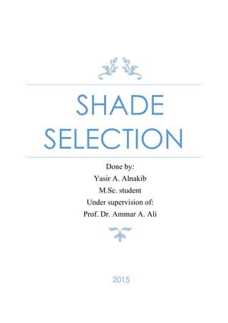
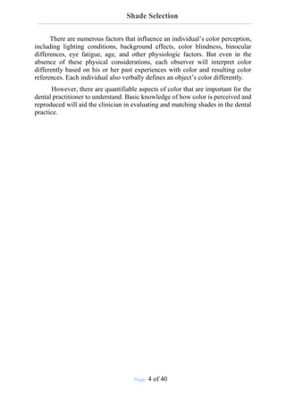
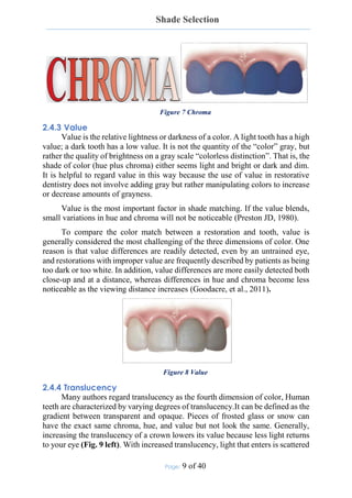
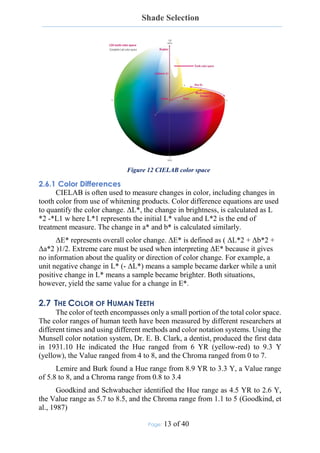
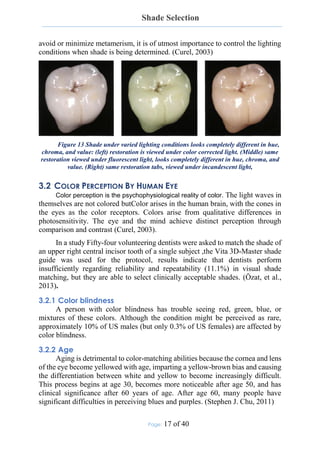
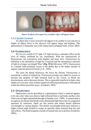
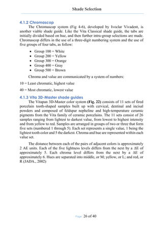
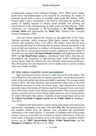
When illuminated, opals and enamel will trans-illuminate the reds and
scatter the blues within its body. This is why enamel appears bluish at the incisal
edge even though it is colorless (Fig. 19 c ) (Bosch, et al., 1995). The opalescent
effects of enamel brighten the tooth and give it optical depth and vitality (Garber,
2000)
.
Figure 19 In vitro examples of light effects exhibited by a natural tooth. Natural
light effects (a), fluorescence (b), opalescence (blue [c] and orange [d])
3.6 REFLECTIONS OF LIGHT
It is important to realize that matching the hue and chroma is sixth or
seventh in importance on the list of things to match when constructing a prosthetic
replacement. You have to be fairly close to someone to detect subtle differences
in hue; yet shape, value, surface texture, luster, and opacity disparities can be seen
from four or five feet away or more. Violating conformity of the unique
characteristics of the natural dentition will cause an unwanted prominence of your
restoration. (Glick, 1994)
These characteristics determine how light is reflected, transmitted, or
scattered thus affecting its hue, chroma, value, and translucency. The appearance
of teeth is mostly determined by how light interacts with the curved and varied
surface. (Glick, 1994)
3.6.1 Surface Texture
A roughened surface texture will not yield as well defined an image and
will scatter the light and the individual wavelengths will all bend differently
yielding a substantially different spectrum returning to the eye. (Obregon, 1981)
Texture can be broken down into subgroups: vertical, horizontal, and
malformations. Vertical surface textures are primarily composed of the heights of
contour of the marginal ridges and the developmental lobes. Perichymata, the
fine transverse wavelike grooves believed to be external manifestations of the
striae of retzius are horizontal textures. The striae or lines of retzius are the result
of the layering manner in which the deposition of enamel takes place.
Malformations are the third textural group and can be from cracks, chips, and
other surface aberrations (Fondriest, 2003).
24. Shade Selection
Page: 23 of 40
Surface texture can be generalized as being heavy, medium, or light. A
rough or heavy surface texture (Fig. 20) will have a lower value because it tends
to diffuse light by reflecting it in many directions and less light returns to the
viewer. A light surface texture has a higher value due to the increased specular
reflection. At eruption, teeth have their roughest surface texture. With age, these
surface features gradually wear. As the wear process continues into the later years
of life, all signs of the perikymata are lost and even the definition of the
developmental lobes is obliterated and the tooth appears smooth with a highly
reflective glassy surface. (Fondriest, 2003)
Figure 20 teeth with heavy surface texture
3.6.2 Luster
Reducing the surface luster of a piece of clear window glass by wet sanding
or etching will produce a frosty white look. As light hits the surface of the etched
glass, it scatters or bends irregularly. This scattering of the light at the surface
causes an increase in opacity. The light isn’t carried off and away from the surface
but rather reflected. As the glass becomes less translucent, the value goes up. The
net effect is more light returns to the viewer as the luster goes down. (Fondriest,
2003)
It is important to note that surface texture and not luster determines specular
reflection. Although the surface luster has been roughened the glass remains flat
and has low texture so it will remain a specular reflector. Polishing the rough
glaze off of a porcelain restoration is a subtle way to lower value by making the
porcelain clearer and more translucent. (Geller, 1983)
3.7 BLEACHING
Most people say they want white teeth. However, the color white is
scientifically described as being completely reflective of all visible wavelengths
of light, which implies an opacity that is undesirable in the dentition. In the
context of esthetic dentistry, white as an ideal tooth color refers to the lightness
or translucency of a tooth or restoration. When teeth are bleached, the relative
lightness (value) of the teeth is increased, making them appear whiter. Therefore,
bleaching does not necessarily involve making the teeth more opaque and
reflective; rather, intrinsic colored pigments are removed, allowing a tooth to
become whiter yet remain highly translucent (Stephen J. Chu, 2011).
25. Shade Selection
Page: 24 of 40
4 CONVENTIONAL SHADE MATCHING
For nearly a century, dental professionals have relied on tooth shade guides
for an “accurate” shade match. Shade guides are sets of physical standards that
are routinely used in dentistry for visual comparison with natural teeth in order to
match color and other optical properties of the target tooth or restoration. The
value of this method depends on the education and training of the clinician
performing shade matching, the quality of the shade guide used, and the quality
of the shade-matching method and conditions.
Traditional shade taking involves matching one or more selected colors
from a range of shade tabs to the teeth adjacent or contralateral to the teeth to be
restored. This serves as a guide to the lab technician fabricating the crown or the
bridge. The more information (and accuracy) that the dentist can provide in the
prescription, the more lifelike the technician’s output can become. Thus the
dentist who provides a drawing of a tooth color map, indicating the various shades
within the tooth and their borders, is more likely to have a positive result than the
dentist who describes the shade as a single generic color (Freedman, 2012).
4.1 SHADE GUIDE SYSTEMS
Tooth shade matching is most frequently performed visually using dental
shade guides. The first shade guide was introduced on the market in 1956 by Vita
Zahnfabrik for the measurement of the color of ceramic systems. Although still
imperfect, it introduced some visual parameters that with some minor
modifications are still routinely used by dental practitioners. The Vitapan
Classical Shade Guide consists of 16 tabs arranged into four groups based on hue
and within the groups according to increasing chroma (also known as A-to-D
arrangement) (Paravina, 2009). The most important issues are related to the need
for and lack of a logical and adequate distribution in part of the color space
encompassed by human teeth. Another quite popular shade guide not
systematically arranged is the Ivoclar-Vivadent Chromascop. As with the Vitapan
Classical, the Chromascop is arranged in groups based on the hue (1=White,
2=Light Yellow, 3=Dark Yellow, 4= Grey and 5=Brown) and within the groups
according to increasing chroma (from 10 to 40). Dental color science then
developed in a manner that minimized the errors in visual color selection (Miller,
1993 and 1994). In the late 1990s the Lab* system was adopted by dentistry and
one of the first clinical results was the development of the Vita 3D Master Shade
Guide.
26. Shade Selection
Page: 25 of 40
4.1.1 Vita Classical
In the Vita Classical shade guide, the tabs are arranged alphabetically
according to hue:
A = Orange
B = Yellow
C = Yellow/Gray
D = Orange/Gray (Brown)
The chroma and value for each hue are communicated by a system of
numbers:
1 = Least chromatic, highest value 4 = Most chromatic, lowest value
Vita Classical (Fig. 21) has been the gold standard for shade matching in
dentistry since it was introduced in 1956. Indeed, the majority of restorative
materials, particularly composite resins, are keyed to it. However, the criticism
of its empiric conception, especially regarding the arrangement of the tabs and
the color distribution, persists even today. The tabs can be arranged according to
value (light to dark) in addition to hue and chroma. While this adds to the shade
guide’s versatility, studies have found inconsistencies as a result of using the
value scale (Paravina, et al., 2001).
Before the VITAPAN Classical Shade Guide system is used for shade
matching, the color tabs should be rearranged from their alphabetic order
(which is how they are packaged) into the indicated value-based order that is
included with every kit and goes from B1 to C4
The patient is asked to smile, and the VITAPAN Classical Shade Guide is
passed in front of the teeth from the darker shades through the middle of the
range to the lighter shades. The tabs of this shading group are brought edge to
edge with the tooth in question in order to narrow down the actual color. The
most appropriate color tab is pulled from the guide. Several readings are
necessary to create a color map of a tooth. (Freedman, 2012)
Figure 21 Vita Classical shade guide system
27. Shade Selection
Page: 26 of 40
4.1.2 Chromascop
The Chromascop system (Fig 4-6), developed by Ivoclar Vivadent, is
another viable shade guide. Like the Vita Classical shade guide, the tabs are
initially divided based on hue, and then further intra-group selections are made.
Chromascop differs in the use of a three-digit numbering system and the use of
five groups of four tabs, as follow:
Group 100 = White
Group 200 = Yellow
Group 300 = Orange
Group 400 = Gray
Group 500 = Brown
Chroma and value are communicated by a system of numbers:
10 = Least chromatic, highest value
40 = Most chromatic, lowest value
4.1.3 Vita 3D-Master shade guides
The Vitapan 3D-Master color system (Fig. 22) consists of 11 sets of fired
porcelain tooth-shaped samples built up with cervical, dentinal and incisal
powders and composed of feldspar nepheline and high-temperature ceramic
pigments from the Vita family of ceramic porcelains. The 11 sets consist of 26
samples ranging from lightest to darkest value, from lowest to highest intensity
and from yellow to red. Samples are arranged in groups of two or three that form
five sets (numbered 1 through 5). Each set represents a single value, 1 being the
lightest tooth color and 5 the darkest. Chroma and hue are represented within each
value set.
The distance between each of the pairs of adjacent colors is approximately
2 AE units. Each of the five lightness levels differs from the next by a ΔE of
approximately 5. Each chroma level differs from the next by a ΔE of
approximately 6. Hues are separated into middle, or M; yellow, or L; and red, or
R (JADA., 2002)
28. Shade Selection
Page: 27 of 40
Figure 22 Toothguide VITA 3D-Master shade system
The VITA 3D-Master Shade Guide is based on a color classification
principle in which 3 dimensions of color, value (brightness), chroma (intensity
of the color), and hue (the color itself), are considered equally so that the
determination of shade can be easily carried out using systematic, consistent
criteria (Paravina, 2009).
The VITA 3D-Master Shade Guide addressed the most important elements
of tooth shade measurement: a scientific color distribution, having a systematic
arrangement of shades within the natural tooth color space and an objective,
numerical measure of color, according to the colorimetric CIELab* order
principle, rather than on the mere observation of the natural tissue aspects , that
can be written as a clear prescription for the laboratory technicians.
Figure 23 Shade tabs distribution in color space: (left) VITA classical, (right) VITA
3D master
29. Shade Selection
Page: 28 of 40
4.1.3.1 Steps of Using the Vita 3D-Master
The first step in using the Vita 3D-Master system is to make sure the tabs
are aligned vertically. Misaligned tabs can be distracting to the operator. The
patient is asked to smile then the Toothguide Vita 3D-Master is used in 3 steps
as follows:
1. Value determination: Shade guide is passed adjacent to the teeth,
going from the darker shades through the intermediate to the lighter
shades. The user selects the value level (from 0 to 5, with 0 being the
lightest [high value] and 5 being the darkest [low value]) that is
closest to the value of the tooth to be matched, and then takes the
medium (M) shade sample from the selected value group
2. Chroma determination: The user selects the color sample from the
M group with the chroma level (from 1 to 3, with 1 being the least
chromatic and 3 being the most chromatic) that is closest to that of
the tooth to be matched.
3. Hue determination: shade than the color sample of the M group
selected in the second step. Now the best-matching shade sample can
be determined and the information recorded in the color
communication form.
Several readings are necessary to create a color map of a tooth. Typically at
least three readings per tooth are suggested, one each for the gingival, middle,
and incisal thirds.
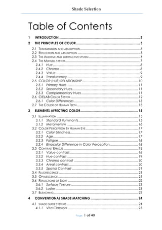
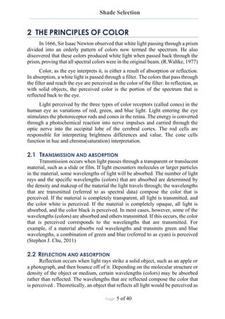
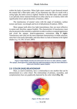
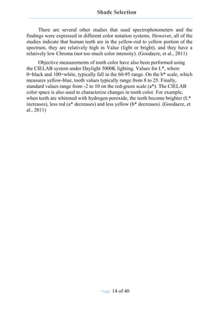
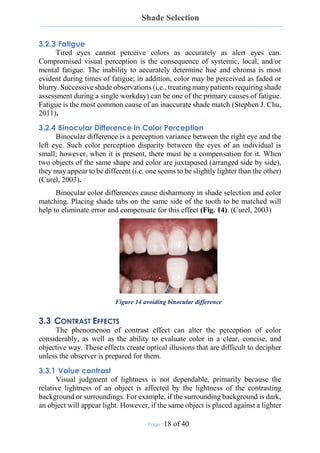
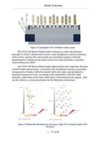
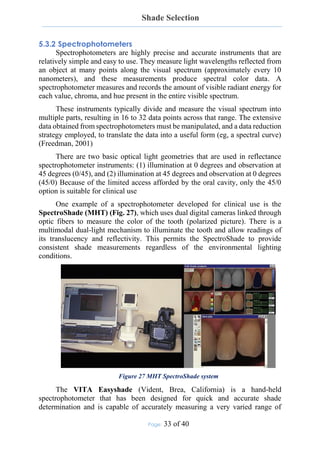
The Linearguide Vita 3D-Master (Fig. 24) has the same shade tabs as the
Toothguide but a different design, and shade matching is reduced to two steps:
1. Value selection: A dark-gray holder, containing only 6 middle tabs
(0M2 to 5M2) is used. The small number of tabs with large color
differences and the linear tab arrangement simplify group selection.
2. Chroma and hue selection: A final selection based on chroma and hue
is made from the initial value group selected.
Figure 24 Linearguide VITA 3D-Master shade guide system
30. Shade Selection
Page: 29 of 40
Its relative simplicity makes Linearguide recommended for a “pick the best
match” approach, while Toothguide is recommended for a “dimension-by-
dimension” approach. It was also found that, overall, Linearguide enables better
shade-matching results and was found to be superior in a subjective evaluation
compared to Toothguide. Linearguide and Toothguide were both found to enable
significantly better (closer) matches compared to Classical. (Paravina, 2009)
The Bleachedguide Vita 3D-Master is the only shade guide developed
specifically for visual evaluation of tooth whitening. Bleachedguide exhibits a
wider color range and more consistent color distribution than the Vita Classical
and other shade guides, such as Trubyte Bioform (Dentsply). In one study, the
progression of lightening in natural teeth was found to be identical to the order
suggested by Bleachedguide (Ontiveros, et al., 2009)
4.2 COMMUNICATION
In the conventional shade-matching system the lab technician takes the
basic information about value and chroma provided by the shade tabs and applies
that information to the ceramic system and effect powders being used. Effective
communication between the lab technician and the clinician is therefore critical
to achieving a successful shade match. (Stephen J. Chu, 2011)
It is recommended that the clinician send photographs along with the shade
tabs as reference. Photography is a valuable means of communication between
the clinician and the lab technician and adds credibility to the shade tab selection.
Once the gingival, body, and incisal shade tabs are selected, photographs of each
of the tabs should be taken next to the tooth to be matched, together with an
aggregate photograph of all three tabs near the dentition. It also makes sense to
photograph the matched tooth next to the two extreme shades (one lighter than
the perceived shade and one darker); this allows the lab technician to get a
concrete sense of the shade and value variation. Finally, photographs should be
taken of the patient’s face and full smile to allow the technician to envision how
the restoration will fit into the patient’s overall appearance. (Avery, 2003)
31. Shade Selection
Page: 30 of 40
5 TECHNOLOGY-BASED SHADE MATCHING
Precise color communication is integral to the development of esthetic
harmony and overall restorative success. While traditional shade-taking
procedures enable some degree of shade information transfer, contemporary
shade-analysis devices allow for standardized, repeatable shade determinations
by placing technology in the role of “observer” in the light-object-observer triad
required for color perception
Several clinical studies have confirmed that computer-assisted shade
analysis is more accurate and more consistent compared with human shade
assessment (Paul, et al., 2002) (Judeh, et al., 2009)
The need for improvement in the accuracy of shade matching was
highlighted by a study that showed that 80% of patients notice a difference in the
shade of their natural teeth compared with their restored teeth (Ishikawa-Nagai,
et al., 1992). Such a widespread lack of accuracy should not be accepted as the
standard; rather, clinicians should strive to improve the esthetic quality of
restorative work.
Advantages of computer-aided shade determination include:
1. No influence of surroundings
2. No influence of lighting
3. Results are reproducible
4. Easy documentation
5. Reliable data transmission
5.1 DEVELOPMENT OF TECHNOLOGY-BASED SHADE SYSTEMS
Advances in technology in the areas of computers, the Internet, and
communication systems have greatly influenced and shaped modern society.
Commensurate with these strides are the advances in contemporary dentistry:
During the past half-decade, the dental profession has experienced the growth of
a new generation of technologies devoted to the analysis, communication, and
verification of shade (Stephen J. Chu, 2011).
The earliest color-measuring device designed specifically for clinical dental
use was a filter colorimeter. The Chromascan (Sterngold) was introduced in the
early 1980s but enjoyed limited success because of its poor design and accuracy
(Goodkind, et al., 1985). Further development was hindered primarily by a lack
of resources and commitment on industry’s side—the market was too small. In
the late 1980s and early 1990s, Seghi and Ishigawa-Nagai published experimental
research using both colorimeters and spectrophotometers (Ishigawa-Nagai,
1994). Several prominent color science experts also tried to objectively quantify
color. Bergen experimented with spectrophotometers and computers in an effort
32. Shade Selection
Page: 31 of 40
to standardize analysis in the profession (Bergen, 1985). Miller used a single-
point-source spectrophotometer in his research for correlating the shades of
extracted natural teeth to those of available shade guide tabs (Miller, 1988).
Preston made a major contribution to the field by identifying the quantity and
quality of lighting required to analyze shade properly and pointing out
inconsistencies in the manufacturing of shade guides and tabs (Preston, 1985).
Yamamoto was instrumental in the development of the Shofu ShadeEye
Chroma Meter and subsequently the Shofu NCC (Natural Color Concept)
System (Yamamoto, 1998).
Chu and Tarnow reported the clinical use and application of the Cortex
Machina prototype, which employed RGB digital camera technology that
inferred color properties (Chu, et al., 2001). The authors found that the more
accurate data provided by technology-based systems allowed technicians at all
levels of skill and experience to produce well-matched restorations. In 2001 and
2002, the first two measurement analysis systems mapping the whole surface of
the tooth were developed: the SpectroShade System (a spectrophotometer) from
MHT and the ShadeVision system (a colorimeter) from X-Rite. Recently, the
development and integration of light-emitting diode (LED) technology into
various dental fields has allowed for more portable, battery-powered devices.
This development has also lowered the cost of shade analysis systems, making
them more readily available.
5.2 SPOT VERSUS COMPLETE-TOOTH MEASUREMENT
Spot measurement devices measure a small area on the tooth surface. The
size or diameter of the optical device aperture (generally 3 mm2) determines how
much of the tooth surface and subsequent shade is measured. The average central
incisor is 80 to 100 mm2; therefore, spot measurement cannot deliver all of the
information necessary to create an overall image. Spot measurement devices
generally require three points of reference each for the gingival, body, and incisal
areas of the tooth (a total of nine reference measurements). This increased number
equates to greater sources of error during image capture as well as increased time
for shade information data capture. Examples of spot measurement devices are
the Vident EasyShade Compact system and the X-Rite Shade-X. (Stephen J.
Chu, 2011)
Complete-tooth measurement systems measure the whole tooth surface area
and provide a topographic color map of the tooth (Fig. 25). The measurement of
the complete surface gives the operator more consistent and reproducible
information of the tooth structure. A drawback of complete-tooth measurement
systems, however, is that their use is limited to anterior teeth because of the size
of the sensor, which does not allow access to the molar region. Examples of these
devices are the MHT SpectroShade Micro, X-Rite ShadeVision, and the
Olympus CrystalEye. (Da Silva, et al., 2008)
33. Shade Selection
Page: 32 of 40
Figure 25 Complete-tooth measurement devices measure the entire surface of the
tooth and provide a detailed color distribution map
5.3 TYPES OF TECHNOLOGY-BASED SHADE SYSTEMS
5.3.1 RGB devices
Most consumer video or digital still cameras acquire red, green, and blue
image information that is utilized to create a color image and are commonly
referred to as RGB devices. Digital cameras and other RGB devices represent the
most basic approach to electronic shade taking and require a certain degree of
subjective shade verification with the human eye. (Stephen J. Chu, 2011)
Various approaches to the translation of this data into useful dental color
information have been used. The problem inherent in the use of these systems is
that they do not control some of the key variables associated with accurate color
determination. Typically, color is synthesized from RGB data according to
various assumptions about the camera and the use of reference materials within
the captured image. The information accuracy (reliability) of RGB devices is
questionable since they are not measurement instruments; rather, they infer color
properties of the captured image. These systems are more helpful for providing
lab technicians with a starting reference point than for visually determining the
shade of a tooth. The ShadeVision system from X-Rite is an example of an RGB
device (Fig. 26) . (Stephen J. Chu, 2011)
Figure 26 The ShadeVision system from X-Rite
34. Shade Selection
Page: 33 of 40
5.3.2 Spectrophotometers
Spectrophotometers are highly precise and accurate instruments that are
relatively simple and easy to use. They measure light wavelengths reflected from
an object at many points along the visual spectrum (approximately every 10
nanometers), and these measurements produce spectral color data. A
spectrophotometer measures and records the amount of visible radiant energy for
each value, chroma, and hue present in the entire visible spectrum.
These instruments typically divide and measure the visual spectrum into
multiple parts, resulting in 16 to 32 data points across that range. The extensive
data obtained from spectrophotometers must be manipulated, and a data reduction
strategy employed, to translate the data into a useful form (eg, a spectral curve)
(Freedman, 2001)
There are two basic optical light geometries that are used in reflectance
spectrophotometer instruments: (1) illumination at 0 degrees and observation at
45 degrees (0/45), and (2) illumination at 45 degrees and observation at 0 degrees
(45/0) Because of the limited access afforded by the oral cavity, only the 45/0
option is suitable for clinical use
One example of a spectrophotometer developed for clinical use is the
SpectroShade (MHT) (Fig. 27), which uses dual digital cameras linked through
optic fibers to measure the color of the tooth (polarized picture). There is a
multimodal dual-light mechanism to illuminate the tooth and allow readings of
its translucency and reflectivity. This permits the SpectroShade to provide
consistent shade measurements regardless of the environmental lighting
conditions.
Figure 27 MHT SpectroShade system
The VITA Easyshade (Vident, Brea, California) is a hand-held
spectrophotometer that has been designed for quick and accurate shade
determination and is capable of accurately measuring a very varied range of
35. Shade Selection
Page: 34 of 40
VITAPAN Classical and VITAPAN 3D-Master shades. Easyshade simplifies the
shade-matching procedure, providing high-quality, predictable, dependable,
totally objective shade determination, resulting in fewer re-shades, fewer color
alterations, and an overall superior esthetic product. (Freedman, 2012)
The Vita Easyshade is one of the latest spectrophotometers available for
clinical use. The instrument's software is programmed to give absolute CIELab*
color values only for natural teeth. Conversely, when assessing the color of a
ceramic, the spectrophotometer provides the differences (Δ values) from color
value presets in the instrument's database. The spectrophotometer data acquisition
is different when reading natural tooth and ceramic also because the instrument
applies different scanning methods on the two diversely structured substrates (JJL
Technology, 2003). Ceramic restorations are generally less than 1.6mm in
thickness and the color layers (dentin and opaque) are from 0.2-0.4 mm under the
enamel porcelain layer. Conversely, inside the teeth, the dentin layer is generally
1.0-1.5 mm from the outer surface (Shillingburg et al., 1991). Furthermore, the
enamel thickness causes a scattering of the penetrating light. Moreover, the
overall thickness of a natural tooth is greater than that of a ceramic tooth.
In 2009 Vita Zahnfabrik comapny introduced onto the market a new shade
taking device, the Easyshade Compact (Fig. 28), which represents an evolution
of the previous one. Thanks to the use of LED technology, the new device became
smaller, wireless and easier to handle. The most important difference between the
two devices is that the Compact has a single spectrophotometer rather than the
two ones belonging to Easyshade standard. In the standard Easyshade the
difference in color reading between natural teeth and ceramic restorations is due
to two different spectrophotometers embedded in the instrument. In the
Easyshade Compact's tip there are two different sets of LEDs that provide
different light according to what is being measured and the reflected light
converges in a single Charge Coupled Device (CCD).
Figure 28 VITA Easyshade Compact (Vident).
36. Shade Selection
Page: 35 of 40
5.3.3 Colorimeters
These instruments approximate the spectral function of the standard
observer’s eye and are engineered to directly measure color as perceived by the
human eye. A colorimeter filters light in three or four areas of the visible spectrum
to determine the color of an object. Properly designed colorimeters such as X-
Rite’s ShadeVision system (Fig. 24) can provide greater data efficiency because
they store only the necessary 3 data points of hue, value, and chroma instead of
the 16 or more data points of reflectance. A colorimeter can deliver color
information accuracy similar to that of spectrophotometers and reduce the data
load time by avoiding the unnecessary color mapping associated with
spectrophotometers.
The ShadeVision system provides simple, reliable shade measurement
information for precise, quantifiable communications between the dental office
and laboratory. The assurance of an accurate shade match is significantly
improved compared with traditional techniques (Stephen J. Chu, 2011).
5.4 RELIABILITY AND ACCURACY
Recent study tested 4 commercially available digital shade matching
devices demonstrated that the Accuracy of devices was as follows: VITA
Easyshade, 92.6%; ShadeVision, 84.8%; SpectroShade, 80.2%; and
ShadeScan, 66.8%. (Seungyee, et al., 2009)
37. Shade Selection
Page: 36 of 40
Table 1 Specifications for currently available technology-based shade systems
5.5 INTERPRETATION METHODS OF DIFFERENT TECHNOLOGIES
As discussed previously, the surface of the tooth has significant impact on
the perceived value of the shade. The smoother (more reflective) the surface is,
the brighter the surface will appear. To overcome this problem, some systems use
filters to adjust for the surface gloss. Shade-matching systems that do not use such
filters often record shades at a value that is too high, which can be very
problematic.
Accuracy of color measurement is also affected by the phenomenon of edge
loss, which occurs because of light lost primarily through the translucent tooth
and ceramic enamel layers. Although algorithms are incorporated into the
software to accommodate for the different light-scattering properties of teeth,
crowns, and shade tabs, it is difficult to fully compensate for these differences,
and this can be a significant source of error.
38. Shade Selection
Page: 37 of 40
Translucency mapping is inadequate in all of the systems. Replication of
tooth translucency remains the most challenging aspect of matching the
appearance of a natural tooth. The transfer of this three-dimensional quality to a
two-dimensional map provides little benefit. Systems that incorporate digital
imaging have the best chance because a high-quality visual is the best that is
currently available.
Positioning of the probe or mouthpiece seems to be critical to the
repeatability of the measurement. In addition, any device that uses a small-
diameter contact probe is limited because it cannot give detailed mapping of color
on the surface and provides only a general base shade of the limited area
measured. The larger mouthpieces are limited to measurements of anterior teeth
because of access. (Brewer, et al., 2004)
39. Shade Selection
Page: 38 of 40
6 REFERENCES
1. AH.Munsell A grammar of color [Book]. - NY : Van Nostrand Reinhold,
1969.
2. Aschheim Kenneth W. Esthetic Dentistry - A Clinical Approach to
Techniques and Materials [Book]. - [s.l.] : Mosby, 2015. - Vol. 3.
3. Avery D. New shade-matching technology: The final piece of the shade
communication puzzle [Journal]. - [s.l.] : J Dent Technol , 2003. - 6 : Vol.
20.
4. Bergen SF. Color in aesthetics. [Journal]. - [s.l.] : N Y State Dent J, 1985. -
Vol. 51.
5. Bosch J. and Coops JC. Tooth color and reflectance as related to light
scattering and enamel hardness. [Journal]. - [s.l.] : J. Dent. Res, 1995. - 1 :
Vol. 74.
6. Brewer JD,, Wee A and R. Seghi Advances in color matching. [Journal]. -
[s.l.] : Dent Clin North Am, 2004. - Vol. 48.
7. Carsten Successful shade matching—What does it take? [Journal]. - [s.l.] :
Compend Contin Educ Dent, 2003. - Vol. 24. - pp. 175–178.
8. Chu SJ. and DP. Tarnow Digital shade analysis and verification: A case
report and discussion [Journal]. - [s.l.] : Pract Periodontics Aesthet Dent,
2001. - Vol. 13.
9. Cornell D. and Winter R. Manipulating light with the refractive index of
an all-ceramic material [Journal]. - [s.l.] : Prac. Proced. Aesthet. Dent,
1999. - 1 : Vol. 8. - pp. 913-917.
10. CP. Sim, AU. Yap and J. Teo Color perception among different dental
personnel. [Journal]. - [s.l.] : Oper Dent, 2001. - Vol. 26. - pp. 435–439.
11. Curel Galip The Science and Art of Porcelain Laminate Veneers [Book]. -
[s.l.] : quintessence, 2003. - Vol. First edition.
12. Da Silva JD, [et al.] Clinical performance of a newly developed
spectrophotometric system on tooth color reproduction [Journal]. - [s.l.] : J
Prosthet Dent , 2008. - Vol. 99.
13. Fondriest james S. Shade matching in restorative dentistry: the science and
strategies. [Journal]. - [s.l.] : Int J Periodontics Restorative Dent, 2003. - 5 :
Vol. 23.
14. Freedman G. Communicating color [Journal]. - [s.l.] : Dent Today, 2001. -
Vol. 20.
40. Shade Selection
Page: 39 of 40
15. Freedman George CONTEMPORARY ESTHETIC DENTISTRY
[Book]. - St. Louis, Missouri : mosby, 2012. - Vol. first edition.
16. Garber D. The Quest for the All-Ceramic Restoration. [Journal]. - [s.l.] :
Quintessence Dental Technology, 2000.
17. Geller W Polishing porcelain makes a crown smoother, more translucent,
and improves the color [Journal]. - [s.l.] : QDT, 1983. - 6 : Vol. 7.
18. Glick K. Color and Shade Selection in Cosmetic Dentistry: Part III
Establishing the Proper Environment and Technique [Journal]. - [s.l.] : J. of
AACD, 1994. - Vol. summer.
19. Goodacre Charles J. and Sagel Paul A. Dental Esthetics in Practice: Part
3 - Understanding Color & Shade Selection. - [s.l.] : dentalcare.com
Continuing Education Course, 2011.
20. Goodkind RJ and Schwabacher WB. Use of fiber-optic colorimeter for in
vivo color measurement of 2830 anterior teeth [Journal]. - [s.l.] : J Prosthet
Dent , 1987. - Vol. 58.
21. Goodkind RJ., K. Keenan and WB. Schwabacher A comparison of
Chromascan and spectophotometric measurements of 100 natural teeth.
[Journal]. - [s.l.] : J Prosth Dent , 1985. - Vol. 53.
22. Ishigawa-Nagai S, Sato RR, Shiraishi A, Ishibashi K. Using a computer
color-matching system in color reproduction of porcelain restorations. Part
3: a newly developed spectrophotometer designed for clinical application.
[Journal]. - [s.l.] : Int J Prosthodont , 1994. - Vol. 7.
23. Ishikawa-Nagai S. [et al.] Using a computer color-matching system in
color reproduction of porcelain restorations. Part 1: Application of CCM to
the opaque layer [Journal]. - [s.l.] : Int J Prosthodont, 1992. - Vol. 5.
24. JADA. Dental Shade Guides. [Journal]. - [s.l.] : J Am Dent Assoc, 2002. -
133.
25. Judeh A. and " A. Al-Wahadni. A comparison between conventional
visual and spectrophotometric methods for shade selection [Journal]. -
[s.l.] : Quintessence international, 2009. - 9 : Vol. 40.
26. McLaren E. Luminescent Veneers [Journal]. - [s.l.] : J.Esthetic Dent,
1997. - 1 : Vol. 9.
27. Miller LL. A scientific approach to shade matching. In: Preston JD (ed).
Perspectives in Dental Ceramics: Proceedings of the Fourth International
Symposium on Ceramics. [Journal]. - [s.l.] : Quintessence, 1988.
41. Shade Selection
Page: 40 of 40
28. Obregon A. Effects of opaque and porcelain surface texture on the color of
ceramometal restorations [Journal]. - [s.l.] : J. Prosth. Dent., 1981. - 3 : Vol.
46.
29. Ontiveros JC and Paravina RD. Color change of vital teeth exposed to
bleaching performed with and without supplementary light. [Journal]. -
[s.l.] : J Dent, 2009. - Vol. 37.
30. Özat, Tuncel İ. and Eroğlu E. Repeatability and reliability of human eye
in visual shade selection [Journal]. - [s.l.] : Journal of Oral Rehabilitation,
2013. - Vol. 40.
31. Paravina RD. Performance assessment of dental shade guides. [Journal]. -
[s.l.] : J Dent , 2009. - Vol. 37.
32. Paravina RD., Powers JM. and Fay RM. Dental color standards: shade
tab arrangement. [Journal]. - [s.l.] : J Esthet Restor Dent , 2001. - Vol. 13.
33. Paul S [et al.] Visual and spectrophotometric shade analysis of human teeth
[Journal]. - [s.l.] : J Dent Res, 2002. - Vol. 81.
34. Preston JD Bergen SF Color science and dental art: a self-teaching
program, [Book]. - St Louis : mosby, 1980.
35. Preston JD. Current status of shade selection and color matching.
[Journal]. - [s.l.] : Quintessence Int, 1985. - Vol. 16.
36. R.Waltke Color in the human dentition [Book]. - NY : Jelenko, 1977.
37. Seungyee Kim, Brewer Jane D. and Davis Elaine L. Reliability and
accuracy of four dental shade-matching devices [Journal]. - [s.l.] : The
Journal of Prosthetic Dentistry, 2009. - 3 : Vol. 101.
38. Sproull RC. Color matching in dentistry, part I. The three-dimensional
nature of color. [Journal]. - [s.l.] : J Prosthet Dent, 1973. - Vol. 29.
39. Stephen J. Chu DMD, MSD, CDT, MDT, Alessandro Devigus, Dr MED
DENT, Rade D. Paravina, DDS, MS, PhD, Adam J. Mieleszko, CDT
Fundamentals of color : shade matching and communication in esthetic
dentistry [Book]. - [s.l.] : Quintessence Pub, 2011. - Vol. 2nd edition.
40. Sundar V. and Amber PL. Opals in nature. [Journal]. - [s.l.] : J. of Dental
Technology, 1999. - 8 : Vol. 16.
41. Yamamoto M. Development of the vintage halo computer color search
system. [Journal]. - [s.l.] : Quintessence Dent Technol, 1998. - Vol. 21.