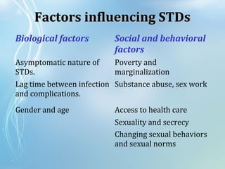
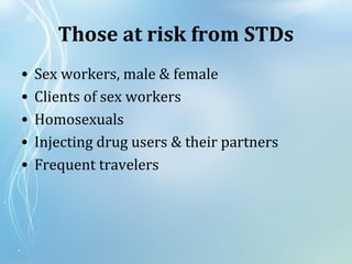
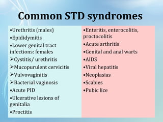
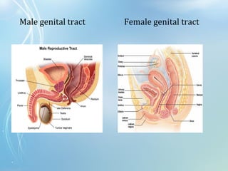
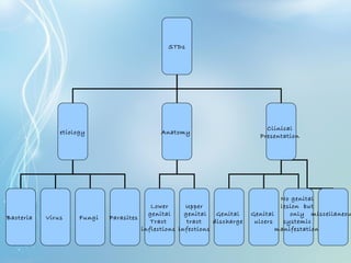
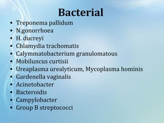
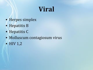
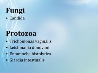
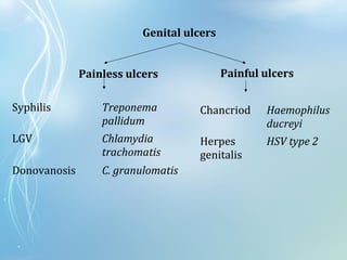
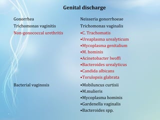
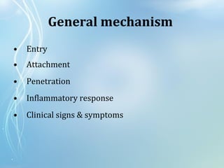
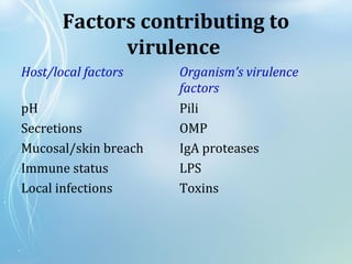
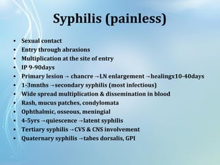
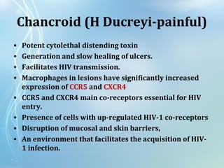
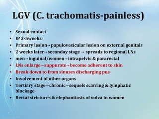
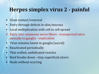
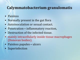
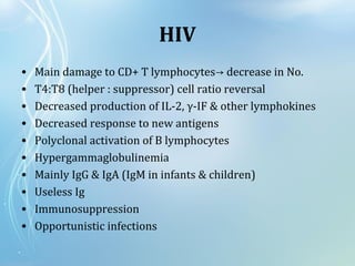
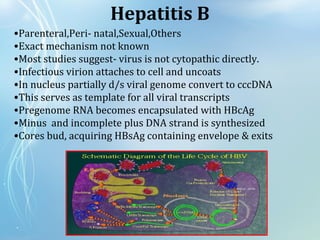
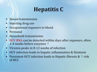
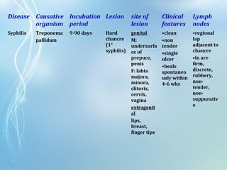
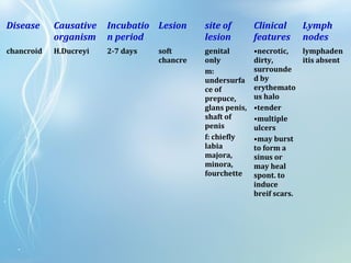
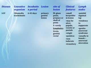
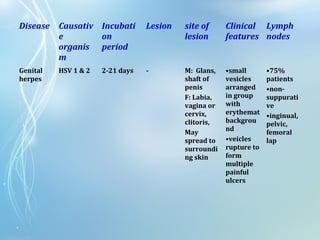
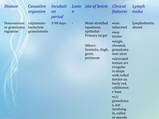
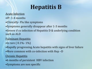
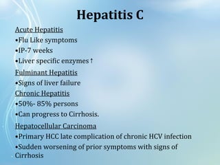
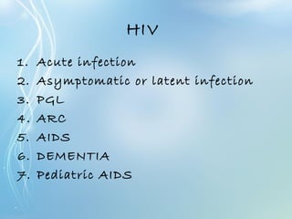
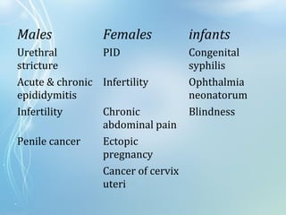
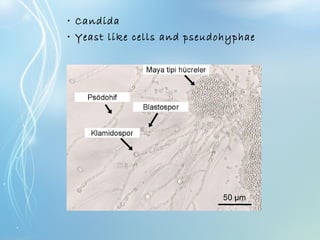
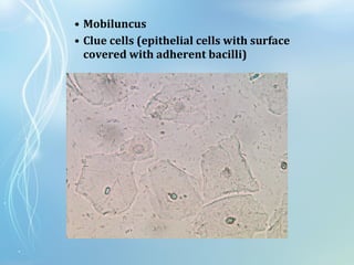
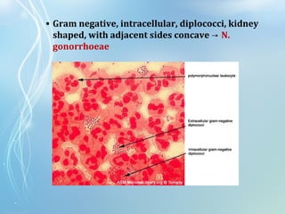
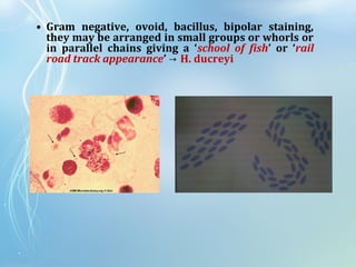
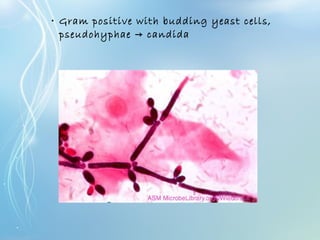
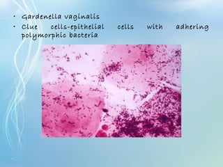
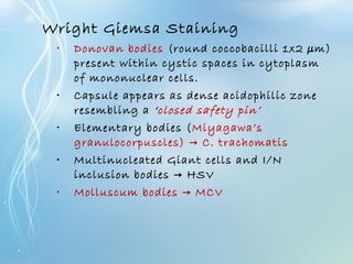
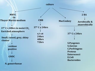
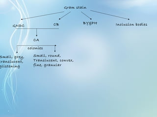
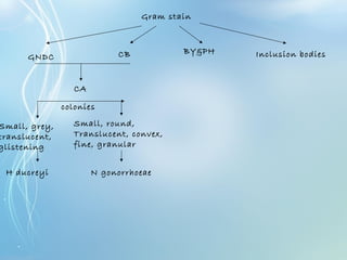
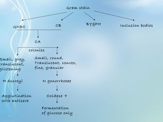
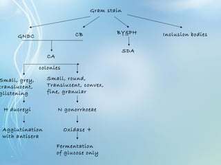
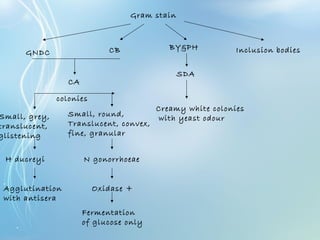
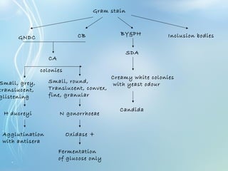
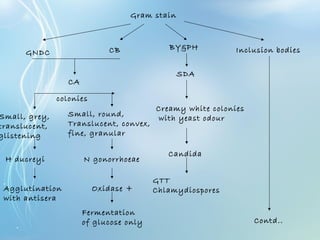
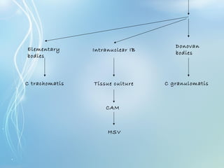
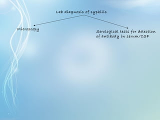
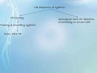
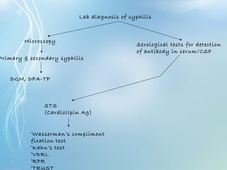
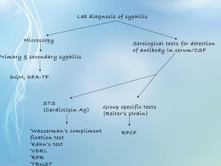
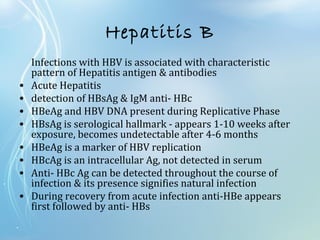
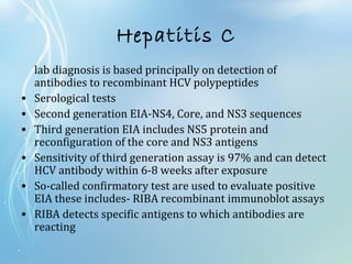
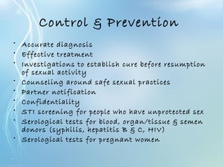
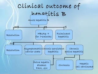
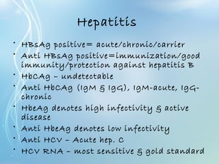
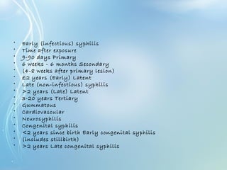
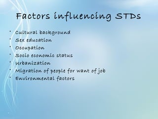
2. The sexually transmitted diseases are a
group of communicable diseases that are
transmitted predominantly by sexual
contact and caused by a wide range of
bacterial, viral, protozoal and fungal agents
3. Biological factors Social and behavioral
factors
Asymptomatic nature of
STDs.
Poverty and
marginalization
Lag time between infection
and complications.
Substance abuse, sex work
Gender and age Access to health care
Sexuality and secrecy
Changing sexual behaviors
and sexual norms
Factors influencing STDsFactors influencing STDs
4. Those at risk from STDs
• Sex workers, male & female
• Clients of sex workers
• Homosexuals
• Injecting drug users & their partners
• Frequent travelers
22. Factors contributing to
virulence
Host/local factors Organism’s virulence
factors
pH Pili
Secretions OMP
Mucosal/skin breach IgA proteases
Immune status LPS
Local infections Toxins
23. Syphilis (painless)
• Sexual contact
• Entry through abrasions
• Multiplication at the site of entry
• IP 9-90days
• Primary lesion → chancre LN enlargement healingx10-40days→ →
• 1-3mnths secondary syphilis (most infectious)→
• Wide spread multiplication & dissemination in blood
• Rash, mucus patches, condylomata
• Ophthalmic, osseous, meningial
• 4-5yrs quiescence latent syphilis→ →
• Tertiary syphilis CVS & CNS involvement→
• Quaternary syphilis tabes dorsalis, GPI→
24. Chancroid (H Ducreyi-painful)
• Potent cytolethal distending toxin
• Generation and slow healing of ulcers.
• Facilitates HIV transmission.
• Macrophages in lesions have significantly increased
expression of CCR5 and CXCR4
• CCR5 and CXCR4 main co-receptors essential for HIV
entry.
• Presence of cells with up-regulated HIV-1 co-receptors
• Disruption of mucosal and skin barriers,
• An environment that facilitates the acquisition of HIV-
1 infection.
25. LGV (C. trachomatis-painless)
• Sexual contact
• IP 3-5weeks
• Primary lesion papulovesicular lesion on external genitals→
• 2 weeks later seconday stage spreads to regional LNs→ →
• men inguinal/women intrapelvic & pararectal→ →
• LNs enlarge suppurate become adherent to skin→ →
• Break down to from sinuses discharging pus
• Involvement of other organs
• Tertiary stage chronic sequels scarring & lymphatic→ →
blockage
• Rectal strictures & elephantiasis of vulva in women
26. Herpes simplex virus 2 - painful
• Close contact/venereal
• Entry through defects in skin/mucosa
• Local multiplication with cell to cell spread
• Entry into cutaneous nerve fibers transported intra-→
axonally to ganglia replication→
• Virus remains latent in ganglia (sacral)
• Reactivated periodically
• Thin walled, umbilicated vesicles
• Roof breaks down tiny superficial ulcers→
• Heals without scarring
27. Calymmatobacterium granulomatis
• Painless
• Normally present in the gut flora
• Autoinoculation or sexual contact.
• Penetration→ inflammatory reaction,
• Destruction of the infected tissue.
• mainly intracellularly inside tissue macrophages
(Donovan bodies).
• Painless papules→ ulcers
• Superinfection
28.
29. HIV
• Main damage to CD+ T lymphocytes decrease in No.→
• T4:T8 (helper : suppressor) cell ratio reversal
• Decreased production of IL-2, γ-IF & other lymphokines
• Decreased response to new antigens
• Polyclonal activation of B lymphocytes
• Hypergammaglobulinemia
• Mainly IgG & IgA (IgM in infants & children)
• Useless Ig
• Immunosuppression
• Opportunistic infections
30. •Parenteral,Peri- natal,Sexual,Others
•Exact mechanism not known
•Most studies suggest- virus is not cytopathic directly.
•Infectious virion attaches to cell and uncoats
•In nucleus partially d/s viral genome convert to cccDNA
•This serves as template for all viral transcripts
•Pregenome RNA becomes encapsulated with HBcAg
•Minus and incomplete plus DNA strand is synthesized
•Cores bud, acquiring HBsAg containing envelope & exits
Hepatitis B
31. Hepatitis C
• Sexual transmission
• Injecting drug use
• Occupational exposure to blood
• Perinatal
• Household transmission
• HCV RNA can be detected within days after exposure, often
1-4 weeks before enzymes ↑
• Viremia peaks in 8-12 weeks of infection
• HCV infection leads to hepatic inflammation & Steatosis
• Persistent HCV infection leads to Hepatic fibrosis & risk↑
of HCC
33. Disease Causative
organism
Incubation
period
Lesion site of
lesion
Clinical
features
Lymph
nodes
Syphilis Treponema
pallidum
9-90 days Hard
chancre
(1°
syphilis)
genital
M:
undersurfa
ce of
prepuce,
penis
F: labia
majora,
minora,
clitoris,
cervix,
vagina
extragenit
al
lips,
breast,
finger tips
•clean
•non
tender
•single
ulcer
•heals
spontaneo
usly within
4-6 wks
•regional
lap
adjacent to
chancre
•ln are
firm,
discrete,
rubbery,
non-
tender,
non-
suppurativ
e
34. Disease Causative
organism
Incubatio
n period
Lesion site of
lesion
Clinical
features
Lymph
nodes
chancroid H.Ducreyi 2-7 days soft
chancre
genital
only
m:
undersurfa
ce of
prepuce,
glans penis,
shaft of
penis
f: chiefly
labia
majora,
minora,
fourchette
•necrotic,
dirty,
surrounde
d by
erythemato
us halo
•tender
•multiple
ulcers
•may burst
to form a
sinus or
may heal
spont. to
induce
breif scars.
lymphaden
itis absent
35. Disease Causative
organism
Incubatio
n period
Lesion site of
lesion
Clinical
features
Lymph
nodes
LGV Chlamydia
trachomatis
6-42 days primary
genital
lesion
M: glans
penis,
prepuce or
shaft of
penis
F: rarely
lymphatics
Groin,
genital,
rectal
•small
herpetifor
m lesion
•may be
ulcer,
papule,
vesicle or
pustule
•single
•painless
•non
indurate
•transitory
•painful
inguinal
lap
•tendency
to
suppurate
•genito-
anorectal
syndrome
•usually in
female –
anorectal
ulceration
36. Disease Causativ
e
organis
m
Incubati
on
period
Lesion site of
lesion
Clinical
features
Lymph
nodes
Genital
herpes
HSV 1 & 2 2-21 days - M: Glans,
shaft of
penis
F: Labia,
vagina or
cervix,
clitoris,
May
spread to
surroundi
ng skin
•small
vesicles
arranged
in group
with
erythemat
backgrou
nd
•veicles
rupture to
form
multiple
painful
ulcers
•75%
patients
•non-
suppurati
ve
•inginual,
pelvic,
femoral
lap
37. Disease Causative
organism
Incubati
on
period
Lesio
n
site of lesion Clinical
features
Lymph
nodes
Donovaniasis
or granuloma
inguinale
calymmato-
bacterium
granulomatis
3-90 days - Moist stratified
squamous
epithelial -
Primary target
Others:
Genitalia, thigh,
groin,
perineum
•non
indurated
•non
tender
•single,
elevated,
granuloma
tous ulcer
•ulcerated
lesions are
irregular
in shape
with rolled
border on
beefy red,
cobbleston
e base
•s/c
granuloma
s, not
involving
ln, called
as pseudo
lymphadenitis
absent
38. Hepatitis B
Acute Infection
•IP- 1-4 months
•Clinically- Flu like symptoms
•Symptoms generally disappear after 1-3 months
•Severe if co infection of Hepatitis D & underlying condition
such as ALD
Fulminant Hepatitis
•Is rare ( 0.1%- 1%)
•Rapidly progressing Acute hepatitis with signs of liver failure
•More common with co-infection with Hep –D
Chronic Hepatitis
•6 months of persistent HBV infection
•Symptoms are non specific
39. Acute Hepatitis
•Flu Like symptoms
•IP-7 weeks
•Liver specific enzymes↑
Fulminant Hepatitis
•Signs of liver failure
Chronic Hepatitis
•50%- 85% persons
•Can progress to Cirrhosis.
Hepatocellular Carcinoma
•Primary HCC late complication of chronic HCV infection
•Sudden worsening of prior symptoms with signs of
Cirrhosis
Hepatitis C
40. HIV
1. Acute infection
2. Asymptomatic or latent infection
3. PGL
4. ARC
5. AIDS
6. DEMENTIA
7. Pediatric AIDS
44. Precautions
1. Use of gloves while taking samples
2. Sample should be collected before start
of antibiotics
3. No antiseptics should be used while
collecting specimen
4. Clean area with gauge soaked in
normal saline
47. Specimen Container Patient preparation Special instructions
Bartholin cyst Anaerobic
transport
Disinfect skin
before collection
Aspirate fluid;
consider chlamydia
& GC culture
Cervix Swab moistened
with Stuart’s or
Amies medium
Remove mucus
before collection
Do not use
lubricant on
speculum; use
viral/chlamydial
transport medium,
if necessary; swab
deeply into cervical
canal
Cul-de-sac Anaerobic
transport
Submit aspirate
Endometrium Anaerobic
transport
Surgical biopsy or
transcervical
aspirate via
sheathed catheter
Urethra Swab moistened
with Stuart’s or
Amies medium
Remove exudate
from urethral
opening
Collect discharge by
massaging urethra
against pubic
symphis or insert
flexible swab 2-4
cm into urethra &
rotate for 2 secs; at
48. Specimen Container Patient preparation Special instructions
Vagina Swab moistened
with Stuart’s or
Amies medium
Remove exudate Swab secretions &
mucus membrane
of vagina
50. Specimen Container Patient preparation Special instructions
Prostate Swab moistened
with Stuart’s or
Amies medium or
sterile screw-cap
tube
Clean glans with
soap & water
Collect secretions
on swab or in tube
Urethra Swab moistened
with Stuart’s or
Amies medium
Insert flexible swab
2-4 cm into urethra
& rotate for 2 secs
or collect discharge
52. Collection of sample to detect
T.pallidum
• Cleanse area around ulcer with
moistened swab
• Remove scab if present
• Squeeze lesion to obtain serous fluid
• Collect drop on cover slip-invert it on
slide
• Deliver immediately
53. Collection of sample to detect
C.granulomatis
• Cleanse area around ulcer with
moistened swab
• Pinch off some tissue from edge or base
of lesion
• Crush between two slides
• Deliver immediately
• If delay-fix with methyl alcohol (1-
2mnts)
56. • T. vaginalis
• Pear shaped, ovoid nucleus at the anterior end, &
cytostome
• 3-5 anterior flagella which are free
• Axostyle runs down the middle of the body and ends
in pointed tail like extremity
• Jerky motility
62. • Gram negative, intracellular, diplococci, kidney
shaped, with adjacent sides concave → N.
gonorrhoeae
63. • Gram negative, ovoid, bacillus, bipolar staining,
they may be arranged in small groups or whorls or
in parallel chains giving a ‘school of fish’ or ‘rail
road track appearance’ → H. ducreyi
64. • Gram positive with budding yeast cells,
pseudohyphae candida→
68. culture
MNYC
Or
Thayer Martin medium
2 BA
MacConkeyCMB
Aerobically &
anaerobically
37° C x 24hrs
37° C x 24hrs
s/c at
24hrs
48hrs
72hrs
37° C x 48hrs in moist CO2
Enriched atmosphere
Small, raised, grey, shiny
clonies
oxidase
positive
GNDC
N. gonorrhoeae
S.Pyogenes
S.Aureus
C.Perfringens
Proteus
Enterococci
E.Coli
Bacteroides
74. Gram stain
GNDC CB BY&PH Inclusion bodies
CA
Small, grey,
translucent,
glistening
Small, round,
Translucent, convex,
fine, granular
colonies
H ducreyi N gonorrhoeae
75. Gram stain
GNDC CB BY&PH Inclusion bodies
CA
Small, grey,
translucent,
glistening
Small, round,
Translucent, convex,
fine, granular
colonies
H ducreyi N gonorrhoeae
Agglutination
with antisera
Oxidase +
Fermentation
of glucose only
76. Gram stain
GNDC CB BY&PH Inclusion bodies
CA
Small, grey,
translucent,
glistening
Small, round,
Translucent, convex,
fine, granular
colonies
H ducreyi N gonorrhoeae
Agglutination
with antisera
Oxidase +
Fermentation
of glucose only
SDA
77. Gram stain
GNDC CB BY&PH Inclusion bodies
CA
Small, grey,
translucent,
glistening
Small, round,
Translucent, convex,
fine, granular
colonies
H ducreyi N gonorrhoeae
Agglutination
with antisera
Oxidase +
Fermentation
of glucose only
SDA
Creamy white colonies
with yeast odour
78. Gram stain
GNDC CB BY&PH Inclusion bodies
CA
Small, grey,
translucent,
glistening
Small, round,
Translucent, convex,
fine, granular
colonies
H ducreyi N gonorrhoeae
Agglutination
with antisera
Oxidase +
Fermentation
of glucose only
SDA
Creamy white colonies
with yeast odour
Candida
79. Gram stain
GNDC CB BY&PH Inclusion bodies
CA
Small, grey,
translucent,
glistening
Small, round,
Translucent, convex,
fine, granular
colonies
H ducreyi N gonorrhoeae
Agglutination
with antisera
Oxidase +
Fermentation
of glucose only
SDA
Creamy white colonies
with yeast odour
Candida
GTT
Chlamydiospores
80. Gram stain
GNDC CB BY&PH Inclusion bodies
CA
Small, grey,
translucent,
glistening
Small, round,
Translucent, convex,
fine, granular
colonies
H ducreyi N gonorrhoeae
Agglutination
with antisera
Oxidase +
Fermentation
of glucose only
SDA
Creamy white colonies
with yeast odour
Candida
GTT
Chlamydiospores
Contd..
85. Lab diagnosis of syphilis
Serological tests for detection
of antibody in serum/CSF
Microscopy
86. Lab diagnosis of syphilis
Primary & secondary syphilis
DGM, DFA-TP
Serological tests for detection
of antibody in serum/CSF
Microscopy
87. Lab diagnosis of syphilis
Primary & secondary syphilis
DGM, DFA-TP
Serological tests for detection
of antibody in serum/CSF
Microscopy
88. Lab diagnosis of syphilis
Primary & secondary syphilis
DGM, DFA-TP
Serological tests for detection
of antibody in serum/CSF
STS
(Cardiolipin Ag)
•Wasserman’s compliment
fixation test
•Kahn’s test
•VDRL
•RPR
•TRUST
Microscopy
89. Lab diagnosis of syphilis
Primary & secondary syphilis
DGM, DFA-TP
Serological tests for detection
of antibody in serum/CSF
STS
(Cardiolipin Ag)
•Wasserman’s compliment
fixation test
•Kahn’s test
•VDRL
•RPR
•TRUST
Group specific tests
(Reiter’s strain)
RPCF
Microscopy
90. Lab diagnosis of syphilis
Primary & secondary syphilis
DGM, DFA-TP
Serological tests for detection
of antibody in serum/CSF
STS
(Cardiolipin Ag)
•Wasserman’s compliment
fixation test
•Kahn’s test
•VDRL
•RPR
•TRUST
Group specific tests
(Reiter’s strain)
RPCF
Specific tests using
pathogenic to treponeme
(Nichol’s strain)
•TPI
•FTA
•FTA-ABS
•TPHA
•EIA
Microscopy
93. Specific tests for HIV
• Ag detection P24 earliest detected→
• Virus isolation
• PCR
• Antibody detection
• Western Blot method confirmation→
94. Hepatitis B
Infections with HBV is associated with characteristic
pattern of Hepatitis antigen & antibodies
• Acute Hepatitis
• detection of HBsAg & IgM anti- HBc
• HBeAg and HBV DNA present during Replicative Phase
• HBsAg is serological hallmark - appears 1-10 weeks after
exposure, becomes undetectable after 4-6 months
• HBeAg is a marker of HBV replication
• HBcAg is an intracellular Ag, not detected in serum
• Anti- HBc Ag can be detected throughout the course of
infection & its presence signifies natural infection
• During recovery from acute infection anti-HBe appears
first followed by anti- HBs
95. Hepatitis C
lab diagnosis is based principally on detection of
antibodies to recombinant HCV polypeptides
• Serological tests
• Second generation EIA-NS4, Core, and NS3 sequences
• Third generation EIA includes NS5 protein and
reconfiguration of the core and NS3 antigens
• Sensitivity of third generation assay is 97% and can detect
HCV antibody within 6-8 weeks after exposure
• So-called confirmatory test are used to evaluate positive
EIA these includes- RIBA recombinant immunoblot assays
• RIBA detects specific antigens to which antibodies are
reacting
96. Skin tests
Frie,s test (C. Trachomatis)
• Ag used : Lygranum
• 0.1ml Ag inj. I.D on ventral aspect of forearm &
• a control prepared from uninfected yolk sac on
other forearm
• 5-5 days nodule (7mm diameter )→
Ducrey’s skin test (H. Ducreyi)
• Killed organism used as Ag
• 0.1ml Ag inj. I/D on forearm
• NS injected in other forearm as control
• erythema+induration (36-48hrs) = positive result
97. Treatment
Discharge Ulcers
Urethral vaginal Transudated
(Syphilis )
Non-indurated
(herpes)
Gonococ
cal
NGU Mucopur
ulent
(TV)
Curd like
(candida
)
Benzathaine pen G
2.4 1.2million
units I/M
ODx10days
Symptomatic
Norflox
800mg
OD
Doxy
100mg
BDX7-
14days
Metro
200mg
TDSx7
days
Co-Tri
ODx6
days
Procaine pen G
1.2million units
I/M ODx1Odays
Acyclovir 200mg
POx5times/dayx7
Procaine
penicilli
n 5.8lac
with
oral
probenci
d
Erythro
500mg
qidx7-
14days
Or
meconaz
ole
Doxy OD if Pen
sensitive
LGV
Erthro 100mg
BDx14days
Ceftraix
one
98. Control & Prevention
• Accurate diagnosis
• Effective treatment
• Investigations to establish cure before resumption
of sexual activity
• Counseling around safe sexual practices
• Partner notification
• Confidentiality
• STI screening for people who have unprotected sex
• Serological tests for blood, organ/tissue & semen
donors (syphilis, hepatitis B & C, HIV)
• Serological tests for pregnant women
104. Lab diagnosis of syphilis
Primary & secondary syphilis
Wet film, DGM, DFA-TP
Serological tests for detection
of antibody in serum/CSF
STS
(Cardiolipin Ag)
•Wasserman’s compliment
fixation test
•Kahn’s test
•VDRL
•RPR
•TRUST
Group specific tests
(Reiter’s strain)
RPCF
Specific antibody
to T palladium
(Nichol’s strain)
•TPI
•FTA
•FTA-ABS
•TPHA
•EIA
Microscopy
105. Gram stain
GNDC CB BY&PH Inclusion bodies
CA
Small, grey,
translucent,
glistening
Small, round,
Translucent, convex,
fine, granular
colonies
H ducreyi N gonorrhoeae
Agglutination
with antisera
Oxidase +
Fermentation
of glucose only
SDA
Creamy white colonies
with yeast odour
Candida
GTT
Chlamydiospores
Contd..
106. Blood
• Position the patient
• Hyperextend patient's arm.
• Apply the tourniquet 3-4 inches above
the selected puncture site.
• Identify vein
• Disinfect with 10% Povidone-Iodine in a
circle of approximately 5 cm in diameter
• After 2 min collect 5-10 ml blood
• Plain vial
107. Clinical outcome of
hepatitis B
Acute hepatitis B
Resolution
HBsAg +
for >6mnths
Fulminant
hepatitis
Resolution
Asymptomatic
carrier state
Chronic persistent
hepatitis
Chronic
active hepatitis
Extra hepatic
disease:
PAN, GN
Cirrhosis
Hepatic
cell carcinoma
9% 1%
50%
90%
108. Hepatitis
• HBsAg positive= acute/chronic/carrier
• Anti HBsAg positive=immunization/good
immunity/protection against hepatitis B
• HbCAg – undetectable
• Anti HbCAg (IgM & IgG), IgM-acute, IgG-
chronic
• HbeAg denotes high infectivity & active
disease
• Anti HbeAg denotes low infectivity
• Anti HCV – Acute hep. C
• HCV RNA – most sensitive & gold standard
109. • Early (infectious) syphilis
• Time after exposure
• 9-90 days Primary
• 6 weeks - 6 months Secondary
• (4-8 weeks after primary lesion)
• £2 years (Early) Latent
• Late (non-infectious) syphilis
• >2 years (Late) Latent
• 3-20 years Tertiary
• Gummatous
• Cardiovascular
• Neurosyphilis
• Congenital syphilis
• <2 years since birth Early congenital syphilis
• (includes stillbirth)
• >2 years Late congenital syphilis
110. Factors influencing STDs
• Cultural background
• Sex education
• Occupation
• Socio economic status
• Urbanization
• Migration of people for want of job
• Environmental factors