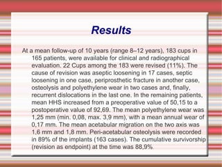
Applications and Efficacies of HA (Hydroxyapatite) American Institute of Medical Sales By: Fred Davis & Jeff Knight
Failure of dual radius hydroxyapatite-coated acetabular cups Many kinds of hydroxyapatite-coated cups were used, with favorable results in short term studies; it was supposed that its use could improve osteointegration of the cup, enhancing the stability and survivorship. The purpose of this study is to analyze the long term behavior of the hemispheric HA coated, Dual Radius Osteonics cup and to discuss the way of failure through the exam of the revised components and of both periacetabular and osteolysis tissue.
Materials and Methods Between 1994 and 1997, at the Department of Orthopedic Sciences of the Insubria University, using the posterolateral approach, were implanted 276 Dual Radius Osteonics ® in 256 patients, with mean age of 63 years.
Results At a mean follow-up of 10 years (range 8–12 years), 183 cups in 165 patients, were available for clinical and radiographical evaluation. 22 Cups among the 183 were revised (11%). The cause of revision was aseptic loosening in 17 cases, septic loosening in one case, periprosthetic fracture in another case, osteolysis and polyethylene wear in two cases and, finally, recurrent dislocations in the last one. In the remaining patients, mean HHS increased from a preoperative value of 50,15 to a postoperative value of 92,69. The mean polyethylene wear was 1,25 mm (min. 0,08, max. 3,9 mm), with a mean annual wear of 0,17 mm. The mean acetabular migration on the two axis was 1,6 mm and 1,8 mm. Peri-acetabular osteolysis were recorded in 89% of the implants (163 cases). The cumulative survivorship (revision as endpoint) at the time was 88,9% .
Conclusion Our study confirms the bad behavior of this type of cup probably related to the design, to the method of HA fixation. The observations carried out on the revised cup confirm these hypotheses but did not clarify if the third body wear could be a further problem. Another interesting aspect is the high incidence of osteolysis, which are often asymptomatic becoming a problem for the surgeon as the patient refuses the possibility of a revision.
Primary human osteoblast  culture on 3D porous collagen- hydroxyapatite  scaffolds. Guy Hilton Research Centre, Institute for Science and Technology in Medicine, Keele  University, Thornburrow  Drive, Hartshill  ST4 7QB, Stoke-on-Trent, United Kingdom.
There is a need in tissue-engineering for 3D scaffolds that mimic the natural extracellular matrix of bone to enhance cell adhesion, proliferation, and differentiation. The scaffold is also required to be degradable. A highly porous scaffold has been developed to incorporate two of the extracellular components found in bone-collagen and hydroxyapatite  (HA). The scaffold's collagen component is an afibrillar monomeric  type I atelocollagen  extracted from foetal  calf's skin. This provided a novel environment for the inclusion of HA powder. Five hundred thousand primary human osteoblasts  were seeded onto 4 mm cubed scaffolds that varied in ratio of HA to collagen. Weight ratios of 1:99, 25:75, 50:50, and 75:25 hydroxyapatite:collagen  ( HA:Collagen ) were analysed .
The scaffolds plus cells were cultured for 21 days. DNA assays and live/dead viability staining demonstrated that all of the scaffolds supported cell proliferation and viability. An alkaline phosphatase  assay showed similar osteoblast  phenotype maintenance on all of the 3D scaffolds analysed  at 21 days. MicroCT  analysis demonstrated an increase in total sample volume (correlating to increase in unmineralised  matrix production). An even distribution of HA throughout the collagen matrix was observed using this technique. Also at 3 weeks, reductions in the percentage of the mineralised  phase of the constructs were seen. These results indicate that each of the ratios of HA/collagen scaffolds have great potential for bone tissue engineering. (c) 2010 Wiley Periodicals, Inc. J Biomed Mater Res Part A, 2010.
CELL-DERIVED MATRIX ENHANCES OSTEOGENIC PROPERTIES OF HYDROXYAPATITE. National O.Bohomolets  Medical University, Department of Oral and Maxillofacial Surgery, Kyiv , Ukraine; Gregory.Tour@ki.se.
The study aimed to evaluate osteogenic  properties of hydroxyapatite  (HA) scaffold combined with extracellular matrix (ECM) derived in vitro from rat primary calvarial osteoblasts  or dermal fibroblasts. The cellular viability, and the ECM deposited onto synthetic HA microparticles  were assessed by MTT, Glycosaminoglycan , and Hydroxyproline  assays as well as immunohistochemistry  and scanning electron microscopy after 21 days of culture. The decellularized  HA-ECM constructs were implanted in critical-sized calvarial  defects of Sprague- Dowley  rats, followed by bone repair and local inflammatory response assessments by histomorphometry  and immunohistochemistry  at 12 weeks postoperatively.
We demonstrated that HA supported cellular adhesion, growth and ECM production in vitro, and the HA-ECM constructs significantly enhanced calvarial  bone repair (p<0.05, Mann-Whitney U test), compared to HA alone, despite the significantly increased number of CD68+ macrophages, and foreign body giant cells (p<0.05, Mann-Whitney U test). Selective accumulation of BSP, OPN and periostin  was observed at the tissue-HA interfaces. In conclusion, in vitro-derived ECM mimics the native bone matrix, enhances the osteogenic  properties of the HA microparticles , and might modulate the local inflammatory response in a bone repair-favorable way. Our findings highlight the ability to produce functional HA-ECM constructs for bone tissue engineering applications.
Biocompatibility of Poly-ε- caprolactone-hydroxyapatite  composite on mouse bone marrow-derived osteoblasts  and endothelial cells By: Fred Davis & Jeff Knight
Received: 17 May 2008 Accepted: 26 February 2009 Published: 26 February 2009   Department of Biomedical Engineering, Wayne State University, Detroit, Michigan, USA 2  Department of Orthopaedic Surgery, Wayne State University, Detroit, Michigan, USA 3  Orthopaedic Research Institute, Via Christi Health System, Department of Biological Sciences, Wichita State University, 1845 Fairmount Street, Wichita, KS 67260, USA
BACKGROUND Tissue-engineered bone may be developed by seeding the cells capable of both osteogenesis  and vascularization  on biocompatible composite scaffolds. The current study investigated the performance of mice bone marrow-derived osteogenic  cells and endothelial cells as seeded on hydroxyapatite  (HA) and poly-ε- caprolactone  (PCL) composite scaffolds.
METHODS Mononuclear cells were induced to osteoblasts  and endothelial cells respectively, which were defined by the expression of osteocalcin , alkaline phosphatase  (ALP), and deposits of calcium-containing crystal for osteoblasts , or by the expression of vascular endothelial growth factor receptor-2 (VEGFR-2) and von Willebrand  factor ( vWF ), and the formation of a capillary network in Matrigel ™ for endothelial cells. Both types of cell were seeded respectively on PCL-HA scaffolds at HA to PCL weight ratio of 1:1, 1:4, or 0:1 and were evaluated using scanning electron microscopy, ALP activity (of osteoblasts ) and nitric oxide production (of endothelial cells) plus the assessment of cell viability.
RESULTS The results indicated that HA led to a positive stimulation of osteoblasts  viability and ALP activity, while HA showed less influence on endothelial cells viability. An elevated nitric oxide production of endothelial cells was observed in HA-containing group.
CONCLUSION Supplement of HA into PCL improved biocompatible for bone marrow-derived osteoblasts  and endothelial cells. The PCL-HA composite integrating with two types of cells may provide a useful system for tissue-engineered bone grafts with vascularization .

Presentation8 16 10[1]

  • 1.
    Applications and Efficaciesof HA (Hydroxyapatite) American Institute of Medical Sales By: Fred Davis & Jeff Knight
  • 2.
    Failure of dualradius hydroxyapatite-coated acetabular cups Many kinds of hydroxyapatite-coated cups were used, with favorable results in short term studies; it was supposed that its use could improve osteointegration of the cup, enhancing the stability and survivorship. The purpose of this study is to analyze the long term behavior of the hemispheric HA coated, Dual Radius Osteonics cup and to discuss the way of failure through the exam of the revised components and of both periacetabular and osteolysis tissue.
  • 3.
    Materials and MethodsBetween 1994 and 1997, at the Department of Orthopedic Sciences of the Insubria University, using the posterolateral approach, were implanted 276 Dual Radius Osteonics ® in 256 patients, with mean age of 63 years.
  • 4.
    Results At amean follow-up of 10 years (range 8–12 years), 183 cups in 165 patients, were available for clinical and radiographical evaluation. 22 Cups among the 183 were revised (11%). The cause of revision was aseptic loosening in 17 cases, septic loosening in one case, periprosthetic fracture in another case, osteolysis and polyethylene wear in two cases and, finally, recurrent dislocations in the last one. In the remaining patients, mean HHS increased from a preoperative value of 50,15 to a postoperative value of 92,69. The mean polyethylene wear was 1,25 mm (min. 0,08, max. 3,9 mm), with a mean annual wear of 0,17 mm. The mean acetabular migration on the two axis was 1,6 mm and 1,8 mm. Peri-acetabular osteolysis were recorded in 89% of the implants (163 cases). The cumulative survivorship (revision as endpoint) at the time was 88,9% .
  • 5.
    Conclusion Our studyconfirms the bad behavior of this type of cup probably related to the design, to the method of HA fixation. The observations carried out on the revised cup confirm these hypotheses but did not clarify if the third body wear could be a further problem. Another interesting aspect is the high incidence of osteolysis, which are often asymptomatic becoming a problem for the surgeon as the patient refuses the possibility of a revision.
  • 6.
    Primary human osteoblast culture on 3D porous collagen- hydroxyapatite scaffolds. Guy Hilton Research Centre, Institute for Science and Technology in Medicine, Keele University, Thornburrow Drive, Hartshill ST4 7QB, Stoke-on-Trent, United Kingdom.
  • 7.
    There is aneed in tissue-engineering for 3D scaffolds that mimic the natural extracellular matrix of bone to enhance cell adhesion, proliferation, and differentiation. The scaffold is also required to be degradable. A highly porous scaffold has been developed to incorporate two of the extracellular components found in bone-collagen and hydroxyapatite (HA). The scaffold's collagen component is an afibrillar monomeric type I atelocollagen extracted from foetal calf's skin. This provided a novel environment for the inclusion of HA powder. Five hundred thousand primary human osteoblasts were seeded onto 4 mm cubed scaffolds that varied in ratio of HA to collagen. Weight ratios of 1:99, 25:75, 50:50, and 75:25 hydroxyapatite:collagen ( HA:Collagen ) were analysed .
  • 8.
    The scaffolds pluscells were cultured for 21 days. DNA assays and live/dead viability staining demonstrated that all of the scaffolds supported cell proliferation and viability. An alkaline phosphatase assay showed similar osteoblast phenotype maintenance on all of the 3D scaffolds analysed at 21 days. MicroCT analysis demonstrated an increase in total sample volume (correlating to increase in unmineralised matrix production). An even distribution of HA throughout the collagen matrix was observed using this technique. Also at 3 weeks, reductions in the percentage of the mineralised phase of the constructs were seen. These results indicate that each of the ratios of HA/collagen scaffolds have great potential for bone tissue engineering. (c) 2010 Wiley Periodicals, Inc. J Biomed Mater Res Part A, 2010.
  • 9.
    CELL-DERIVED MATRIX ENHANCESOSTEOGENIC PROPERTIES OF HYDROXYAPATITE. National O.Bohomolets Medical University, Department of Oral and Maxillofacial Surgery, Kyiv , Ukraine; Gregory.Tour@ki.se.
  • 10.
    The study aimedto evaluate osteogenic properties of hydroxyapatite (HA) scaffold combined with extracellular matrix (ECM) derived in vitro from rat primary calvarial osteoblasts or dermal fibroblasts. The cellular viability, and the ECM deposited onto synthetic HA microparticles were assessed by MTT, Glycosaminoglycan , and Hydroxyproline assays as well as immunohistochemistry and scanning electron microscopy after 21 days of culture. The decellularized HA-ECM constructs were implanted in critical-sized calvarial defects of Sprague- Dowley rats, followed by bone repair and local inflammatory response assessments by histomorphometry and immunohistochemistry at 12 weeks postoperatively.
  • 11.
    We demonstrated thatHA supported cellular adhesion, growth and ECM production in vitro, and the HA-ECM constructs significantly enhanced calvarial bone repair (p<0.05, Mann-Whitney U test), compared to HA alone, despite the significantly increased number of CD68+ macrophages, and foreign body giant cells (p<0.05, Mann-Whitney U test). Selective accumulation of BSP, OPN and periostin was observed at the tissue-HA interfaces. In conclusion, in vitro-derived ECM mimics the native bone matrix, enhances the osteogenic properties of the HA microparticles , and might modulate the local inflammatory response in a bone repair-favorable way. Our findings highlight the ability to produce functional HA-ECM constructs for bone tissue engineering applications.
  • 12.
    Biocompatibility of Poly-ε-caprolactone-hydroxyapatite composite on mouse bone marrow-derived osteoblasts and endothelial cells By: Fred Davis & Jeff Knight
  • 13.
    Received: 17 May 2008 Accepted:26 February 2009 Published: 26 February 2009   Department of Biomedical Engineering, Wayne State University, Detroit, Michigan, USA 2  Department of Orthopaedic Surgery, Wayne State University, Detroit, Michigan, USA 3  Orthopaedic Research Institute, Via Christi Health System, Department of Biological Sciences, Wichita State University, 1845 Fairmount Street, Wichita, KS 67260, USA
  • 14.
    BACKGROUND Tissue-engineered bonemay be developed by seeding the cells capable of both osteogenesis and vascularization on biocompatible composite scaffolds. The current study investigated the performance of mice bone marrow-derived osteogenic cells and endothelial cells as seeded on hydroxyapatite (HA) and poly-ε- caprolactone (PCL) composite scaffolds.
  • 15.
    METHODS Mononuclear cellswere induced to osteoblasts and endothelial cells respectively, which were defined by the expression of osteocalcin , alkaline phosphatase (ALP), and deposits of calcium-containing crystal for osteoblasts , or by the expression of vascular endothelial growth factor receptor-2 (VEGFR-2) and von Willebrand factor ( vWF ), and the formation of a capillary network in Matrigel ™ for endothelial cells. Both types of cell were seeded respectively on PCL-HA scaffolds at HA to PCL weight ratio of 1:1, 1:4, or 0:1 and were evaluated using scanning electron microscopy, ALP activity (of osteoblasts ) and nitric oxide production (of endothelial cells) plus the assessment of cell viability.
  • 16.
    RESULTS The resultsindicated that HA led to a positive stimulation of osteoblasts viability and ALP activity, while HA showed less influence on endothelial cells viability. An elevated nitric oxide production of endothelial cells was observed in HA-containing group.
  • 17.
    CONCLUSION Supplement ofHA into PCL improved biocompatible for bone marrow-derived osteoblasts and endothelial cells. The PCL-HA composite integrating with two types of cells may provide a useful system for tissue-engineered bone grafts with vascularization .