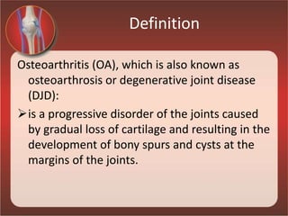
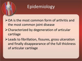
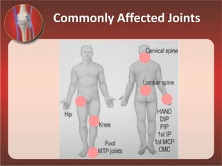
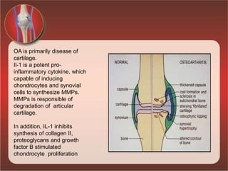
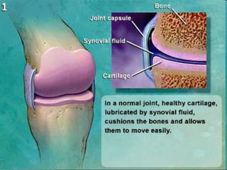
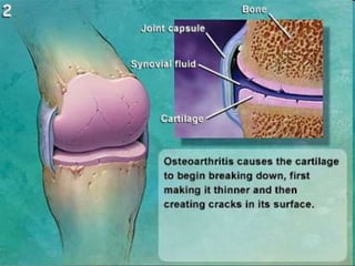
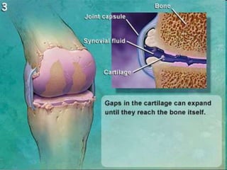
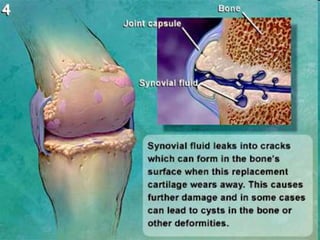
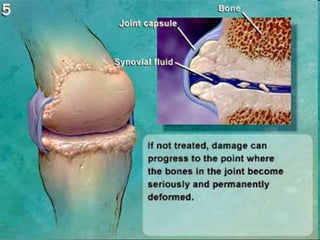
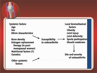
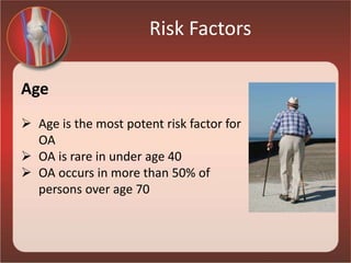
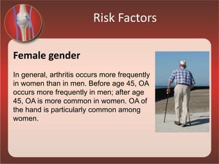
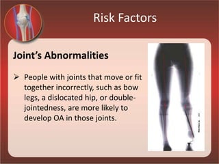
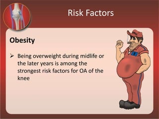
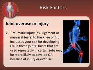
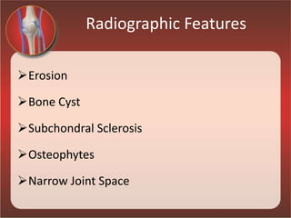
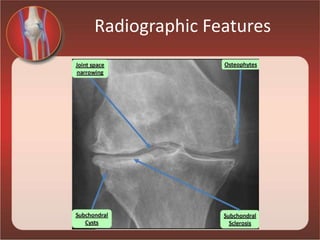
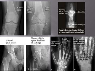
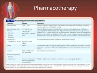
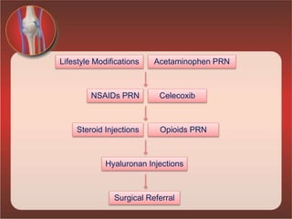
Osteoarthritis
Definition
Osteoarthritis (OA), which is also known as
osteoarthrosis or degenerative joint disease
(DJD):
is a progressive disorder of the joints caused
by gradual loss of cartilage and resulting in the
development of bony spurs and cysts at the
margins of the joints.
Epidemiology
OA is the most common form of arthritis and
the most common joint disease
Characterized by degeneration of articular
cartilage
Leads to fibrillation, fissures, gross ulceration
and finally disappearance of the full thickness
of articular cartilage
Commonly Affected Joints
Uncommonly Affected Joints
• Shoulder
• Wrist
• Elbow
• Metacarpophalangeal joint
• TMJ
• SI
• Ankle
Joint Protective Mechanisms
Joint Capsule
Ligaments
Muscle
Sensory afferents
Underlying bone
OA is primarily disease of
cartilage.
Il-1 is a potent pro-
inflammatory cytokine, which
capable of inducing
chondrocytes and synovial
cells to synthesize MMPs.
MMPs is responsible of
degradation of articular
cartilage.
In addition, IL-1 inhibits
synthesis of collagen II,
proteoglycans and growth
factor B stimulated
chondrocyte proliferation
Risk Factors
Age
 Age is the most potent risk factor for
OA
 OA is rare in under age 40
 OA occurs in more than 50% of
persons over age 70
Risk Factors
Female gender
In general, arthritis occurs more frequently
in women than in men. Before age 45, OA
occurs more frequently in men; after age
45, OA is more common in women. OA of
the hand is particularly common among
women.
Risk Factors
Hereditary Gene defect
A defect in one of the genes
responsible for the cartilage
component collagen can cause
deterioration of cartilage.
Risk Factors
Joint’s Abnormalities
 People with joints that move or fit
together incorrectly, such as bow
legs, a dislocated hip, or double-
jointedness, are more likely to
develop OA in those joints.
Risk Factors
Obesity
 Being overweight during midlife or
the later years is among the
strongest risk factors for OA of the
knee
Risk Factors
Joint overuse or injury
 Traumatic injury (ex. Ligament or
meniscal tears) to the knee or hip
increases your risk for developing
OA in these joints. Joints that are
used repeatedly in certain jobs may
be more likely to develop OA
because of injury or overuse
Sources of pain
Increase hemostatic pressure within bone rise
in OA
Bone marrow edema
Synovial inflammation
Joint effusions
Osteophytes
Symptoms & Signs
 Pain increases with activity and worse at night.
 Morning Stiffness less than 30 mins
 Swelling
 Deformities
 Joint instability
 Loss of function
 Neurological signs if spine Involved.
 Crepitus
 Muscle wasting
Diagnosis – Blood Tests
No Blood tests are routinely indicated for
workup of patients with OA unless
symptoms & signs suggest inflammatory
arthritis.
Diagnosis – Synovial Fluid
Examination of the synovial fluid is more
helpful diagnostically than an X-Ray.
Synovial Fluid in OA:
WBC<1000/mm3
Clear color
High Viscosity
Diagnosis – Radiography
X-Rays are indicated to evaluate chronic hand
pain and hip pain thought to be due to OA
For knee pain, X-Ray should be obtained if
symptoms or signs are not typical of OA or
Pain persists after inauguration of effective
treatment
Radiographic Features
Erosion
Bone Cyst
Subchondral Sclerosis
Osteophytes
Narrow Joint Space
Radiographic Features
Differential Diagnosis
Rheumatoid Arthritis
Gout
CPPD (Calcium pyrophosphate crystal
deposition disease)
Septic Joint
Polymyalgia Rheumatica
DISH
Management of OA
GOALs:
Educate patient about disease & management
Improve function
Control pain
Alter disease process and its consequences
Management of OA
Treatment of OA:
Pharmacotherapy
Non Pharmacotherapy
Patients with mild and intermittent symptoms
may need only Non Pharmacotherapy
Patients with ongoing, disabling pain are likely
to need both Non Pharmacotherapy and
Pharmacotherapy
Non Pharmacotherapy
NonPharmacotherapy
Reduce Load on the
joint
Avoid unnecessary stress
Increase muscle strength
Use of walking stick
Weight loss
Exercise
Correction of Malalignment
Pharmacotherapy
Lifestyle Modifications Acetaminophen PRN
NSAIDs PRN
Opioids PRN
Celecoxib
Steroid Injections
Hyaluronan Injections
Surgical Referral
Thank you

Osteoarthritis

  • 1.
  • 2.
    Definition Osteoarthritis (OA), whichis also known as osteoarthrosis or degenerative joint disease (DJD): is a progressive disorder of the joints caused by gradual loss of cartilage and resulting in the development of bony spurs and cysts at the margins of the joints.
  • 3.
    Epidemiology OA is themost common form of arthritis and the most common joint disease Characterized by degeneration of articular cartilage Leads to fibrillation, fissures, gross ulceration and finally disappearance of the full thickness of articular cartilage
  • 4.
  • 5.
    Uncommonly Affected Joints •Shoulder • Wrist • Elbow • Metacarpophalangeal joint • TMJ • SI • Ankle
  • 6.
    Joint Protective Mechanisms JointCapsule Ligaments Muscle Sensory afferents Underlying bone
  • 7.
    OA is primarilydisease of cartilage. Il-1 is a potent pro- inflammatory cytokine, which capable of inducing chondrocytes and synovial cells to synthesize MMPs. MMPs is responsible of degradation of articular cartilage. In addition, IL-1 inhibits synthesis of collagen II, proteoglycans and growth factor B stimulated chondrocyte proliferation
  • 14.
    Risk Factors Age  Ageis the most potent risk factor for OA  OA is rare in under age 40  OA occurs in more than 50% of persons over age 70
  • 15.
    Risk Factors Female gender Ingeneral, arthritis occurs more frequently in women than in men. Before age 45, OA occurs more frequently in men; after age 45, OA is more common in women. OA of the hand is particularly common among women.
  • 16.
    Risk Factors Hereditary Genedefect A defect in one of the genes responsible for the cartilage component collagen can cause deterioration of cartilage.
  • 17.
    Risk Factors Joint’s Abnormalities People with joints that move or fit together incorrectly, such as bow legs, a dislocated hip, or double- jointedness, are more likely to develop OA in those joints.
  • 18.
    Risk Factors Obesity  Beingoverweight during midlife or the later years is among the strongest risk factors for OA of the knee
  • 19.
    Risk Factors Joint overuseor injury  Traumatic injury (ex. Ligament or meniscal tears) to the knee or hip increases your risk for developing OA in these joints. Joints that are used repeatedly in certain jobs may be more likely to develop OA because of injury or overuse
  • 20.
    Sources of pain Increasehemostatic pressure within bone rise in OA Bone marrow edema Synovial inflammation Joint effusions Osteophytes
  • 21.
    Symptoms & Signs Pain increases with activity and worse at night.  Morning Stiffness less than 30 mins  Swelling  Deformities  Joint instability  Loss of function  Neurological signs if spine Involved.  Crepitus  Muscle wasting
  • 22.
    Diagnosis – BloodTests No Blood tests are routinely indicated for workup of patients with OA unless symptoms & signs suggest inflammatory arthritis.
  • 23.
    Diagnosis – SynovialFluid Examination of the synovial fluid is more helpful diagnostically than an X-Ray. Synovial Fluid in OA: WBC<1000/mm3 Clear color High Viscosity
  • 24.
    Diagnosis – Radiography X-Raysare indicated to evaluate chronic hand pain and hip pain thought to be due to OA For knee pain, X-Ray should be obtained if symptoms or signs are not typical of OA or Pain persists after inauguration of effective treatment
  • 25.
    Radiographic Features Erosion Bone Cyst SubchondralSclerosis Osteophytes Narrow Joint Space
  • 26.
  • 28.
    Differential Diagnosis Rheumatoid Arthritis Gout CPPD(Calcium pyrophosphate crystal deposition disease) Septic Joint Polymyalgia Rheumatica DISH
  • 29.
    Management of OA GOALs: Educatepatient about disease & management Improve function Control pain Alter disease process and its consequences
  • 30.
    Management of OA Treatmentof OA: Pharmacotherapy Non Pharmacotherapy Patients with mild and intermittent symptoms may need only Non Pharmacotherapy Patients with ongoing, disabling pain are likely to need both Non Pharmacotherapy and Pharmacotherapy
  • 31.
    Non Pharmacotherapy NonPharmacotherapy Reduce Loadon the joint Avoid unnecessary stress Increase muscle strength Use of walking stick Weight loss Exercise Correction of Malalignment
  • 32.
  • 33.
    Lifestyle Modifications AcetaminophenPRN NSAIDs PRN Opioids PRN Celecoxib Steroid Injections Hyaluronan Injections Surgical Referral
  • 34.