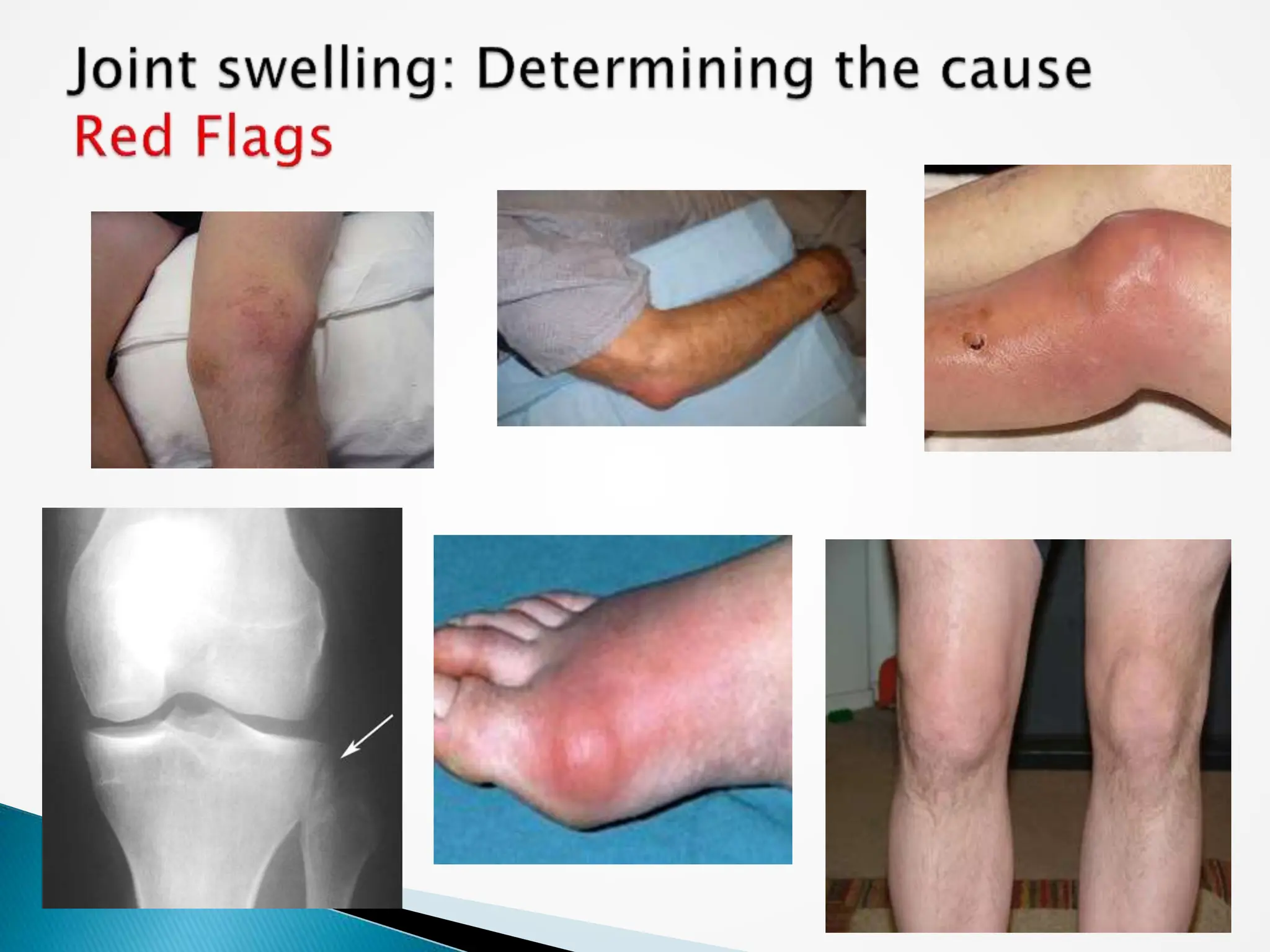
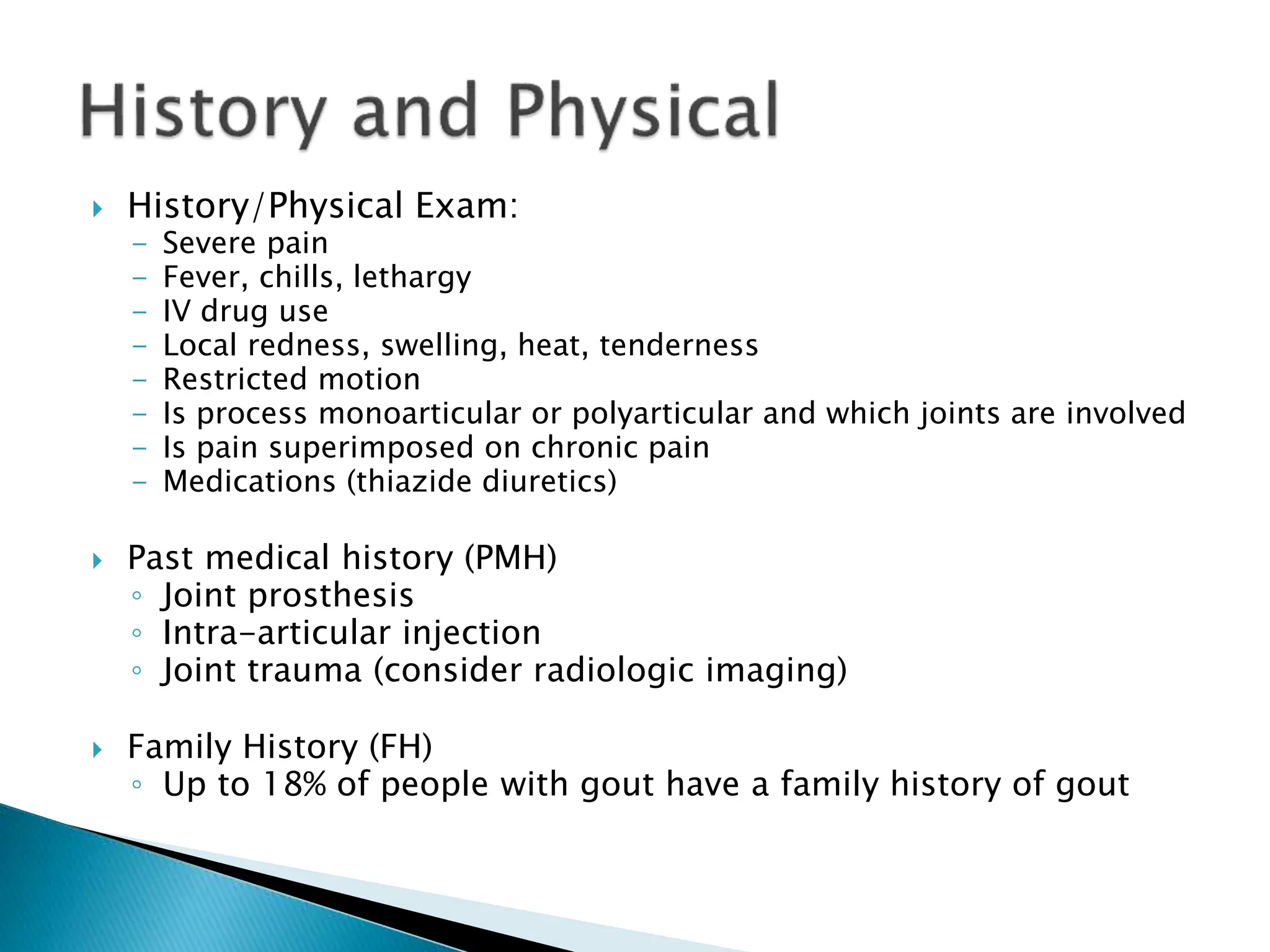
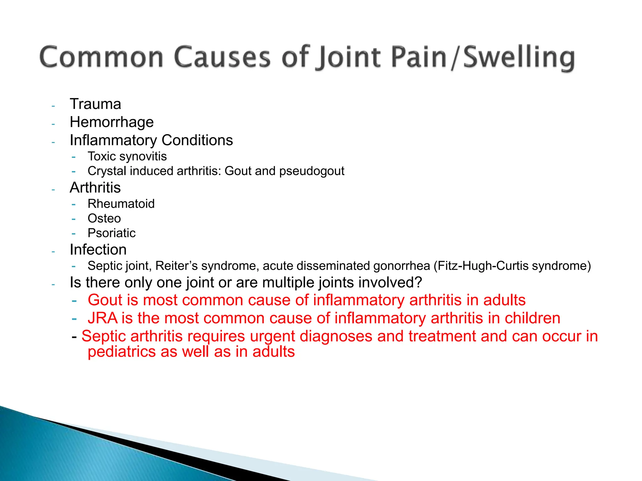
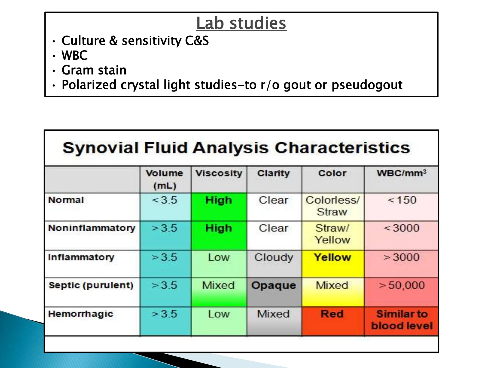
2. Please read chapters 40-41 in Campo and
Lafferty procedure book
3. To discuss the evaluation and management of
joint pain
To describe the most common etiologies for joint
pain and how to differentiate them
To demonstrate the proper techniques for
performing arthrocentesis and trigger point
injections for diagnostic and therapeutic purposes
4.
5. History/Physical Exam:
- Severe pain
- Fever, chills, lethargy
- IV drug use
- Local redness, swelling, heat, tenderness
- Restricted motion
- Is process monoarticular or polyarticular and which joints are involved
- Is pain superimposed on chronic pain
- Medications (thiazide diuretics)
Past medical history (PMH)
◦ Joint prosthesis
◦ Intra-articular injection
◦ Joint trauma (consider radiologic imaging)
Family History (FH)
◦ Up to 18% of people with gout have a family history of gout
6. - Trauma
- Hemorrhage
- Inflammatory Conditions
- Toxic synovitis
- Crystal induced arthritis: Gout and pseudogout
- Arthritis
- Rheumatoid
- Osteo
- Psoriatic
- Infection
- Septic joint, Reiter’s syndrome, acute disseminated gonorrhea (Fitz-Hugh-Curtis syndrome)
- Is there only one joint or are multiple joints involved?
- Gout is most common cause of inflammatory arthritis in adults
- JRA is the most common cause of inflammatory arthritis in children
- Septic arthritis requires urgent diagnoses and treatment and can occur in
pediatrics as well as in adults
8. To rule out an infectious etiology
◦ Synovial fluid evaluation (joint aspiration)
◦ CBC with diff, CRP, ESR and blood cultures
◦ Consider a plain film to assess baseline joint status
For suspected gout/pseudogout
◦ Synovial fluid analysis of crystals
◦ CBC, uric acid level: 10% of patients with gout will
have normal serum uric acid levels
9. Indications for arthrocentesis
Need to obtain synovial fluid for diagnosis
Drainage of hemarthrosis when conservative
management is unsuccessful
Instillation of local analgesic and anti-inflammatory
agents into a joint
Contraindications
Soft tissue infection overlying proposed site of
aspiration (absolute contraindication)
Uncooperative patient (relative contraindication)
Known coagulopathy or receiving anticoagulant
therapy (relative contraindication)
10.
11.
12.
13. Advantages of Ultrasound-guided injections
Precise Needle placement into joints for aspiration or
injection
Injection into tendon sheaths or bursa
Aspiration and injection of ganglion cysts
Ultrasound important tool in diagnosing and treating
baker’s cyst
In a clinical setting, a Baker’s cyst can mimic
the presentation of DVT – US can aide in diagnosis/Tx
14. Obtain 2-10 mL of synovial fluid in purple or green top
tube, 1 mL in red and 1 mL in gray top
Normal synovial fluid is viscous and will “string”
Synovial fluid analysis includes: Protein and Glucose,
Cell count and differential, Gm stain and C&S, crystals
◦ Normal synovial fluid is clear, straw-colored containing fewer
than 200 white cells per microliter, of which less than 25% are
polymorphonuclear neutrophils
◦ Serum glucose > 40 mg/dL in synovial fluid suggests infection
◦ Normal synovial fluid protein is 1/3 that of serum
◦ Urate crystals are diagnostic for gout
◦ Calcium crystals are diagnostic for pseudogout
15. Normal Traumatic Inflammatory Infected
Clarity Clear Clear to slightly
turbid
Slightly turbid to
turbid
Turbid to very
turbid
Viscosity Very viscous Mildly reduced Moderately
reduced
Greatly reduced
WBC/mm3 13-180 300-3000 3000-50,000 15,000-
>200,000
% polys 6.5
(0-25)
0-30 40-80 50-100
Glucose (diff
vs serum)
<10mg/
100mL less
than plasma
<20mg/
100mL less than
plasma
10-40mg/
100mL
less than plasma
>40mg/
100mL less than
plasma
Mucin clot Good Good-Fair Fair-Poor Poor to very poor
Crystals None None +/- none
Differential
diagnosis
DJD, trauma,
osteo-
chondromatosis
Rheumatic fever
Lupus, Gout
Pseudogout
Reiter’s syndrome
RA, Psoriatic
arthritis,
Ankylosing
spondylitis
Septic joint
TB arthritis
GC arthritis
19. Acute attack:
◦ NSAIDs are first-line treatment
Maximum doses for first 3 days, then taper over 4 days
Ibuprofen: 600-800 mg PO tid
◦ Colchicine is second line for acute attack in patient’s unable to tolerate NSAIDs
Should be used within 12-24 hrs of onset of attack
Dosing: 1mg followed by 0.5 mg q2h until absence of symptoms or GI side effects
◦ Joint aspiration
◦ Corticosteroids
Methylprednisolone 40 mg IM/IV every day for 3-4 days
Prednisone: 40 mg PO every day fro 3-4 days and then taper over 7-14 days
Chronic Suppressive Therapy:
◦ Xanthine oxidase inhibitors: Allopurinol considered first line treatment for chronic
management
Dosing: start at 100 mg daily and adjust every 2-4 weeks until goal of serum uric acid is <6 mg/dL for 3-6
mo.
◦ Uricosuric agents: Probenacid considered second line treatment for chronic
management
Dosing: Start at 250 mg PO bid and gradually increase to 500-2000 mg PO until desired SUA
22. Slipped capital femoral epiphysis (SCFE)
Legg-Calve-Perthes (aeseptic necrosis)
Toxic synovitis
◦ Common cause of hip pain in children before
puberty (4-10 yrs). It is a transient inflammatory
arthritis. Its cause is not known, but boys are
affected more often than girls (4 to 1).
◦ Associated with fever, hip, thigh or knee pain
◦ CBC, ESR, CRP, US of joint, x-ray, MRI, joint
aspiration
23. Avascular necrosis of hip in pediatrics
Risk factors: age less than 4, male gender
Reiter’s syndrome: A “reactive” arthritis with a high male to female ratio (20:1). The primary infectious site is usually urogenital or occasionally intestinal. Classic infection is Chlamydia urethritis.
90% of patients possess the HLA-B27 antigen.
Diagnosis of Reiter’s syndrome should be used only for patients who have classic triad (uveitis, urethritis, and arthritis).
The arthritis usually involves the large joints of the lower limbs. The spine is also involved frequently, especially the sacroiliac joints.
The urethritis should be appropriately diagnosed (culture) and treated with antibiotics Doxycycline 100 mg BID x 10 days. The painful arthritis can be treated with NSAIDs, etc.
Recurrences often occur even after treatment, and many patients develop chronic joint disease.
Gonococcal arthritis
Disseminated gonococcal infection (DGI)
Last reviewed: June 9, 2011.
Gonococcal arthritis is inflammation of a joint (usually just one) due to a gonorrhea infection.
See also: Non-gonococcal bacterial arthritis
Causes, incidence, and risk factors
Gonococcal arthritis is an infection of a joint. It occurs in people who have gonorrhea caused by the bacteria Neisseria gonorrhoeae. It affects women more often than men, and it is most common among sexually active adolescent girls.
Two forms of gonococcal arthritis exist:
One involves skin rashes and many joints, usually large joints such as the knee, wrist, and ankle
The second, less common form involves the spread of the bacteria through the blood (disseminated gonococcemia), which leads to infection of a single joint
Symptoms
Fever
Lower abdominal pain, may have liver tenderness and enlargement
Joint pain for 1 to 4 days
Pain in the hands or wrists due to tendon inflammation
Pain or burning during urination
Single joint pain
Skin rash (sores are slightly raised, pink to red, and may later contain pus or appear purple)
Signs and tests
Blood cultures should be checked in all cases of possible gonococcal arthritis.
http://www.ncbi.nlm.nih.gov/pubmedhealth/PMH0001489/
CRP is more sensitive than ESR in acute inflammation (Colombet, et al, 2010, American Journal of Medicine). ESR elevates as a result of fibrinogen entering the blood causing RBCs sticking together = ESR. Elevated ESR indicates an inflammatory process has been going on for at least several days. CRP is synthesized in the liver. Its physiologic role is to bind to phosphocholine expressed on the surface of dead or dying (apoptosis) cells in order to activate the complement/immune system, which enhances phagocytosis by macrophages. Levels of CRP begin to rise within 2 hours of an insult, and has a half-life of about 18 hours. The rapid action of CRP makes it a participant in the acute or first phase of the inflammatory process, which is why it is often called an "acute-phase protein."
An arthrocentesis is the most important diagnostic procedure for evaluation of the acutely inflamed joint
Normal Uric acid levels are 2.4-6.0 mg/dL (female) and 3.4-7.0 mg/dL (male). Hyperuricemia > 11 may cause risk of renal stones and renal impairment
Trigger points are discrete, focal, hyperirritable spots located in a taut band of skeletal muscle. Trigger-point injection has been shown to be one of the most effective treatment modalities to inactivate trigger points and provide prompt relief of symptoms.
Ganglion or synovial cysts are common, non-neoplastic soft tissue tumors that may occur in any joint, but most often occur on or around joints and tendon sheaths in the hands and feet
Legg-Calve-Perthes disease (LCPD) is avascular necrosis of the proximal femoral head resulting from compromise of the tenuous blood supply to this area. LCPD usually occurs in children aged 4-10 years. The disease has an insidious onset and may occur after an injury to the hip. In the vast majority of instances, the disorder is unilateral.
The incidence rate in boys (13.35 per 100,000) is higher than in girls (8.07 per 100,000). When compared with white children, black children have a higher incidence rate at 3.94 times, and Hispanic children have a 2.54 times higher incidence rate. SCFE mainly occurs between the ages of 10 and 16 years. 20% of patients have bilateral involvement at the time of presentation. The risk of SCFE is increased in children who are obese, as well as in children with other medical issues such as hypothyroidism, low growth hormone level, pituitary tumors, craniopharyngioma, Down syndrome, renal osteodystrophy, and adiposogenital syndrome. A slight downward trend has occurred for average ages over several years, with some data finding the average age for boys at 12.7 years and girls at 11.2 years. This change could be due to the phenomenon of children maturing at a younger age.