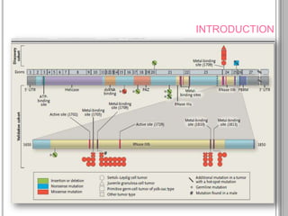
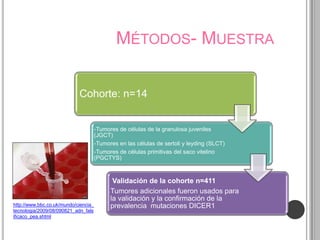
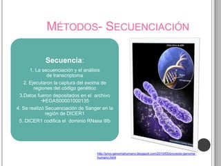
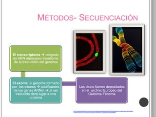
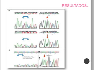
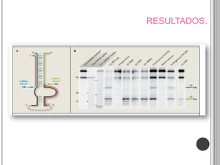
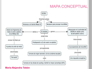
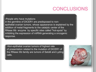
RECURRENT SOMATIC DICER1
                        MUTATIONS
         IN NONEPITHELIAL OVARIAN
                         CANCERS
          ALIREZA HERAVI-MOUSSAVI, PH.D., MICHAEL S. ANGLESIO, PH.D.,
S.-W. GRACE CHENG, PH.D., JANINE SENZ, B.SC., WINNIE YANG, B.SC., ET AL


                                      Mariana Tamayo Correa ID: 000192750
                               María Alejandra Tobón Arango. ID: 000192513
                                         Third Semester – Molecular biology
                                          Universidad Pontificia Bolivariana
                                                                       2012
INTRODUCTION
                                            Cancer is the uncontrolled
                                            growth of abnormal cells in the
                                            body. Cancerous cells are also
Cells are the building
                                            called malignant cells
blocks of living things.


 Cancer grows out of       Cancer can develop in
  normal cells in the       almost any organ or
        body                       tissue


  Cancer appears to
occur when the growth      It can also occur when
 of cells in the body is   cells forget how to die.
out of control and cells   Cell doesn't apoptosis
   divide too quickly.
INTRODUCTION

There are many
causes of cancers
INTRODUCTION


-Malignant tumor that grows in any part of
the ovary. Often in the epithelium.

-Generally, no symptoms.

-Sixth most common malignancy in women.

-High mortality.

-It is more common in women over 50 years.

-It is diagnosed in advanced stages.

-More common in nulliparous women.
INTRODUCTION


DICER1 gene encodes a protein synthesis that
bears his name, DICER1. It is present in many
organisms as Dicer.

Dicer: Is an endoribonuclease of the RNase III
family that cleaves double-stranded RNA
(dsRNA) and pre-microRNA (miRNA) into short
double-stranded RNA fragments.
INTRODUCTION


DICER1 mutations are common in non-
epithelial ovarian cancer, these mutations do not
affect completely the activity of DICER1, except, the
alteration in a kind of specific cells which is a novel
mechanism that disrupts
the microRNA processing that will cause cancer.


These mutations were restricted to the
codons encoding the metal binding sites within
the RNase IIIb catalytic centers, the enzyme
complex from which the DICER1 takes part.
GENERAL OBJECTIVE.

Demonstrate experimentally that DICER1 mutations in
the RNase family IIIb, are largely related to non-
epithelial ovarian tumors predominantly
in Sertolli cells and Leydig, that such mutations are
attributed to the addition of metal fragments in the
catalytic center of RNase IIIb, and that such changes do
not affect completely the function of DICER1, the
impacts are oncogenic only in certain types of cells like
ovarian epithelial cells.
INTRODUCTION
MÉTODOS- MUESTRA

                            Cohorte: n=14


                                      -Tumores de células de la granulosa juveniles
                                      (JGCT)
                                      -Tumores en las células de sertoli y leyding (SLCT)
                                      -Tumores de células primitivas del saco vitelino
                                      (PGCTYS)


                                             Validación de la cohorte n=411
                                            Tumores adicionales fueron usados para
                                            la validación y la confirmación de la
http://www.bbc.co.uk/mundo/ciencia_         prevalencia mutaciones DICER1
tecnologia/2009/08/090821_adn_fals
ificaco_pea.shtml
MÉTODOS- SECUENCIACIÓN

            Secuencia:
     1. La secuenciación y el análisis
             de transcriptoma
   2. Ejecutaron la captura del exoma de
        regiones del código genético
 3.Datos fueron depositados en el archivo
           EGAS00001000135
4. Se realizó Secuenciación de Sanger en la
              región de DICER1
 5. DICER1 codifica el dominio RNasa IIIb


                                    http://proy-genomahumano.blogspot.com/2010/05/proyecto-genoma-
                                    humano.html
MÉTODOS- SECUENCIACIÓN


El transcriptoma  conjunto
de ARN mensajero resultante
de la traducción del genoma


El exoma  genoma formado
por los exones  codificantes               Los datos fueron depositados
 de los genes ARNm  al ser                  en el archivo Europeo del
  traducido dara lugar a una                     Genoma-Fenoma
           proteina


                                http://noticias.lainformacion.com/ciencia-y-tecnologia/ciencias-general/definen-el-transcriptoma-completo-de-la-cianobacteria-
                                anabaena-decisiva-en-la-fijacion-del-nitrogeno-atmosferico_dhZQytF9OU5WqyElI9eIM3/
MÉTODOS- SECUENCIACIÓN
                                                      - Nucleótidos
                         4.Nucleótidos didesoxi    modificados que han
 Secuenciación                                      perdido el grupo
                         (ddATP, ddTTP, ddCTP
    Sanger                     y ddGTP)             hidroxilo 3' de la
                                                     desoxirribosa


análisis más detallado                              5. Nucleótidos 
 de la estructura del         3. Cebador o          incorporarse a la
 ADN averiguar la         "primer“, suele ser       cadena de ADN
    secuencia de                marcado            naciente, no se une
     nucleótidos.                                   por el extremo 3¨


                           2. Un enzima que       6. Una vez incorporado
  1. El ADN molde o
                          replique el ADN        un nucleótido didesoxi
segmento de ADN que
                         ADN Polimerasa I del      se termina la síntesis
 se desea secuenciar
                            bacteriofago T4.       de la cadena de ADN.
MÉTODOS - MARCAJE
          MARCAJE CON ISOTOPOS:


    • Incubacion de la proteina DICER1
    • 5'-32phosphate (32P) es un isótopo
1     radioactivo de fósforo


    • Etiquetaron sustrato de ARN de oligonucleotidos
    • Oligonucleótido secuencia corta
2     de ADN o ARN, 50 PB pares de bases o menos.


    • se analizaron los productos de escisión por medio
      de electroforesis en gel y fosforimaginacion.
3
MÉTODOS - MARCAJE

Técnica de electroforesis en gel:
separación de moléculas basadas en el
tamaño y la carga, que pueden hacerse
pasar a través de un gel hecho de agar o de
poliacrilamida.


          Las moléculas se dispersan en el gel y
          este se coloca en una cámara de
          electroforesis, que luego se conecta a
          una fuente de alimentación


          http://www.taringa.net/posts/ciencia-educacion/9792687/Aprende-de-
          Electroforesis-y-hacela-vos-mismo_.html
MÉTODOS - MARCAJE

Cuando la corriente eléctrica se aplica, las
moléculas más grandes se mueven lentamente
a través del gel mientras que las moléculas
más pequeñas se mueven más rápido


  http://javeriana.edu.co/Facultades/Ciencias/neurobioquimica/libros/celular/electroforesis.html
MÉTODOS - MARCAJE


    PHOSPHORIMAGING-
  FOSFORIMAGINACION:
Autorradiografía estándar que
 es mucho más precisa en la
cuantificación de la cantidad de
radiactividad en una sustancia.


                              1. http://quimicoclinico.wordpress.com/2008/02/03/toma-de-muestras-
                              sanguinea-venosa-y-gases-arteriales/
                              2. http://escritoriodocentes.educ.ar/datos/catalizadores.html
RESULTADOS.
RESULTADOS.
DISCUSSION
  AUTHORS             PUBLICATION              AGREEMENT
                                            OR DISAGREEMENT


                 A survey of cancer
                 cell lines showed that 4 of
Slade I, et al                               AGREEMENT
                 781 had truncating
                 changes in DICER1. 34
                 Somatic mutations
                 in DICER1 (albeit not the
Berger MF, et    hot-spot mutations
                                              AGREEMENT
al.              described here) have been
                 reported in a single patient
                 with prostate cancer. 35
DISCUSSION
  AUTHORS            PUBLICATION             AGREEMENT
                                          OR DISAGREEMENT


                 Among 823 patients with a AGREEMENT
                 variety of primitive tumors,
                 there was a low prevalence
Slade I, et al
                 of     germline     DICER1
                 mutations that are likely to
                 cause loss of function.34
                 Recent studies suggest
                 that miRNA* species may
Yang JS, Guo     be important in gene
                                          AGREEMENT
L, et al         regulation   rather than
                 simply being an inert
                 strand.38,39
MAPA CONCEPTUAL


Mariana Tamayo Correa
MAPA CONCEPTUAL


María Alejandra Tobón
CONCLUSIONS


                                                            The identification
                                                       of specific sequences of
                                                             gene mutations
                                                       associated with cancer is
                                                     important for epidemiological
                                                     studies to determine the early
                                                     detection of mutation in order
                                                     to find appropriate treatment.


http://www.bubbleinfo.com/2011/10/19/hot-new-idea/
CONCLUSIONS

                                                        Mutations in DICER1(Hot
                                                     spot)in Nonepithelial Ovarian
                                                       Cancers are highly frecuent
                                                      (60%) these mutations are in
                                                          primitive tumors and
                                                           embryonal tumors
                                                     suggests involvement of deve
                                                           loping cells, these
                                                       mutations are rare incancer
                                                                patients.


http://www.bubbleinfo.com/2011/10/19/hot-new-idea/
CONCLUSIONS
-People who have mutations
in the germline of DICER1 are predisposed to non-
epithelial ovarian tumors, whose appearance is explained by the
addition of metal fragments to the catalytic center of the
RNase IIIb enzyme by specific sites called "hot-spots" by
inhibiting the expression of miRNA generating a oncogenic
response.


 -Non-epithelial ovarian tumors of highest rate
 of presentation related to the mutation of DICER1 of
 the RNase IIIb family are tumors of Setolli and Lyding
 cells.
BIBLIOGRAFÍA


http://by168w.bay168.mail.live.com/default.aspx#!/mail/InboxLight.as
px?n=1723511564!n=731457103&fid=1&fav=1&mid=59a66730-
591e-11e1-94f2-002264c154b4&fv=1

-http://es.wikipedia.org/wiki/Northern_blot

-http://www.cancer.org/Espanol/cancer/ovario/Guiadetallada/

-http://www.ovationsforthecure.org/indexAwareness.php

-http://www.ambrygen.com/tests/dicer1-syndrome
Seminario biologia

Seminario biologia

  • 1.
    RECURRENT SOMATIC DICER1 MUTATIONS IN NONEPITHELIAL OVARIAN CANCERS ALIREZA HERAVI-MOUSSAVI, PH.D., MICHAEL S. ANGLESIO, PH.D., S.-W. GRACE CHENG, PH.D., JANINE SENZ, B.SC., WINNIE YANG, B.SC., ET AL Mariana Tamayo Correa ID: 000192750 María Alejandra Tobón Arango. ID: 000192513 Third Semester – Molecular biology Universidad Pontificia Bolivariana 2012
  • 2.
    INTRODUCTION Cancer is the uncontrolled growth of abnormal cells in the body. Cancerous cells are also Cells are the building called malignant cells blocks of living things. Cancer grows out of Cancer can develop in normal cells in the almost any organ or body tissue Cancer appears to occur when the growth It can also occur when of cells in the body is cells forget how to die. out of control and cells Cell doesn't apoptosis divide too quickly.
  • 3.
  • 4.
    INTRODUCTION -Malignant tumor thatgrows in any part of the ovary. Often in the epithelium. -Generally, no symptoms. -Sixth most common malignancy in women. -High mortality. -It is more common in women over 50 years. -It is diagnosed in advanced stages. -More common in nulliparous women.
  • 5.
    INTRODUCTION DICER1 gene encodesa protein synthesis that bears his name, DICER1. It is present in many organisms as Dicer. Dicer: Is an endoribonuclease of the RNase III family that cleaves double-stranded RNA (dsRNA) and pre-microRNA (miRNA) into short double-stranded RNA fragments.
  • 6.
    INTRODUCTION DICER1 mutations arecommon in non- epithelial ovarian cancer, these mutations do not affect completely the activity of DICER1, except, the alteration in a kind of specific cells which is a novel mechanism that disrupts the microRNA processing that will cause cancer. These mutations were restricted to the codons encoding the metal binding sites within the RNase IIIb catalytic centers, the enzyme complex from which the DICER1 takes part.
  • 7.
    GENERAL OBJECTIVE. Demonstrate experimentallythat DICER1 mutations in the RNase family IIIb, are largely related to non- epithelial ovarian tumors predominantly in Sertolli cells and Leydig, that such mutations are attributed to the addition of metal fragments in the catalytic center of RNase IIIb, and that such changes do not affect completely the function of DICER1, the impacts are oncogenic only in certain types of cells like ovarian epithelial cells.
  • 8.
  • 9.
    MÉTODOS- MUESTRA Cohorte: n=14 -Tumores de células de la granulosa juveniles (JGCT) -Tumores en las células de sertoli y leyding (SLCT) -Tumores de células primitivas del saco vitelino (PGCTYS) Validación de la cohorte n=411 Tumores adicionales fueron usados para la validación y la confirmación de la http://www.bbc.co.uk/mundo/ciencia_ prevalencia mutaciones DICER1 tecnologia/2009/08/090821_adn_fals ificaco_pea.shtml
  • 10.
    MÉTODOS- SECUENCIACIÓN Secuencia: 1. La secuenciación y el análisis de transcriptoma 2. Ejecutaron la captura del exoma de regiones del código genético 3.Datos fueron depositados en el archivo EGAS00001000135 4. Se realizó Secuenciación de Sanger en la región de DICER1 5. DICER1 codifica el dominio RNasa IIIb http://proy-genomahumano.blogspot.com/2010/05/proyecto-genoma- humano.html
  • 11.
    MÉTODOS- SECUENCIACIÓN El transcriptoma conjunto de ARN mensajero resultante de la traducción del genoma El exoma  genoma formado por los exones  codificantes Los datos fueron depositados de los genes ARNm  al ser en el archivo Europeo del traducido dara lugar a una Genoma-Fenoma proteina http://noticias.lainformacion.com/ciencia-y-tecnologia/ciencias-general/definen-el-transcriptoma-completo-de-la-cianobacteria- anabaena-decisiva-en-la-fijacion-del-nitrogeno-atmosferico_dhZQytF9OU5WqyElI9eIM3/
  • 12.
    MÉTODOS- SECUENCIACIÓN - Nucleótidos 4.Nucleótidos didesoxi modificados que han Secuenciación perdido el grupo (ddATP, ddTTP, ddCTP Sanger y ddGTP) hidroxilo 3' de la desoxirribosa análisis más detallado 5. Nucleótidos  de la estructura del 3. Cebador o incorporarse a la ADN averiguar la "primer“, suele ser cadena de ADN secuencia de marcado naciente, no se une nucleótidos. por el extremo 3¨ 2. Un enzima que 6. Una vez incorporado 1. El ADN molde o replique el ADN  un nucleótido didesoxi segmento de ADN que ADN Polimerasa I del se termina la síntesis se desea secuenciar bacteriofago T4. de la cadena de ADN.
  • 13.
    MÉTODOS - MARCAJE MARCAJE CON ISOTOPOS: • Incubacion de la proteina DICER1 • 5'-32phosphate (32P) es un isótopo 1 radioactivo de fósforo • Etiquetaron sustrato de ARN de oligonucleotidos • Oligonucleótido secuencia corta 2 de ADN o ARN, 50 PB pares de bases o menos. • se analizaron los productos de escisión por medio de electroforesis en gel y fosforimaginacion. 3
  • 14.
    MÉTODOS - MARCAJE Técnicade electroforesis en gel: separación de moléculas basadas en el tamaño y la carga, que pueden hacerse pasar a través de un gel hecho de agar o de poliacrilamida. Las moléculas se dispersan en el gel y este se coloca en una cámara de electroforesis, que luego se conecta a una fuente de alimentación http://www.taringa.net/posts/ciencia-educacion/9792687/Aprende-de- Electroforesis-y-hacela-vos-mismo_.html
  • 15.
    MÉTODOS - MARCAJE Cuandola corriente eléctrica se aplica, las moléculas más grandes se mueven lentamente a través del gel mientras que las moléculas más pequeñas se mueven más rápido http://javeriana.edu.co/Facultades/Ciencias/neurobioquimica/libros/celular/electroforesis.html
  • 16.
    MÉTODOS - MARCAJE PHOSPHORIMAGING- FOSFORIMAGINACION: Autorradiografía estándar que es mucho más precisa en la cuantificación de la cantidad de radiactividad en una sustancia. 1. http://quimicoclinico.wordpress.com/2008/02/03/toma-de-muestras- sanguinea-venosa-y-gases-arteriales/ 2. http://escritoriodocentes.educ.ar/datos/catalizadores.html
  • 17.
  • 18.
  • 19.
    DISCUSSION AUTHORS PUBLICATION AGREEMENT OR DISAGREEMENT A survey of cancer cell lines showed that 4 of Slade I, et al AGREEMENT 781 had truncating changes in DICER1. 34 Somatic mutations in DICER1 (albeit not the Berger MF, et hot-spot mutations AGREEMENT al. described here) have been reported in a single patient with prostate cancer. 35
  • 20.
    DISCUSSION AUTHORS PUBLICATION AGREEMENT OR DISAGREEMENT Among 823 patients with a AGREEMENT variety of primitive tumors, there was a low prevalence Slade I, et al of germline DICER1 mutations that are likely to cause loss of function.34 Recent studies suggest that miRNA* species may Yang JS, Guo be important in gene AGREEMENT L, et al regulation rather than simply being an inert strand.38,39
  • 21.
  • 22.
  • 23.
    CONCLUSIONS The identification of specific sequences of gene mutations associated with cancer is important for epidemiological studies to determine the early detection of mutation in order to find appropriate treatment. http://www.bubbleinfo.com/2011/10/19/hot-new-idea/
  • 24.
    CONCLUSIONS Mutations in DICER1(Hot spot)in Nonepithelial Ovarian Cancers are highly frecuent (60%) these mutations are in primitive tumors and embryonal tumors suggests involvement of deve loping cells, these mutations are rare incancer patients. http://www.bubbleinfo.com/2011/10/19/hot-new-idea/
  • 25.
    CONCLUSIONS -People who havemutations in the germline of DICER1 are predisposed to non- epithelial ovarian tumors, whose appearance is explained by the addition of metal fragments to the catalytic center of the RNase IIIb enzyme by specific sites called "hot-spots" by inhibiting the expression of miRNA generating a oncogenic response. -Non-epithelial ovarian tumors of highest rate of presentation related to the mutation of DICER1 of the RNase IIIb family are tumors of Setolli and Lyding cells.
  • 26.