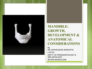
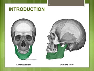
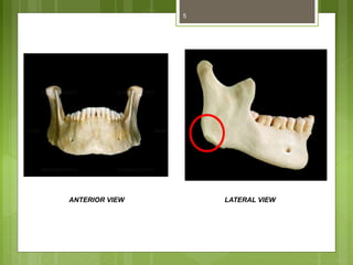
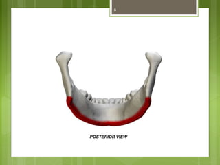
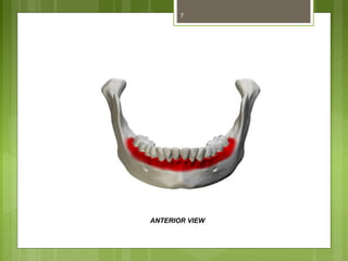
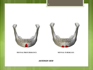
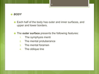
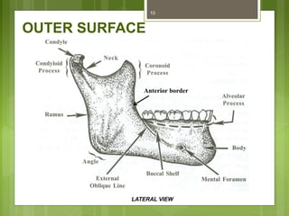
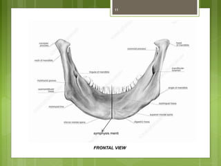
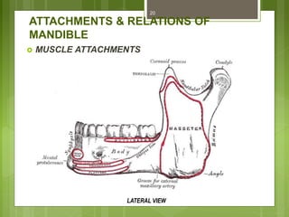
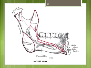
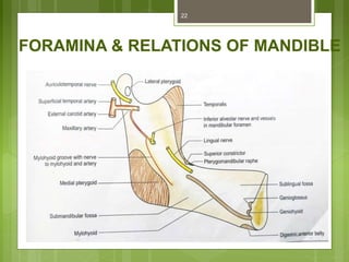
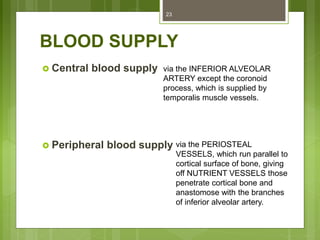
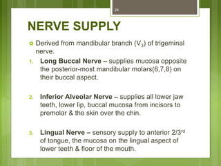
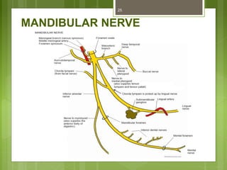
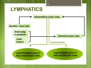
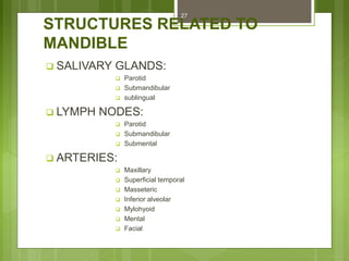
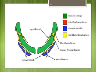
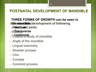
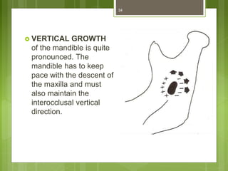
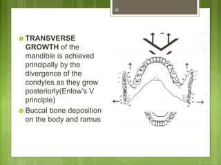
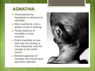
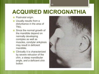
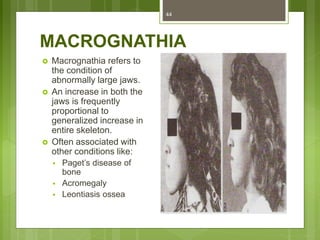
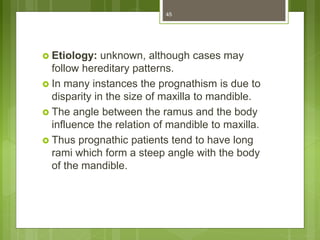
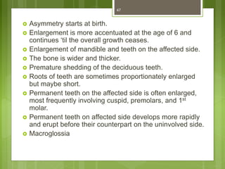
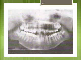
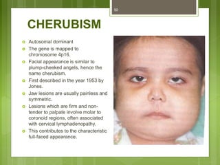
This document provides an overview of the mandible, including its anatomy, development, growth, age-related changes, and anatomical considerations. It describes the mandible's body, ramus, coronoid process, condylar process, attachments, foramina, blood supply, and related structures. It discusses the mandible's prenatal development from Meckel's cartilage and endochondral bone formation. It also addresses the postnatal development and growth of the mandible's various parts, as well as theories of mandibular growth. Common anatomical variations and conditions involving the mandible are described.
Trigeminal nerve maxillary nerve and clinical implicationDr Ravneet Kour
The document discusses the maxillary branch of the trigeminal nerve and its clinical implications. It begins by describing the basic anatomy of neurons, nerves and cranial nerves. It then focuses on the trigeminal nerve as the fifth cranial nerve, describing its nuclei, ganglion and three main branches - the ophthalmic, maxillary and mandibular nerves. Most of the document details the anatomy and branches of the maxillary nerve, including those in the pterygopalatine fossa, orbit, infraorbital canal and face. It concludes by discussing three clinical implications - trigeminal neuralgia, herpes zoster ophthalmicus and Wallenberg syndrome.
The document provides an overview of the facial artery, including its origin from the external carotid artery, course through the neck and face, branches, variations, clinical significance, and applied anatomy. The facial artery supplies structures of the superficial face like skin and muscles. It has cervical and facial parts. In the neck it passes beneath muscles and through the submandibular gland before curving over the mandible. Its branches include those supplying muscles, glands, lips and nose. Variations and its role in reconstructive procedures are discussed.
The document discusses the facial artery, which arises from the external carotid artery. It has both cervical and facial parts. The cervical part runs upwards in the neck, allowing movement of neck structures. It gives off branches like the ascending palatine and tonsillar arteries. The facial part enters the face by piercing the mandible. In the face, it gives branches like the inferior and superior labial arteries and terminates by anastomosing with the ophthalmic artery. The document also discusses the common carotid artery and its branches.
This document discusses condylar fractures of the mandible. It begins by providing background on condylar fracture development, anatomy, surgical anatomy, blood supply, nerve supply and muscle attachments. It then covers the etiology, associated injuries, mechanisms of injury and various classification systems for condylar fractures. The document outlines the diagnosis process including history, clinical examination and radiographic imaging. It concludes by discussing treatment approaches, focusing on the aims of surgery and indications for conservative versus surgical management.
Recent and Latest Advances in Oral and Maxillofacial surgery, Dr. Lidetu Afew...LIDETU AFEWORK
Every one should update himself according to the recent advances in every single profession/department. These are some of advancements We got in OMFS. We have also some latest advances and future advances under study that is going to be released in near future. BE HIGHTECH HIGH QUALITY UPDATED AND INFORMED PROFESSION.
The anatomy of the nerve supply of the head and neck has many significant applications in maxillofacial surgery. Understanding these important anatomic relations- variations enables surgeons to perform the surgical procedures safely. Knowledge of these concepts helps us to recognize the problems and complications as and when they occur and manage them accordingly.
The document discusses the lymphatic drainage of the head and neck region. It begins by describing the development, functions, and components of the lymphatic system. It then details the specific lymphatic drainage pathways and lymph nodes of the head and neck region. There are both superficial and deep lymph nodes that drain different areas and connect via lymphatic vessels and trunks to eventually drain into the right lymphatic duct or thoracic duct and return lymph to systemic circulation.
Different flap designs used for the management of impacted wisdommohamedamr94
This document discusses different flap designs used for surgical removal of impacted third molars. It describes several types of mucosal and mucoperiosteal flaps including envelope, triangular, modified triangular, comma-shaped, and Szmyd flaps. Key principles for flap design are outlined such as ensuring adequate access and blood supply while avoiding vital structures. Factors like bone exposure, flap position, and limitations of different techniques are compared. The literature review evaluates studies on primary wound healing comparing conventional versus modified flap designs.
Applied surgical anatomy of facial nerve in oral and maxillofacial surgeryShalini Bhatia
The document describes the anatomy and clinical considerations related to the facial nerve. It begins by detailing the various branches of the facial nerve and their course through the parotid gland and across the face. It then discusses landmarks used during surgery to identify the nerve. Several causes of facial nerve paralysis are listed including Bell's palsy, Ramsay Hunt syndrome, Melkersson-Rosenthal syndrome, and complications from procedures like IANB. Classification systems for nerve injuries are also summarized.
The document discusses naso-orbito-ethmoidal (NOE) fractures, which involve the central upper midface region. It describes the anatomy and classification of NOE fractures. Markowitz classification system categorizes NOE fractures into 5 types based on the status of the central bony fragment and involvement of the medial canthal tendon. Type I and II fractures involve a single or displaced central fragment with an intact tendon. Type III fractures have comminution beneath the tendon. Imaging such as CT is important for diagnosis.
This document provides an overview of maxilla anatomy and development. It discusses:
- The development of the maxilla from the first branchial arch during weeks 4-8 of gestation, including how the maxillary process, palatal shelves, and tongue form.
- Features of the adult maxilla, including its four surfaces and processes. It houses the maxillary sinus and articulates with several cranial bones.
- Age-related changes like a more vertical diameter in adults and absorption in older individuals.
- Considerations for periodontal and implant procedures related to anatomical structures like nerves, vessels and muscle attachments in the maxilla.
The document provides information on the growth and development of the maxilla. It begins with definitions of growth and development. It then discusses prenatal and postnatal growth of the maxilla, including formation from the first pharyngeal arch and development of related structures like the palate. The document outlines the anatomy of the maxilla including its surfaces, processes, sinuses and articulations. It notes age-related changes and clinical and prosthodontic considerations for treating developmental anomalies and edentulous patients.
This document provides an overview of Lefort I osteotomy, including:
- A brief history describing the development of maxillary osteotomy techniques.
- Anatomical considerations and the biologic basis for maxillary osteotomies, which have shown adequate blood supply can be maintained.
- Indications for Lefort I osteotomy include altering vertical dimension, anteroposterior movements, and surgical expansion of the maxilla.
- Types of Lefort I osteotomies are described, including classic, quadrangular, and segmental variations. Postoperative management and potential complications are also outlined.
This document provides an overview of the arterial supply of the head and neck. It begins with the embryological development of the aortic arches, which give rise to many major arteries. It then discusses the histology of arteries and describes the major arteries originating from the common carotid, external carotid, and internal carotid arteries. These include the lingual, facial, maxillary, and occipital arteries. It provides details on the branches, course, and anatomical relationships of these arteries.
The major salivary glands are the parotid, submandibular, and sublingual glands. The parotid gland is the largest and is located below and in front of the ear. The submandibular gland is beneath the jawbone and the sublingual gland is beneath the floor of the mouth. Each gland has specific blood supply, nerve innervation and ducts through which saliva passes. Imaging modalities like ultrasound and CT are useful for evaluating salivary gland disorders. Surgical procedures like gland excision require careful dissection to preserve structures like nerves and ducts.
This document discusses various maxillary osteotomies and orthognathic surgery procedures. It describes common maxillary deformities and the evaluation and planning process. Several maxillary osteotomy techniques are outlined, including segmental, total, Le Fort I, II, and III osteotomies. Key steps for each technique like incisions, osteotomy cuts, down fracture, and fixation are summarized. The roles of presurgical orthodontics and postsurgical orthodontics are also briefly discussed.
Condylar sag is defined as an immediate or late change in the position of the condyle in the glenoid fossa after orthognathic surgery, leading to an undesirable change in occlusion. It can occur after procedures like BSSO, IVRO, and Lefort I osteotomy. Risk factors include incorrect vectoring during condylar positioning and incomplete bone splits. Condylar sag is classified as central or peripheral, and can be diagnosed intraoperatively by examining changes in occlusion. Preventative measures include rigid fixation and intraoperative patient awakening to detect changes.
Venous drainage of head , neck and faceBhavesh Dhoke
The document summarizes the anatomy of head and neck veins. It describes the structure and layers of veins. It then discusses various superficial and deep veins of the head and neck region including the internal jugular vein, external jugular vein, anterior jugular vein, facial vein, pterygoid plexus, and venous sinuses. It notes that venous drainage of the head and neck terminates in the internal jugular vein. The document also briefly mentions diseases of veins.
Anatomy of mandible and its importance in implant placementDr Rajeev singh
This document discusses the anatomy and importance of the mandible in implant placement. It begins by defining the mandible and its embryological development. It then describes the osteology and features of the body, rami, processes, borders and attachments in detail. It discusses the blood supply, nerve supply and growth of the mandible postnatally. Finally, it explains the applied anatomy of the mandible and importance of anatomical structures like the mandibular foramen, inferior alveolar canal, mental foramen and nerve, and mandibular incisive canal in safe implant placement.
Dr. Ahmed M. Adawy, Professor Emeritus, Dep. Oral & Maxillofacial Surgery. Former Dean, Faculty of Dental Medicine
Al-Azhar University. There are four pairs of air sinuses making the boundaries of the nasal cavity. Maxillary sinus is the largest air cell. Anatomy and physiology of the maxillary sinus are given. Maxillary sinusitis is an inflammation of the sinus. Odontogenic causes represent nearly 30% of the etiology. Clinical and radiographic examinations are discussed together with treatment plan.
Oro-antral fistula is a rare complication of surgery at the posterior maxillary region. Several techniques for closure are presented. Additionally, information about sinus lift procedure is given.
The document discusses the development of the face from the 4th week of embryonic development. It describes how the face develops from three prominences - the frontonasal process and right and left maxillary and mandibular processes. It provides details on how various structures are formed from the fusion and development of these prominences, including the upper lip, lower lip, cheek, nose, palate and muscles of the face. The document also briefly discusses some common developmental anomalies of the face like cleft lip and cleft palate.
The document discusses the development of the face and its structures from early embryonic development through the formation of the branchial arches and facial prominences. It describes how the frontonasal, maxillary, and mandibular processes merge in the midline to form structures of the face, including the lips, nose, and palate. The development of specific structures like the nose, paranasal sinuses, jaws, and palate are then examined in more detail. The role of the branchial arches and Meckel's cartilage in mandibular development is also summarized.
1) The common carotid artery bifurcates into the internal and external carotid arteries at around the level of the thyroid cartilage in most individuals.
2) The external carotid artery gives off branches that supply the head and neck regions including the facial, lingual, occipital, posterior auricular, and superficial temporal arteries.
3) These arteries and their branches form distinct vascular territories called angiosomes that supply specific segments of the head and neck with blood. Surgeons use these angiosome concepts for planning complex tissue flaps.
Post natal growth of maxilla and mandibleDrArti Sharma
This document discusses postnatal growth of the maxilla and mandible. It defines growth, development, and the different phases of postnatal growth. For the maxilla, it describes the key growth mechanisms including endosteal and periosteal growth, cortical drift, the "V" principle, and counterparts in other structures. Growth occurs primarily in width early, then length, and lastly height. For the mandible, it discusses growth from birth to 1 year involving the ramus, condyle and body, and remodeling that occurs after age 1. Matrix and intramatrix rotation influence mandibular growth. Anomalies that can affect growth are also summarized.
The document provides an overview of the surgical anatomy of the mandible. It discusses the parts and features of the mandible, including the body, rami, coronoid and condylar processes. It details the growth and development of the mandible from the prenatal period through adulthood. Key anatomical structures are described, such as ligaments, muscles, nerves, blood vessels and lymph nodes associated with the mandible. Clinical considerations for surgical procedures involving the mandible are also mentioned.
The mandible develops from the first pharyngeal arch. Meckel's cartilage appears around the 6th week as a template for mandibular development. Ossification begins in membrane covering Meckel's cartilage, forming the body of the mandible around the mental and incisive nerves. Endochondral ossification forms the condylar process, mental region, and coronoid process. Postnatally, the mandible undergoes significant growth mediated by genetic and functional factors to accommodate the dentition and masticatory muscles.
This document provides definitions of growth and development and discusses the prenatal and postnatal development of the maxilla. It defines growth as an increase in size and development as progress towards maturity. It describes early embryonic events and development of the face between 4-8 weeks of gestation. Postnatally, it explains that growth of the maxilla occurs through displacement, remodeling, and growth at sutures in the transverse, anteroposterior, and vertical dimensions. It highlights several key factors that influence maxillary growth including the lacrimal suture, maxillary tuberosity, nasal airway, palatal remodeling, and orbital growth.
A detailed description of pre-natal and post-natal development of the mandible, with a brief description of theories of growth, for education purposes by Post Graduate students of Orthodontics and Dentofacial Orthopaedics
This document provides an overview of the mandible, including its development, anatomy, age-related changes, and clinical applications. It discusses how the mandible develops from the first pharyngeal arch and ossifies through both intramembranous and endochondral bone formation. The anatomy of the mandible is described in detail, including its various parts and structures. Age-related changes to the mandible from birth through adulthood and old age are also reviewed. Finally, the document discusses some applied clinical aspects of the mandible relating to dislocations, fractures, and considerations for surgery.
This document provides an overview of the mandible, including its development, anatomy, age-related changes, and clinical applications. It discusses how the mandible develops from the first pharyngeal arch and ossifies through both intramembranous and endochondral bone formation. The anatomy of the mandible is described in detail, including its various parts and surrounding structures. Age-related changes to the mandible from birth through adulthood and old age are also reviewed. Finally, the document discusses some applied clinical aspects of the mandible relating to dislocations, fractures, and considerations for surgery.
prenatal and post natal growth of mandiblemahesh kumar
The document discusses the prenatal and postnatal development of the mandible. Key points include:
1) The mandible initially develops from Meckel's cartilage during prenatal development and undergoes intramembranous and endochondral ossification.
2) Postnatally, the mandible grows at the condylar cartilage, posterior border of the ramus, and alveolar ridges. Growth occurs through remodeling and apposition of bone.
3) Mandibular growth is influenced by functional matrices like muscles and teeth which cause regional changes through resorption and displacement as the mandible grows in a downward and forward direction like an "expanding V".
The document discusses the development of the mandible from early gestation through adulthood. It begins with the formation of pharyngeal arches in the embryo and the development of Meckel's cartilage as the primary cartilage. Intramembranous and endochondral ossification then form the mandibular bone, guided by secondary cartilages like the condylar and coronoid cartilages. Postnatally, the mandible grows through remodeling and positional changes driven by functional needs. Several theories of mandibular growth are also summarized.
This document discusses the prenatal and postnatal growth and development of the mandible. It begins with an overview of the anatomy of the mandible. During prenatal development, the mandible originates from Meckel's cartilage in the first pharyngeal arch. Ossification begins with intramembranous bone formation followed by endochondral ossification at sites like the condyle. Postnatally, the mandible grows through bone remodeling at sites like the ramus, condyle and chin, leading to changes in shape with age. Developmental defects can restrict this growth, resulting in a narrow mandible with an inwardly rotated ramus.
The mandible is the largest and strongest bone of the face. It develops from the first pharyngeal arch and consists of a body with alveolar, inferior and mylohyoid borders, and a ramus with coronoid and condylar processes. The mandible undergoes both prenatal and postnatal growth, with the condyle showing considerable activity as the mandible grows downward and forward. Key parts include the body, ramus, coronoid process, and condylar process. The mandible articulates with the temporal bone at the temporomandibular joint.
This document summarizes the prenatal development and postnatal growth of the mandible. It begins with an overview of the formation of pharyngeal arches during embryonic development, including the mandibular arch which gives rise to the lower jaw. Meckel's cartilage provides a template for mandibular growth. Ossification begins in the mandible through intramembranous and endochondral bone formation. After birth, various regions such as the ramus, body, angle, and condyle continue growing through bone deposition and resorption to accommodate the erupting teeth and enlarging muscles. Growth generally ceases around age 20.
The mandible is the largest and strongest bone of the face. It develops from the first pharyngeal arch and consists of a body and bilateral rami. The body forms the lower jaw and contains the tooth sockets on its superior border. Each ramus has multiple surfaces and borders including the coronoid and condylar processes. Important anatomical landmarks include the mental foramen, mandibular canal, and angle of the mandible. The facial artery and branches of the trigeminal nerve course close to the mandible and must be protected during surgeries in this region.
The Indian Dental Academy is the Leader in continuing dental education , training dentists in all aspects of dentistry and
offering a wide range of dental certified courses in different formats.for more details please visit
www.indiandentalacademy.com
The document discusses the temporomandibular joint (TMJ), providing definitions and describing its key components, development, movements, age-related changes, and clinical applications. The TMJ is a synovial joint that allows hinge-like opening and closing of the mouth. It has unique features like an articular disc and fibrocartilage covering. Development occurs in three stages from weeks 7-17. The joint faces clinical issues like ankylosis, pain disorders, and limited mobility with age. Surgical treatments aim to create gaps and prevent re-fusion for improved function.
TMJ ANKYLOSIS of the Jaw and its clinical significanciesVamshi392572
This document provides an overview of temporomandibular joint (TMJ) ankylosis, including its embryology, anatomy, causes, pathogenesis, classification systems, clinical features, investigations, and management. Some key points:
- TMJ ankylosis is a bony or fibrous adhesion of the joint components that limits mouth opening. It is commonly caused by trauma or infection.
- Following trauma, haemarthrosis leads to organization of the intra-capsular hematoma and bone formation, resulting in ankylosis.
- Classification systems describe the extent of ankylosis based on the area of bony fusion. Management involves aggressive resection, coronoidectomy, inter
This document provides an overview of dental implants, including:
1. It describes the history and development of dental implants beginning with Brånemark's pioneering work in osseointegration in the 1950s and 1960s.
2. It classifies and describes different types of dental implants based on their placement, materials used, and treatment options provided, including root form, blade, cylindrical, screw-shaped, and subperiosteal implants.
3. It discusses the biological aspects and design considerations of dental implants, focusing on macrodesign including implant geometry, and microdesign including surface characteristics and modifications that enhance bone apposition.
This document provides an overview of periodontal plastic surgeries, specifically focusing on gingival recession and methods to increase the width of attached gingiva. It begins with definitions and classifications of gingival recession. Objectives, indications, and concepts like the tissue barrier are discussed. Decision trees and criteria for selecting techniques are presented. Main techniques to increase the width of attached gingiva like free gingival grafts and connective tissue grafts are described, including variations and the use of alternative donor tissues. Healing processes are summarized. The document provides foundational information on periodontal plastic surgery procedures for gingival recession and attached gingiva augmentation.
This document discusses endo-perio lesions, which involve both the dental pulp and surrounding periodontium. It describes the various types of lesions, including primary endodontic, primary periodontal, endo-secondary perio, and perio-secondary endo lesions. It also discusses diagnostic factors, treatment approaches, and case studies involving endo-perio lesions from 2014 to 2020. The optimal treatment is described as endodontic therapy preceding periodontal treatment to support healing, with regenerative procedures and adjuncts like ozone gas showing promise.
This document provides an overview of regenerative periodontal surgery techniques. It discusses the historical concepts of periodontal regeneration including bone grafts, guided tissue regeneration (GTR), and the emerging field of tissue engineering. Key cellular mediators and signaling molecules that can promote periodontal regeneration are described, including platelet-derived growth factor, bone morphogenetic proteins, insulin-like growth factor, and enamel matrix derivative. The document also reviews the different cell types involved in periodontal regeneration, including dental pulp stem cells, periodontal ligament stem cells, dental follicle progenitor cells, and dental epithelial stem cells. The criteria for achieving true periodontal regeneration and methods to guide cell differentiation and maturation are also summarized.
This document discusses risk factors for periodontal disease. It begins by introducing periodontitis as a ubiquitous disease with mixed microbial etiology. It then discusses the need to identify risk factors to inform public health measures and risk assessment. The document categorizes risk factors as modifiable (such as smoking, diabetes, stress) and non-modifiable (such as age, gender, genetics). Specific microorganisms and biomarkers associated with increased risk, like P. gingivalis and C-reactive protein, are also mentioned. Smoking is discussed in depth as a major established modifiable risk factor for periodontal disease.
Research methodology & principles and pitfalls of a clinical trial designDr Antarleena Sengupta
The document discusses principles and pitfalls of clinical trial design in research methodology. It covers topics like the objectives of research, types of research methodology including qualitative and quantitative methods, principles of clinical trial design, and potential pitfalls in clinical trial design. The document emphasizes that research methodology involves systematically solving research problems through logical steps like problem formulation, hypothesis formulation, sampling, data collection, data presentation, analysis and interpretation.
The document discusses factors responsible for failures in periodontal therapy. It identifies failures that can occur during the pre-therapeutic, therapeutic, and post-therapeutic phases of treatment. Pre-therapeutic failures include incorrect patient selection, incomplete diagnosis, and improper prognosis. Therapeutic failures involve issues with nonsurgical treatments like scaling and root planing as well as surgical procedures. Post-therapeutic failures relate to inadequate maintenance by the patient after treatment. Both dentist and patient-related factors can contribute to failures at each treatment phase.
This document discusses gingival enlargement and its classification and management. It begins by defining gingival enlargement and discussing its classification according to etiological factors, location, and degree. It then covers various indices used to measure gingival enlargement. The document discusses inflammatory enlargement, drug-induced gingival overgrowth, idiopathic enlargement, and enlargements associated with systemic diseases. Management techniques for different types of gingival enlargement such as scaling, surgery, and changing medications are presented.
This document discusses the role of viruses in periodontal diseases. It begins with an introduction to viruses and their evolution. It then covers the Baltimore classification system for viruses and viral components. The document discusses viral replication and the host immune response. It examines specific virus families like HIV, herpesviruses, and papillomaviruses in relation to periodontal diseases. It concludes with a section on herpetic gingivostomatitis.
This document discusses neutrophil disorders and their relationship to periodontal diseases. It begins with an introduction on the role of neutrophils in the innate immune system and periodontal diseases. It then describes various quantitative and qualitative neutrophil disorders. Quantitative disorders discussed include chronic benign neutropenia, cyclic neutropenia, congenital neutropenia, agranulocytosis, and Felty's syndrome. Qualitative disorders result from defects in neutrophil functions like rolling, adhesion, chemotaxis, phagocytosis, and intracellular killing. The document examines the oral complications that can result from various neutrophil disorders like gingivitis, periodontitis, and bone loss.
Necrotizing ulcerative gingivitis (NUG) is an acute, painful infection of the gums caused by an interaction between plaque bacteria like fusiform bacillus and spirochetes and the host immune response. It is characterized by necrosis and sloughing of gum tissue, presenting as punched-out ulcerations covered by a pseudomembrane. Diagnosis is based on clinical findings of painful ulcers with pseudomembrane, fetid odor, and potentially fever and lymphadenopathy. Treatment focuses on mechanical plaque removal and antibiotics to eliminate pathogenic bacteria.
This document discusses the effects of smoking on periodontal health. It covers epidemiological evidence that smoking increases the risk of periodontitis 2-5 times and is a major risk factor. The toxic chemicals in tobacco such as nicotine, carbon monoxide, and tar are discussed. These chemicals can impair the immune response and increase periodontal pathogens, leading to inflammation and tissue destruction. Clinical signs of periodontitis are made worse in smokers, such as increased attachment and bone loss. Smoking is also a major risk factor for oral cancer. The document examines the effects of smoking on gingival blood flow, wound healing, and the complications it can cause for periodontal therapy.
JUNCTIONAL EPITHELIUM IN HEALTH & DISEASE-- REGENERATION FOLLOWING SURGERYDr Antarleena Sengupta
This document provides an overview of junctional epithelium, including its structure, development, functions, and clinical significance. Some key points:
- Junctional epithelium forms a collar around the tooth and attaches the gingiva. It is derived from reduced enamel epithelium during tooth development.
- It has a stratified squamous non-keratinizing structure and rapidly turns over cells. The innermost layer of cells directly attach to the tooth surface.
- It plays roles in passive eruption of teeth, acts as a barrier in gingivitis, and its conversion to pocket epithelium is a hallmark of periodontitis development. Loss of its attachment can lead to pocket formation and inflammation.
The document discusses gingival crevicular fluid (GCF), including its history of study over 50 years, mechanisms and factors affecting its production, methods of collection, composition, and clinical significance. GCF is a serum-like fluid found in the gingival sulcus that can be assessed to provide diagnostic information about periodontal health and disease. The document outlines the anatomy of the gingival crevice and epithelium, as well as various methods that have been used to collect and analyze GCF components.
This document provides an overview of blood, its components, coagulation, and bleeding disorders. It discusses the main components of blood including erythrocytes, leukocytes, platelets, and plasma. It covers coagulation of blood and the formation of thrombi and emboli. Finally, it summarizes different bleeding disorders and clotting disorders.
This document provides an overview of local anaesthesia. It discusses the history of local anaesthetics from cocaine to lidocaine. It describes the properties, theories of action, classifications, composition, and pharmacology of local anaesthetics. The key modes of action are blocking sodium channels to prevent nerve impulse conduction. Local anaesthetics reversibly bind to specific receptor sites on sodium channels to inhibit sodium influx and nerve depolarization. Complications can include both local tissue toxicity and systemic effects.
Osteoporosis - Definition , Evaluation and Management .pdfJim Jacob Roy
Osteoporosis is an increasing cause of morbidity among the elderly.
In this document , a brief outline of osteoporosis is given , including the risk factors of osteoporosis fractures , the indications for testing bone mineral density and the management of osteoporosis
Cell Therapy Expansion and Challenges in Autoimmune DiseaseHealth Advances
There is increasing confidence that cell therapies will soon play a role in the treatment of autoimmune disorders, but the extent of this impact remains to be seen. Early readouts on autologous CAR-Ts in lupus are encouraging, but manufacturing and cost limitations are likely to restrict access to highly refractory patients. Allogeneic CAR-Ts have the potential to broaden access to earlier lines of treatment due to their inherent cost benefits, however they will need to demonstrate comparable or improved efficacy to established modalities.
In addition to infrastructure and capacity constraints, CAR-Ts face a very different risk-benefit dynamic in autoimmune compared to oncology, highlighting the need for tolerable therapies with low adverse event risk. CAR-NK and Treg-based therapies are also being developed in certain autoimmune disorders and may demonstrate favorable safety profiles. Several novel non-cell therapies such as bispecific antibodies, nanobodies, and RNAi drugs, may also offer future alternative competitive solutions with variable value propositions.
Widespread adoption of cell therapies will not only require strong efficacy and safety data, but also adapted pricing and access strategies. At oncology-based price points, CAR-Ts are unlikely to achieve broad market access in autoimmune disorders, with eligible patient populations that are potentially orders of magnitude greater than the number of currently addressable cancer patients. Developers have made strides towards reducing cell therapy COGS while improving manufacturing efficiency, but payors will inevitably restrict access until more sustainable pricing is achieved.
Despite these headwinds, industry leaders and investors remain confident that cell therapies are poised to address significant unmet need in patients suffering from autoimmune disorders. However, the extent of this impact on the treatment landscape remains to be seen, as the industry rapidly approaches an inflection point.
Basavarajeeyam is an important text for ayurvedic physician belonging to andhra pradehs. It is a popular compendium in various parts of our country as well as in andhra pradesh. The content of the text was presented in sanskrit and telugu language (Bilingual). One of the most famous book in ayurvedic pharmaceutics and therapeutics. This book contains 25 chapters called as prakaranas. Many rasaoushadis were explained, pioneer of dhatu druti, nadi pareeksha, mutra pareeksha etc. Belongs to the period of 15-16 century. New diseases like upadamsha, phiranga rogas are explained.
Muktapishti is a traditional Ayurvedic preparation made from Shoditha Mukta (Purified Pearl), is believed to help regulate thyroid function and reduce symptoms of hyperthyroidism due to its cooling and balancing properties. Clinical evidence on its efficacy remains limited, necessitating further research to validate its therapeutic benefits.
Local Advanced Lung Cancer: Artificial Intelligence, Synergetics, Complex Sys...Oleg Kshivets
Overall life span (LS) was 1671.7±1721.6 days and cumulative 5YS reached 62.4%, 10 years – 50.4%, 20 years – 44.6%. 94 LCP lived more than 5 years without cancer (LS=2958.6±1723.6 days), 22 – more than 10 years (LS=5571±1841.8 days). 67 LCP died because of LC (LS=471.9±344 days). AT significantly improved 5YS (68% vs. 53.7%) (P=0.028 by log-rank test). Cox modeling displayed that 5YS of LCP significantly depended on: N0-N12, T3-4, blood cell circuit, cell ratio factors (ratio between cancer cells-CC and blood cells subpopulations), LC cell dynamics, recalcification time, heparin tolerance, prothrombin index, protein, AT, procedure type (P=0.000-0.031). Neural networks, genetic algorithm selection and bootstrap simulation revealed relationships between 5YS and N0-12 (rank=1), thrombocytes/CC (rank=2), segmented neutrophils/CC (3), eosinophils/CC (4), erythrocytes/CC (5), healthy cells/CC (6), lymphocytes/CC (7), stick neutrophils/CC (8), leucocytes/CC (9), monocytes/CC (10). Correct prediction of 5YS was 100% by neural networks computing (error=0.000; area under ROC curve=1.0).
8 Surprising Reasons To Meditate 40 Minutes A Day That Can Change Your Life.pptxHolistified Wellness
We’re talking about Vedic Meditation, a form of meditation that has been around for at least 5,000 years. Back then, the people who lived in the Indus Valley, now known as India and Pakistan, practised meditation as a fundamental part of daily life. This knowledge that has given us yoga and Ayurveda, was known as Veda, hence the name Vedic. And though there are some written records, the practice has been passed down verbally from generation to generation.
Does Over-Masturbation Contribute to Chronic Prostatitis.pptxwalterHu5
In some case, your chronic prostatitis may be related to over-masturbation. Generally, natural medicine Diuretic and Anti-inflammatory Pill can help mee get a cure.
ABDOMINAL TRAUMA in pediatrics part one.drhasanrajab
Abdominal trauma in pediatrics refers to injuries or damage to the abdominal organs in children. It can occur due to various causes such as falls, motor vehicle accidents, sports-related injuries, and physical abuse. Children are more vulnerable to abdominal trauma due to their unique anatomical and physiological characteristics. Signs and symptoms include abdominal pain, tenderness, distension, vomiting, and signs of shock. Diagnosis involves physical examination, imaging studies, and laboratory tests. Management depends on the severity and may involve conservative treatment or surgical intervention. Prevention is crucial in reducing the incidence of abdominal trauma in children.
9. BODY
Each half of the body has outer and inner surfaces, and
upper and lower borders.
The outer surface presents the following features:
1. The symphysis menti
2. The mental protuberance
3. The mental foramen
4. The oblique line
9
12. INNER SURFACE has the following
features:
Mylohyoid Line
Submandibular fossa
Sublingual fossa
Superior and Inferior genial tubercles
Mylohyoid groove
12
15. LOWER BORDER
15
The lower border of the mandible is also called the base. Near
the midline the base shows an oval depression called the
digastric fossa.
16. RAMUS
16
ANTERIOR VIEW LATERAL VIEW
• LATERAL SURFACE of ramus is flat and bears a
number of oblique ridges.
U
L
P
A
17. MEDIAL SURFACE of ramus has the following:
Mandibular foramen
Lingula
Mylohyoid groove
Mandibular notch
Angle of mandible
17
Lingula
Submandibular fossaMylohyoid
groove
Mandibular notch
18. Coronoid process
Condyloid process
Head
Neck
Pterygoid fovea
18
Superior view Superior view
Pterygoid fovea
Neck
23. BLOOD SUPPLY
Central blood supply
Peripheral blood supply
23
via the INFERIOR ALVEOLAR
ARTERY except the coronoid
process, which is supplied by
temporalis muscle vessels.
via the PERIOSTEAL
VESSELS, which run parallel to
cortical surface of bone, giving
off NUTRIENT VESSELS those
penetrate cortical bone and
anastomose with the branches
of inferior alveolar artery.
24. NERVE SUPPLY
Derived from mandibular branch (V3) of trigeminal
nerve.
1. Long Buccal Nerve – supplies mucosa opposite
the posterior-most mandibular molars(6,7,8) on
their buccal aspect.
2. Inferior Alveolar Nerve – supplies all lower jaw
teeth, lower lip, buccal mucosa from incisors to
premolar & the skin over the chin.
3. Lingual Nerve – sensory supply to anterior 2/3rd
of tongue, the mucosa on the lingual aspect of
lower teeth & floor of the mouth.
24
26. LYMPHATICS
26
Submandibular lymph nodes
Submental lymph nodes
Jugulo-Omohyoid group of
deep cervical lymph nodes
Jugulo-Digastric group of
deep cervical lymph nodes
Mandible + lower teeth
Small wedge
in symphysis
Lower
incisors Extremely posterior
29. PRENATAL DEVELOPMENT OF
MANDIBLE
MECKEL’S CARTILAGE
Derived from 1st branchial arch
41st-45th day IU
Extends from cartilaginous otic capsule to
midline(symphysis)
Provides template for guiding growth of mandible
MANDIBULAR DIVISION, TRIGEMINAL NERVE(V3)→
first structure to develop
2 ossification centres: 1 for each half; arises 6th wk. IU
29
30. Ossifying membrane located lateral to the
Meckel’s Cartilage
Spreads below & around IAN and incisive
branch and upwards to form a trough to
accommodate developing tooth buds
Dorsally and ventrally spreads to form body and
ramus of mandible
Ossification continues→ Meckel’s cartilage
surrounded by bone→ invaded by bone→
ossification stops at lingula
30
Continued growth into middle ear : develops
auditory ossicles
To sphenoid bone to
form a remnant of
Meckel’s cartilage
Sphenomandibular ligament
31. ENDOCHONDRAL BONE FORMATION
THE CONDYLAR PROCESS:
5th wk. IU
10th- 14th wk. IU
THE CORONOID PROCESS:
10th – 14th wk. IU
Accessory cartilage gets incorporated into
expanding ramus; disappears before birth.
31
Area of mesenchymal condensation
seen above ventral part of the
developing mandible
Develops into a cone-shaped
cartilage → replaced by mid-fetal
life; upper end persists into
adulthood
Secondary cartilage of coronoid
process grows in response to
developing temporalis muscle.
32. MENTAL REGION:
2 small cartilages appear on either side of
symphysis
7th wk. IU
1st yr. postnatal
32
Formation of mental ossicles
Incorporated in intramembranous
ossification
Complete ossification
33. POSTNATAL DEVELOPMENT OF MANDIBLE
Divided into development of following
functional parts:
Ramus
Corpus or Body of mandible
Angle of the mandible
Lingual tuberosity
Alveolar process
Chin
Condyle
Coronoid process
33
THREE FORMS OF GROWTH can be seen in
the mandible:
o Vertical
o Transverse
o rotational
34. VERTICAL GROWTH
of the mandible is quite
pronounced. The
mandible has to keep
pace with the descent of
the maxilla and must
also maintain the
interocclusal vertical
direction.
34
35. TRANSVERSE
GROWTH of the
mandible is achieved
principally by the
divergence of the
condyles as they grow
posteriorly(Enlow’s V
principle)
Buccal bone deposition
on the body and ramus
35
36. In ROTATIONAL
GROWTH, the matrix
surrounding the
mandible acts to
moderate the shape
changes of the bone
rotating with it.
36
37. THEORIES OF GROWTH
ENLOW’S EXPANDING ‘V’ PRINCIPLE
ENLOW’S COUNTERPART PRINCIPLE
37
States that the growth of any given facial/cranial part
relates specifically to other structural and geometric
counterparts in the face and cranium.
The growth, movement &
enlargement of these
bones occur towards the
wide ends of the ‘V’ as a
result of differential
deposition & selective
resorption.
40. AGNATHIA
Characterized by
hypoplasia or absence of
mandible.
More commonly, only a
portion of jaw is missing.
Partial absence of
mandible is more
common.
Entire mandible on one
side may be missing or
more frequently, only the
condyle or the entire
ramus.
Bilateral agenesis of
condyles and ramus have
also been reported.
40
41. MICROGNATHIA
Means small jaw, either the maxilla or
mandible may be involved.
True micrognathia is classified as:
Congenital
Acquired
41
42. CONGENITAL MICROGNATHIA
Etiology:
I. Idiopathic
II. Assoc’d. with congenital heart disease
III. Pierre-Robin Syndrome
Follows a hereditary pattern.
Agenesis of condyles results in true
micrognathia.
Such cases may be due to posterior
positioning of the mandible with regard to the
skull or to a steep mandibular angle resulting
in apparent retrusion of mandible.
42
43. ACQUIRED MICROGNATHIA
Postnatal origin.
Usually results from a
disturbance in the area of
TMJ.
Since the normal growth of
the mandible depend on
normally developing
condyles as well as
muscles, condylar ankylosis
may result in deficient
mandible.
Clinically it is characterized
by severe retrusion of the
chin, a steep mandibular
angle, and a deficient chin
button
43
44. MACROGNATHIA
Macrognathia refers to
the condition of
abnormally large jaws.
An increase in both the
jaws is frequently
proportional to
generalized increase in
entire skeleton.
Often associated with
other conditions like:
Paget’s disease of
bone
Acromegaly
Leontiasis ossea
44
45. Etiology: unknown, although cases may
follow hereditary patterns.
In many instances the prognathism is due to
disparity in the size of maxilla to mandible.
The angle between the ramus and the body
influence the relation of mandible to maxilla.
Thus prognathic patients tend to have long
rami which form a steep angle with the body
of the mandible.
45
46. FACIAL HEMIHYPERTROPHY
One of the rare
developmental disorder.
Asymmetric over growth of
one or more body parts.
Represents hyperplasia
rather than hypertrophy.
It is of 3 types, namely:
Simple hyperplasia
Complex hyperplasia
Hemifacial hyperplasia
F:M > 2:1, often affecting on
right side.
46
47. Asymmetry starts at birth.
Enlargement is more accentuated at the age of 6 and
continues ‘til the overall growth ceases.
Enlargement of mandible and teeth on the affected side.
The bone is wider and thicker.
Premature shedding of the deciduous teeth.
Roots of teeth are sometimes proportionately enlarged
but maybe short.
Permanent teeth on the affected side is often enlarged,
most frequently involving cuspid, premolars, and 1st
molar.
Permanent teeth on affected side develops more rapidly
and erupt before their counterpart on the uninvolved side.
Macroglossia
47
49. PAGET’S DISEASE
Characterized by
excessive growth and
abnormal remodeling of
bone.
Results in bones which are
weak, enlarged and
extensively vascularized.
Etiology: unknown, there
may be evidence of
genetic link.
Possible etiologic factors:
viral infections
Inflammatory cause
Autoimmune connective
tissue
49
o Recognized most commonly after
the age of 50 years.
o Its prevalence increases with age.
o Male:female> 1:1
o Jaws are involved more commonly.
o The most common complaint is
bone pain.
o This pain is perceived as dull
aching pain deep below the soft
tissues.
o It may persist or exacerbate during
the night.
o The involved bone becomes warm
to the touch due to increased
vascularity
50. CHERUBISM
Autosomal dominant
The gene is mapped to
chromosome 4p16.
Facial appearance is similar to
plump-cheeked angels, hence the
name cherubism.
First described in the year 1953 by
Jones.
Jaw lesions are usually painless and
symmetric.
Lesions which are firm and non-
tender to palpate involve molar to
coronoid regions, often associated
with cervical lymphadenopathy.
This contributes to the characteristic
full-faced appearance.
50
51. CHERUBISM- RADIOGRAPHIC
APPEARANCE
Bilateral multilocular
radiolucencies in the
posterior mandible.
These lesions tend to
show varying degree of
remission and involution
after puberty.
There maybe
displacement, rotation of
the teeth.
Premature exfoliation,
delayed eruption.
51
52. EXOSTOSES
Normal anatomic
variation.
Hinders removal of
plaque by patient.
May have to be removed
to improve the prognosis
of neighbouring teeth.
Most common in lingual
area of canine and
premolars, above
mylohyoid muscle.
Also found on
buccal/labial surfaces of
mandibular teeth.
52
53. ANATOMIC SPACES
Several anatomic spaces or compartments
are found close to the operative field of
periodontal & implant surgery sites.
Contain loose connective tissue– easily
distended by hemorrhage, inflammatory fluid,
and infection.
Surgical invasion of these areas may result in
dangerous hemorrhage (intraoperative) or
infections (postoperative) & should be
carefully avoided.
53
54. ANATOMIC SPACES
54
A. SUBMENTAL SPACE: between mylohyoid muscle
superiorly and platysma inferiorly. Infection results in
swelling of the region; more dangerous as it proceeds
posteriorly.
B. MASTICATOR SPACE: contains masseter, pterygoid
(lat. and med.), tendon of insertion of temporalis, ramus
and posterior mandible. Infection leads to swelling of
face, severe trismus and pain.
C. SUBLINGUAL SPACE: below the oral mucosa in
anterior part of floor of mouth. Infection of this space
raises floor of mouth, displacing the tongue, resulting in
pain & difficulty swallowing.
D. SUBMANDIBULAR SPACE: external to sublingual
space below mylohyoid and hyoglossus muscle.
Contains the submandibular gland and connected with
sublingual space. Infections lead to obliterated
submandibular line+ pain in swallowing.
55. SURGICAL CONSIDERATIONS
Surgical trauma (pressure, manipulation and
postsurgical swelling) to the mental nerve
produces paresthesia of lip– recovers slowly.
Partial/complete cutting of the nerve can
result in permanent paresthesia/dysesthesia.
55
56. SURGICAL CONSIDERATIONS
In partially/completely edentulous patients,
disappearance of alveolar portion brings
mandibular canal and mental foramen closer
to superior border.
In such patients, during evaluation for
placement of implants, the distance
between canal and superior surface of the
bone as well as location of mental foramen
must be carefully determined to avoid surgical
injury to the nerve.
56
57. SURGICAL CONSIDERATIONS
The lingual nerve lies close to the surface of
the oral mucosa in the third molar area and
goes deeper as it travels forward.
It can be damaged during anesthetic
injections (and during extraction
procedures).
It can be injured when a periodontal partial-
thickness flap is raised in third molar region
or when releasing incisions are made in the
area.
57
58. SURGICAL CONSIDERATIONS
The alveolar process, which provides the supporting
bone to the teeth, has a narrower distal curvature than
the body of mandible, creating a flat surface in the
posterior area between the teeth and the anterior border
of the ramus.
This results in the formation of external oblique ridge,
which runs downward and forward to region of
second/first molar, creating a shelflike bony area.
Resective osseous therapy may be difficult in this area
because of the amount of bone that must be removed
distally toward ramus to achieve resection of a
periodontal osseous defect on the distal aspect of
mandibular 2nd/3rd molar.
58
59. SURGICAL CONSIDERATIONS
Distal flap procedures in distal to the last molar
can be performed effectively only if there exists
sufficient space.
59
60. CONCLUSION
Familiarity with the location and appearance
of the mental nerve reduces likelihood of
injury to the nerve.
Determining the amount of available bone is
critical for placement of implants.
60
61. REFERENCES
1. B D Chaurasia’s Human Anatomy vol. 3
2. Human Embryology, Inderbir Singh
3. Clinical Periodontology by Carranza, 10th edition,
vol 2
4. Shafer’s Textbook of Oral Pathology
5. Contemporary Orthodontics by W R Profitt
6. Handbook of Local Anesthesia, S F Malamed
7. Image references:
Wikimedia Commons
Gray’s anatomy, 41st edition
61