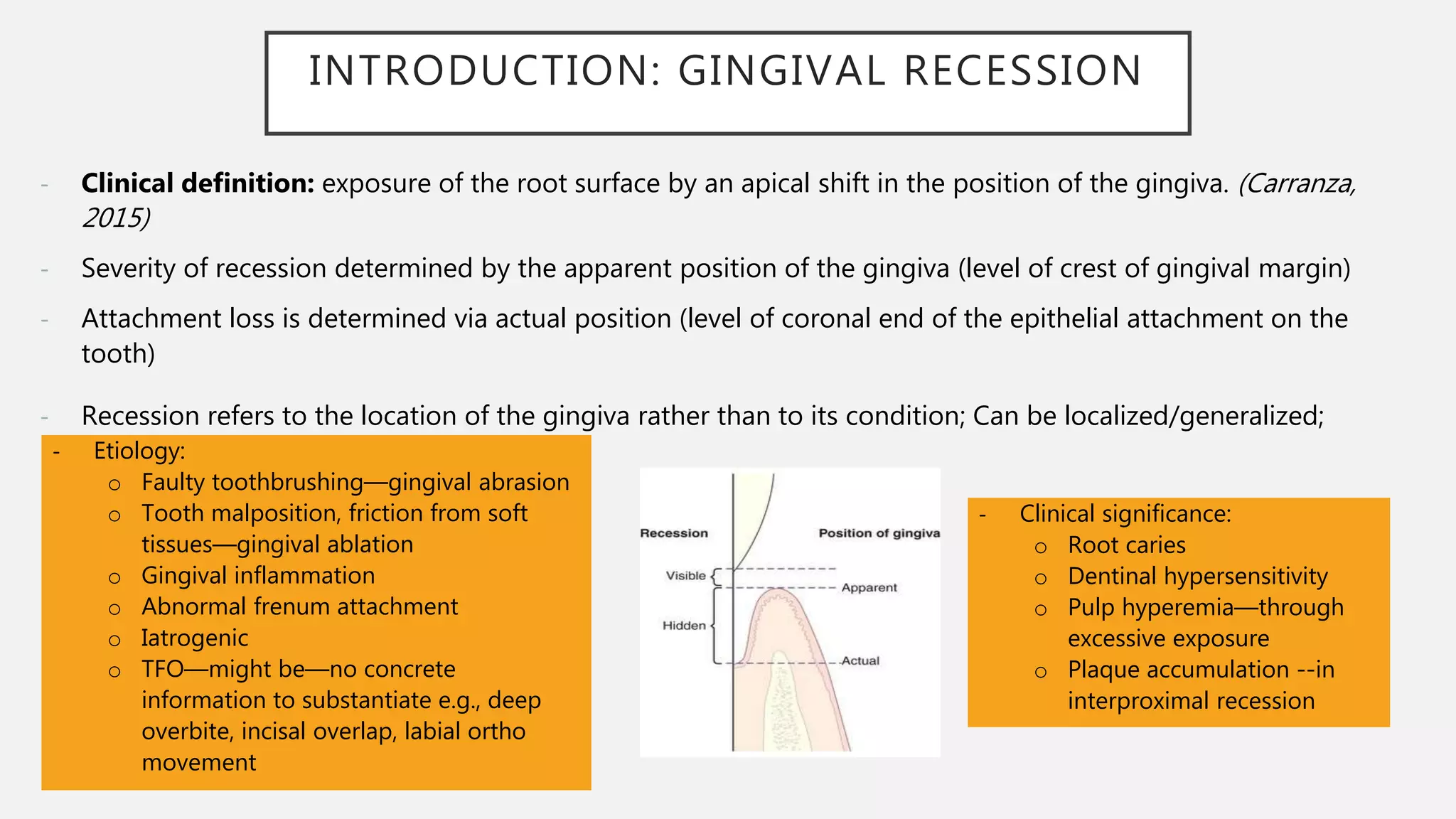
This document provides an overview of periodontal plastic surgeries, specifically focusing on gingival recession and methods to increase the width of attached gingiva. It begins with definitions and classifications of gingival recession. Objectives, indications, and concepts like the tissue barrier are discussed. Decision trees and criteria for selecting techniques are presented. Main techniques to increase the width of attached gingiva like free gingival grafts and connective tissue grafts are described, including variations and the use of alternative donor tissues. Healing processes are summarized. The document provides foundational information on periodontal plastic surgery procedures for gingival recession and attached gingiva augmentation.
This document discusses various surgical techniques for preserving the interdental papilla during periodontal regeneration procedures. It describes the conventional papilla preservation flap technique introduced by Takei in 1985, as well as several modifications including the modified papilla preservation flap, simplified papilla preservation flap, interproximal tissue maintenance technique, and whale's tail technique. The advantages and disadvantages of each technique are summarized. A novel entire papilla preservation technique introduced in 2015 is also outlined, which aims to completely preserve the interdental papilla.
Scaling and root planing (SRP) is a non-surgical treatment for periodontitis that aims to remove dental plaque and calculus from tooth surfaces. It involves scaling to remove deposits and root planing to smooth root surfaces. The goals are to eliminate periodontitis by removing irritants and restoring a healthy environment for tissue healing. The long-term effectiveness depends on factors like patient compliance, disease severity, and anatomical challenges. Overhanging restorations can interfere with cleaning and disturb the ecological balance, allowing disease-causing bacteria to proliferate.
This document discusses periodontal flaps, which are sections of gingiva surgically separated from underlying tissues to provide access to bone and roots. It defines different types of flaps classified by bone exposure, placement, and papilla design. Indications and contraindications for various flaps are outlined. Procedures for modified Widman, undisplaced, apically displaced, and regenerative flaps are described. Distal molar surgery techniques and use of periodontal packs are also summarized.
1. Osseous surgery involves modifying the alveolar bone support of teeth and includes techniques like osteoplasty and ostectomy. It aims to eliminate pockets and correct unphysiological bone architecture.
2. Factors in selecting a technique include the amount and location of bone loss, root trunk length, and anatomical limitations. Techniques range from non-resective procedures like osteoplasty to resective procedures like ostectomy.
3. Outcomes of osseous surgery generally include pocket elimination and establishment of physiological bone contours and architecture, though some bone loss from remodeling is expected in the range of 0.06mm to 1.2mm.
This document discusses furcation involvement, including classifications, diagnosis, treatment options, and prognosis. It notes that furcation involvement indicates advanced periodontitis and poorer prognosis. Treatment depends on the grade of involvement and may include nonsurgical therapy like scaling and root planing, surgical approaches like furcation plasty, regenerative techniques like GTR, or extraction. Prognosis is best for grade I and II furcations treated nonsurgically or with furcation plasty, and poorer for grade III and IV furcations. Long-term success requires eliminating plaque, establishing anatomy to facilitate cleaning, and preventing further attachment loss.
ROS is a substractive method of having positive bone architecture. it includes osteotomy and ostectomy procedures. osteotomy is to remove non supporting bone and ostectomy is to remove supporting bone for having positive bony architecture. there is definitive osseous surgery and compromise osseous surgery. transgingival probing is a method of determining osseous topography. various hand and rotary instruments are use for this procedure.
REFERENCES TAKEN FROM CARRANZA'S TEXTBOOK OF CLINICAL PERIODONTOLOGY AND LINDHE'S TEXTBOOK OF CLINICAL PERIODONTOLOGY AND IMPLANT DENTISTRY. CONTAINS ENOUGH AND MORE DETAILS OF THIS TOPIC FOR BDS STUDENTS.HOPE THIS PRESENTATION WILL HELP U GAIN SOME KNOWLEDGE ABOUT PERIODONTAL PLASTIC AND ESTHETIC DENTISTRY.
Emdogain is a gel containing enamel matrix proteins that has been shown to regenerate hard and soft tissues lost to periodontal disease. It works by attracting mesenchymal cells to the root surface, promoting attachment, proliferation, and differentiation which results in new cementum, bone, and periodontal ligament formation. Over 20 years of clinical studies involving over 2 million patients have demonstrated its effectiveness and safety in treating intra-bony and gingival recession defects.
mucogingival surgery or plastic surgery of muco-gingival tissue is a surgical procedure targeted to correct and eliminate anatomic, developmental and traumatic alterations of gingiva.
The modified Widman flap is a periodontal surgical technique used to obtain access to the root surface and allow for intimate postoperative adaptation of healthy connective tissue and epithelium to the root surface. Key aspects include:
1. Incisions are made internally and intracrevicularly to minimize tissue loss and gingival shrinkage. Vertical releasing incisions are usually not used.
2. The goal is access for root debridement rather than pocket elimination. Minimal flap elevation of 1-2mm is done to access root surfaces.
3. It is indicated for mild to moderate periodontitis with pocket depths up to 6mm and minimal inflammation. Primary intention healing occurs.
This document outlines general principles of periodontal surgery. It discusses indications and contraindications for periodontal surgery, as well as patient preparation, anesthesia, tissue management, hemostasis, dressings, and post-operative care. The document provides details on outpatient versus hospital periodontal surgery and describes various surgical instruments used. It emphasizes the importance of careful planning, thorough debridement, and hemostasis during periodontal surgery to facilitate healing.
Periodontal flap surgeries by Dr. JerryDeepesh Mehta
The document discusses periodontal flap surgeries, which involve procedures to treat periodontitis by eliminating harmful bacteria and reducing disease progression. It provides a historical overview of developments in flap surgery techniques from the 1800s to present. It then describes the objectives, indications, contraindications, critical zones, types of incisions, flap types, and techniques for various pocket therapies like gingivectomy, Widman flap, and apically repositioned flap. The goal of periodontal flap surgeries is to access roots and bone, improve visibility and effectiveness of cleaning, and modify osseous defects to treat periodontal disease.
This document discusses mucogingival surgery and periodontal plastic surgery procedures. It begins with definitions and terminology related to these procedures. The main objectives are to correct issues with attached gingiva, shallow vestibule, and aberrant frenum. Common procedures discussed include gingival augmentation using free gingival grafts to increase the width and thickness of gingiva. Free gingival grafts involve obtaining a partial thickness graft from the palate donor site and suturing it to the recipient site to increase the zone of attached gingiva. Variations on this technique include accordion and strip methods. The healing process of free gingival grafts is also summarized.
This document discusses biological width, which refers to the dimensions of soft tissue attached to the tooth coronal to the alveolar bone crest. It defines biological width as the connective tissue attachment (1.07mm on average) plus the epithelial attachment (0.97mm on average), totaling 2.04mm. It discusses factors that can lead to biological width violation like subgingival restoration margins and its signs. Methods to evaluate and correct biological width violations like bone sounding, surgical crown lengthening, and forced tooth eruption are also described. The importance of respecting biological width is emphasized in restorative and implant dentistry.
The document discusses the history and principles of periodontal plastic surgery, which aims to correct anatomical defects of the gingiva, alveolar mucosa, or bone through procedures like gingival augmentation and root coverage. Various surgical techniques are described for increasing attached gingiva, such as free gingival autografts, free connective tissue autografts, and apically positioned flaps. The healing process and indications for different procedures are also reviewed.
This document summarizes various gingival surgical techniques including gingival curettage. It discusses the history and development of gingival curettage, defines the technique, and outlines the rationale, procedure, indications, contraindications and healing process. Key pioneers in developing gingival curettage techniques are mentioned. Variations of gingival curettage including the excisional new attachment procedure and use of ultrasonic instruments are also summarized.
A brief description of all topics to recent advances,SDD, host modulation and diabetes, host modulation in smokers, chemically modified tetracyclines, bisphosphonates
Resective osseous surgery involves removing tooth-supporting alveolar bone to reshape it and eliminate periodontal pockets. It is indicated for inconsistent bone margins, reverse bone architecture, one-wall defects, and other bone irregularities. The surgery involves using burs and chisels to perform vertical grooving of thick bone, blending any bone ledges on tooth roots, flattening interproximal bone in narrow areas, and gradualizing marginal bone. Flaps are closed to cover the new bony margins. Post-operative maintenance like cleaning and antibiotics is needed to allow healing over 2-3 weeks.
This document discusses gingival recession, including its definitions, classifications, etiology, factors affecting treatment outcomes, and treatments. It provides an overview of several classification systems for gingival recession, including those proposed by Sullivan and Atkins, Miller, Mahajan, Cairo, and Ashish Kumar. Miller's classification is the most widely used but has limitations, so modifications have been suggested. The document also proposes a new comprehensive classification system that aims to address the limitations of previous systems.
This document discusses various flap techniques used in periodontal surgery. It defines flaps as sections of gingiva and mucosa surgically separated from underlying tissues to provide access to bone and roots. Full and partial thickness flaps are classified based on the depth of tissue reflection. Techniques include the modified Widman flap, undisplaced flap, apically displaced flap, papilla preservation flap, and techniques for distal molar surgery. Healing after flap surgery is described in stages from initial clot formation to establishment of new connective tissue attachment after 4 weeks.
Periodontitis is a complex infection initiated by bacteria –tissue destruction.
Host: the organism from which a parasite obtains its nourishment/ an individual who receives a graft
Modulation: the alteration of function or status of something in response to a stimulus or an altered physical or chemical environment
Full Mouth Disinfection (FMD) is a treatment approach that involves scaling and root planing of all teeth in one or two visits to eliminate periodontal pathogens. The goals of FMD are to prevent reinfection of treated sites by untreated sites or other oral niches harboring pathogens. FMD originally included scaling, root planing, chlorhexidine treatment, and prolonged chlorhexidine use. Over time, variations have been developed including replacing chlorhexidine, supplementing with antibiotics or probiotics, and combining with photodynamic therapy. FMD aims to provide more effective periodontal treatment than the standard approach of scaling and root planing in quadrants over multiple visits.
This document discusses the anatomy, measurement, and clinical significance of the attached gingiva. It notes that the attached gingiva extends from the base of the gingival sulcus to the mucogingival junction. The normal width is 3-4.5mm in the maxillary anterior region but narrower in other areas. Inadequate width can facilitate subgingival plaque formation. Methods to measure width and increase width through surgery are described. The importance of keratinized, attached tissue for resisting mechanical irritation and stabilizing the gingival margin is emphasized.
The document discusses the history and techniques of periodontal flap surgery. It describes various flap designs like the Widman flap from 1918, Neumann flap from 1920, and modified Widman flap from 1974. It discusses the objectives of surgical flaps like pocket elimination, preservation of attached gingiva, and access to underlying bone. Key aspects like incision types, critical probing depths, and zones examined in pocket surgery are summarized. The document provides an overview of periodontal flap surgery.
This document discusses various techniques for root coverage procedures in periodontal therapy. It begins with definitions of gingival recession and Miller's classification system. It then describes techniques such as free gingival autografts, pedicle grafts, coronally advanced flaps, subepithelial connective tissue grafts, guided tissue regeneration, and pouch and tunnel techniques. For each technique, it provides background information and outlines the surgical steps. The goal of these procedures is to cover exposed root surfaces and enhance esthetic and functional outcomes.
This document provides information on periodontal plastic and aesthetic surgery procedures. It discusses the objectives of these procedures which include creating an adequate zone of attached gingiva and eliminating muscle pulls. It describes various gingival augmentation techniques used to widen attached gingiva including pedicle flaps and free soft tissue grafts. The document also discusses that while a minimal width of gingiva can maintain health, gingival augmentation may be indicated in situations involving planned orthodontic treatment, subgingival restorations, or patient discomfort.
Reconstructive periodontal surgery aims to treat deep pockets which have not be reduced after non surgical periodontal therapy. periodontal regenerative procedures mainly include the use of modified flap techniques , use of bone grafts and newer gene therapies. Biologic mediators play key role in the regeneration process. Guided tissue regeneration and Guided Bone regeneration are commonly used methods for periodontal regeneration. Minimally invasive surgical techniques are preferred surgical methods for treating deep infrabony pockets
Coronal advanced flap in combination with a connective tissue graft. Is the t...MD Abdul Haleem
Coronal advanced flap in combination with a connective tissue graft. Is the thickness of the flap a predictor for root coverage? - A prospective clinical study.
Department of Periodontology and Oral Implantology.
"A Journal Club Presentation"
This document summarizes techniques to increase the width of attached gingiva. It defines attached gingiva and discusses its importance for periodontal health. Several surgical techniques are described to widen attached gingiva, including free gingival autografts, free connective tissue autografts, pedicle autografts, and guided tissue regeneration. The conventional and modified apically repositioned flap techniques are compared in a clinical study measuring changes in attached gingiva width, gingival recession, sulcus depth, and clinical attachment level over 6 months. The study found the modified technique caused less postoperative gingival recession while achieving similar increases in attached gingiva width.
GINGIVAL SURGICAL TECHNIQUES IN PERIODONTOLOGYSupriya Bhat
This document provides an overview of different gingival surgical techniques including gingival curettage, gingivectomy, and gingivoplasty. It discusses the history, rationale, indications, contraindications, procedures, healing processes, and clinical appearances for each technique. Gingival curettage involves scraping the gingival pocket wall to remove diseased soft tissue, while gingivectomy is the excision of gingiva to eliminate supra bony pockets. Different techniques for performing these surgeries include using curettes, electrosurgery, lasers, or chemosurgery. Proper application of these techniques aims to reduce inflammation and promote new tissue attachment and bone regeneration.
This document discusses soft tissue augmentation techniques for ridge defects. It describes various classification systems for soft tissue phenotypes and ridge defects. Common techniques include pedicle grafts like the roll flap procedure and free grafts like subepithelial connective tissue grafts. Factors like tissue thickness, defect size and location must be considered to select the appropriate technique. The goal is to increase keratinized tissue volume and quality for esthetics and prosthetic outcomes.
Journal Club On Subepithelial Connective Tissue GraftAssociated with Apicoec...Shilpa Shiv
Journal Club On Subepithelial Connective Tissue GraftAssociated with Apicoectomy andRoot-End Fillings in the Treatment ofDeep Localized Gingival Recession withApex Root Exposure
The document discusses various techniques for gingival retraction including mechanical, chemomechanical, and surgical methods. Mechanical methods include using a rubber dam or copper band to displace gingiva. Plain cord techniques involve pushing gingiva with cotton cords. Chemomechanical techniques combine chemical agents like epinephrine, aluminum sulfate gel, or ferric sulfate with cord packing to aid retraction and control bleeding. Selection of retraction materials depends on effectiveness, lack of toxicity, and minimal tissue damage.
The document compares and contrasts coronally advanced flap (CAF) and laterally positioned flap (LPF) periodontal plastic surgery techniques. CAF involves advancing the gingival margin over exposed root surfaces and has been shown to significantly reduce recession and clinical attachment level gain, though results are dependent on biotype and brushing habits. CAF alone may be associated with greater apical relapse over time compared to techniques using connective tissue grafts. LPF involves rotating a pedicle flap from an adjacent tooth to cover recession and has also demonstrated recession reduction and clinical attachment gain, with more maintenance of results long term compared to CAF alone. Both techniques have advantages for less experienced clinicians but are less effective than connective tissue grafting
Periodontal plastic surgery is defined as the surgical procedures performed to correct deformities of the gingiva or alveolar mucosa. It includes widening of attached gingiva,
deepening of shallow vestibules, resection of the aberrant frena, depigmentation of gingiva.In all of these procedures, blood supply is the most significant concern and must be the underlying issue for all decisions regarding the individual surgical procedure.
The document discusses various mucogingival surgical procedures including:
- Widening the zone of attached gingiva through techniques like free soft tissue autografts and apically displaced flaps.
- Root coverage procedures like laterally displaced flaps, double papilla flaps, coronally repositioned flaps, and subepithelial connective tissue grafts.
- Removal of aberrant frenums through frenal attachment excision when they interfere with oral hygiene or periodontal health.
The document provides detailed descriptions of techniques, indications, advantages/disadvantages of each procedure over multiple pages with diagrams.
This document outlines the phases of periodontal therapy, including:
1) Preliminary phase focusing on emergencies and extractions.
2) Nonsurgical phase involving plaque control, non-surgical treatments like scaling and root planing.
3) Surgical phase using various periodontal surgeries and other treatments like implants and endodontics.
4) Restorative phase for final restorations and prosthodontics.
5) Maintenance phase for long-term supportive periodontal therapy.
Surgical periodontal therapy aims to eliminate pathologic changes, create a stable periodontium, and promote regeneration through techniques like pocket reduction surgeries and correction of anatomic defects
This document discusses various surgical periodontal therapy techniques. It describes procedures like gingivectomy, modified Widman flap, and curettage. It outlines their indications, techniques, healing processes, and effectiveness based on studies. Surgical techniques aim to improve access, regenerate structures, and maintain periodontal health. While many techniques are effective, some like curettage provide no additional benefits over nonsurgical scaling and root planing alone. The best approach depends on the individual patient's periodontal case.
The document discusses Minimally Invasive Surgical Techniques (MIST) in periodontal surgery. It provides an overview of the history and rationale for MIPS, describing key features such as smaller incisions, limited flap elevation using sharp dissection only, use of microscopes and microinstruments, and microsuturing techniques. MIPS has advantages like reduced trauma, improved wound stability and closure, and less post-operative discomfort. The document reviews studies supporting the efficacy and effectiveness of MIPS and recent developments in the approach, including use of a videoscope and potential future applications of robot-assisted surgery.
Similar to Perioplastic surgeries- width of attached gingiva (20)
This document provides an overview of dental implants, including:
1. It describes the history and development of dental implants beginning with Brånemark's pioneering work in osseointegration in the 1950s and 1960s.
2. It classifies and describes different types of dental implants based on their placement, materials used, and treatment options provided, including root form, blade, cylindrical, screw-shaped, and subperiosteal implants.
3. It discusses the biological aspects and design considerations of dental implants, focusing on macrodesign including implant geometry, and microdesign including surface characteristics and modifications that enhance bone apposition.
This document discusses endo-perio lesions, which involve both the dental pulp and surrounding periodontium. It describes the various types of lesions, including primary endodontic, primary periodontal, endo-secondary perio, and perio-secondary endo lesions. It also discusses diagnostic factors, treatment approaches, and case studies involving endo-perio lesions from 2014 to 2020. The optimal treatment is described as endodontic therapy preceding periodontal treatment to support healing, with regenerative procedures and adjuncts like ozone gas showing promise.
This document provides an overview of regenerative periodontal surgery techniques. It discusses the historical concepts of periodontal regeneration including bone grafts, guided tissue regeneration (GTR), and the emerging field of tissue engineering. Key cellular mediators and signaling molecules that can promote periodontal regeneration are described, including platelet-derived growth factor, bone morphogenetic proteins, insulin-like growth factor, and enamel matrix derivative. The document also reviews the different cell types involved in periodontal regeneration, including dental pulp stem cells, periodontal ligament stem cells, dental follicle progenitor cells, and dental epithelial stem cells. The criteria for achieving true periodontal regeneration and methods to guide cell differentiation and maturation are also summarized.
This document discusses risk factors for periodontal disease. It begins by introducing periodontitis as a ubiquitous disease with mixed microbial etiology. It then discusses the need to identify risk factors to inform public health measures and risk assessment. The document categorizes risk factors as modifiable (such as smoking, diabetes, stress) and non-modifiable (such as age, gender, genetics). Specific microorganisms and biomarkers associated with increased risk, like P. gingivalis and C-reactive protein, are also mentioned. Smoking is discussed in depth as a major established modifiable risk factor for periodontal disease.
Research methodology & principles and pitfalls of a clinical trial designDr Antarleena Sengupta
The document discusses principles and pitfalls of clinical trial design in research methodology. It covers topics like the objectives of research, types of research methodology including qualitative and quantitative methods, principles of clinical trial design, and potential pitfalls in clinical trial design. The document emphasizes that research methodology involves systematically solving research problems through logical steps like problem formulation, hypothesis formulation, sampling, data collection, data presentation, analysis and interpretation.
The document discusses factors responsible for failures in periodontal therapy. It identifies failures that can occur during the pre-therapeutic, therapeutic, and post-therapeutic phases of treatment. Pre-therapeutic failures include incorrect patient selection, incomplete diagnosis, and improper prognosis. Therapeutic failures involve issues with nonsurgical treatments like scaling and root planing as well as surgical procedures. Post-therapeutic failures relate to inadequate maintenance by the patient after treatment. Both dentist and patient-related factors can contribute to failures at each treatment phase.
This document discusses gingival enlargement and its classification and management. It begins by defining gingival enlargement and discussing its classification according to etiological factors, location, and degree. It then covers various indices used to measure gingival enlargement. The document discusses inflammatory enlargement, drug-induced gingival overgrowth, idiopathic enlargement, and enlargements associated with systemic diseases. Management techniques for different types of gingival enlargement such as scaling, surgery, and changing medications are presented.
This document discusses the role of viruses in periodontal diseases. It begins with an introduction to viruses and their evolution. It then covers the Baltimore classification system for viruses and viral components. The document discusses viral replication and the host immune response. It examines specific virus families like HIV, herpesviruses, and papillomaviruses in relation to periodontal diseases. It concludes with a section on herpetic gingivostomatitis.
This document discusses neutrophil disorders and their relationship to periodontal diseases. It begins with an introduction on the role of neutrophils in the innate immune system and periodontal diseases. It then describes various quantitative and qualitative neutrophil disorders. Quantitative disorders discussed include chronic benign neutropenia, cyclic neutropenia, congenital neutropenia, agranulocytosis, and Felty's syndrome. Qualitative disorders result from defects in neutrophil functions like rolling, adhesion, chemotaxis, phagocytosis, and intracellular killing. The document examines the oral complications that can result from various neutrophil disorders like gingivitis, periodontitis, and bone loss.
Necrotizing ulcerative gingivitis (NUG) is an acute, painful infection of the gums caused by an interaction between plaque bacteria like fusiform bacillus and spirochetes and the host immune response. It is characterized by necrosis and sloughing of gum tissue, presenting as punched-out ulcerations covered by a pseudomembrane. Diagnosis is based on clinical findings of painful ulcers with pseudomembrane, fetid odor, and potentially fever and lymphadenopathy. Treatment focuses on mechanical plaque removal and antibiotics to eliminate pathogenic bacteria.
This document discusses the effects of smoking on periodontal health. It covers epidemiological evidence that smoking increases the risk of periodontitis 2-5 times and is a major risk factor. The toxic chemicals in tobacco such as nicotine, carbon monoxide, and tar are discussed. These chemicals can impair the immune response and increase periodontal pathogens, leading to inflammation and tissue destruction. Clinical signs of periodontitis are made worse in smokers, such as increased attachment and bone loss. Smoking is also a major risk factor for oral cancer. The document examines the effects of smoking on gingival blood flow, wound healing, and the complications it can cause for periodontal therapy.
JUNCTIONAL EPITHELIUM IN HEALTH & DISEASE-- REGENERATION FOLLOWING SURGERYDr Antarleena Sengupta
This document provides an overview of junctional epithelium, including its structure, development, functions, and clinical significance. Some key points:
- Junctional epithelium forms a collar around the tooth and attaches the gingiva. It is derived from reduced enamel epithelium during tooth development.
- It has a stratified squamous non-keratinizing structure and rapidly turns over cells. The innermost layer of cells directly attach to the tooth surface.
- It plays roles in passive eruption of teeth, acts as a barrier in gingivitis, and its conversion to pocket epithelium is a hallmark of periodontitis development. Loss of its attachment can lead to pocket formation and inflammation.
The document discusses gingival crevicular fluid (GCF), including its history of study over 50 years, mechanisms and factors affecting its production, methods of collection, composition, and clinical significance. GCF is a serum-like fluid found in the gingival sulcus that can be assessed to provide diagnostic information about periodontal health and disease. The document outlines the anatomy of the gingival crevice and epithelium, as well as various methods that have been used to collect and analyze GCF components.
This document provides an overview of blood, its components, coagulation, and bleeding disorders. It discusses the main components of blood including erythrocytes, leukocytes, platelets, and plasma. It covers coagulation of blood and the formation of thrombi and emboli. Finally, it summarizes different bleeding disorders and clotting disorders.
This document provides an overview of local anaesthesia. It discusses the history of local anaesthetics from cocaine to lidocaine. It describes the properties, theories of action, classifications, composition, and pharmacology of local anaesthetics. The key modes of action are blocking sodium channels to prevent nerve impulse conduction. Local anaesthetics reversibly bind to specific receptor sites on sodium channels to inhibit sodium influx and nerve depolarization. Complications can include both local tissue toxicity and systemic effects.
This document provides an overview of the mandible, including its anatomy, development, growth, age-related changes, and anatomical considerations. It describes the mandible's body, ramus, coronoid process, condylar process, attachments, foramina, blood supply, and related structures. It discusses the mandible's prenatal development from Meckel's cartilage and endochondral bone formation. It also addresses the postnatal development and growth of the mandible's various parts, as well as theories of mandibular growth. Common anatomical variations and conditions involving the mandible are described.
NVBDCP.pptx Nation vector borne disease control programSapna Thakur
NVBDCP was launched in 2003-2004 . Vector-Borne Disease: Disease that results from an infection transmitted to humans and other animals by blood-feeding arthropods, such as mosquitoes, ticks, and fleas. Examples of vector-borne diseases include Dengue fever, West Nile Virus, Lyme disease, and malaria.
Recomendações da OMS sobre cuidados maternos e neonatais para uma experiência pós-natal positiva.
Em consonância com os ODS – Objetivos do Desenvolvimento Sustentável e a Estratégia Global para a Saúde das Mulheres, Crianças e Adolescentes, e aplicando uma abordagem baseada nos direitos humanos, os esforços de cuidados pós-natais devem expandir-se para além da cobertura e da simples sobrevivência, de modo a incluir cuidados de qualidade.
Estas diretrizes visam melhorar a qualidade dos cuidados pós-natais essenciais e de rotina prestados às mulheres e aos recém-nascidos, com o objetivo final de melhorar a saúde e o bem-estar materno e neonatal.
Uma “experiência pós-natal positiva” é um resultado importante para todas as mulheres que dão à luz e para os seus recém-nascidos, estabelecendo as bases para a melhoria da saúde e do bem-estar a curto e longo prazo. Uma experiência pós-natal positiva é definida como aquela em que as mulheres, pessoas que gestam, os recém-nascidos, os casais, os pais, os cuidadores e as famílias recebem informação consistente, garantia e apoio de profissionais de saúde motivados; e onde um sistema de saúde flexível e com recursos reconheça as necessidades das mulheres e dos bebês e respeite o seu contexto cultural.
Estas diretrizes consolidadas apresentam algumas recomendações novas e já bem fundamentadas sobre cuidados pós-natais de rotina para mulheres e neonatos que recebem cuidados no pós-parto em unidades de saúde ou na comunidade, independentemente dos recursos disponíveis.
É fornecido um conjunto abrangente de recomendações para cuidados durante o período puerperal, com ênfase nos cuidados essenciais que todas as mulheres e recém-nascidos devem receber, e com a devida atenção à qualidade dos cuidados; isto é, a entrega e a experiência do cuidado recebido. Estas diretrizes atualizam e ampliam as recomendações da OMS de 2014 sobre cuidados pós-natais da mãe e do recém-nascido e complementam as atuais diretrizes da OMS sobre a gestão de complicações pós-natais.
O estabelecimento da amamentação e o manejo das principais intercorrências é contemplada.
Recomendamos muito.
Vamos discutir essas recomendações no nosso curso de pós-graduação em Aleitamento no Instituto Ciclos.
Esta publicação só está disponível em inglês até o momento.
Prof. Marcus Renato de Carvalho
www.agostodourado.com
Adhd Medication Shortage Uk - trinexpharmacy.comreignlana06
The UK is currently facing a Adhd Medication Shortage Uk, which has left many patients and their families grappling with uncertainty and frustration. ADHD, or Attention Deficit Hyperactivity Disorder, is a chronic condition that requires consistent medication to manage effectively. This shortage has highlighted the critical role these medications play in the daily lives of those affected by ADHD. Contact : +1 (747) 209 – 3649 E-mail : sales@trinexpharmacy.com
Local Advanced Lung Cancer: Artificial Intelligence, Synergetics, Complex Sys...Oleg Kshivets
Overall life span (LS) was 1671.7±1721.6 days and cumulative 5YS reached 62.4%, 10 years – 50.4%, 20 years – 44.6%. 94 LCP lived more than 5 years without cancer (LS=2958.6±1723.6 days), 22 – more than 10 years (LS=5571±1841.8 days). 67 LCP died because of LC (LS=471.9±344 days). AT significantly improved 5YS (68% vs. 53.7%) (P=0.028 by log-rank test). Cox modeling displayed that 5YS of LCP significantly depended on: N0-N12, T3-4, blood cell circuit, cell ratio factors (ratio between cancer cells-CC and blood cells subpopulations), LC cell dynamics, recalcification time, heparin tolerance, prothrombin index, protein, AT, procedure type (P=0.000-0.031). Neural networks, genetic algorithm selection and bootstrap simulation revealed relationships between 5YS and N0-12 (rank=1), thrombocytes/CC (rank=2), segmented neutrophils/CC (3), eosinophils/CC (4), erythrocytes/CC (5), healthy cells/CC (6), lymphocytes/CC (7), stick neutrophils/CC (8), leucocytes/CC (9), monocytes/CC (10). Correct prediction of 5YS was 100% by neural networks computing (error=0.000; area under ROC curve=1.0).
Here is the updated list of Top Best Ayurvedic medicine for Gas and Indigestion and those are Gas-O-Go Syp for Dyspepsia | Lavizyme Syrup for Acidity | Yumzyme Hepatoprotective Capsules etc
Basavarajeeyam is a Sreshta Sangraha grantha (Compiled book ), written by Neelkanta kotturu Basavaraja Virachita. It contains 25 Prakaranas, First 24 Chapters related to Rogas& 25th to Rasadravyas.
8 Surprising Reasons To Meditate 40 Minutes A Day That Can Change Your Life.pptxHolistified Wellness
We’re talking about Vedic Meditation, a form of meditation that has been around for at least 5,000 years. Back then, the people who lived in the Indus Valley, now known as India and Pakistan, practised meditation as a fundamental part of daily life. This knowledge that has given us yoga and Ayurveda, was known as Veda, hence the name Vedic. And though there are some written records, the practice has been passed down verbally from generation to generation.
Muktapishti is a traditional Ayurvedic preparation made from Shoditha Mukta (Purified Pearl), is believed to help regulate thyroid function and reduce symptoms of hyperthyroidism due to its cooling and balancing properties. Clinical evidence on its efficacy remains limited, necessitating further research to validate its therapeutic benefits.
ABDOMINAL TRAUMA in pediatrics part one.drhasanrajab
Abdominal trauma in pediatrics refers to injuries or damage to the abdominal organs in children. It can occur due to various causes such as falls, motor vehicle accidents, sports-related injuries, and physical abuse. Children are more vulnerable to abdominal trauma due to their unique anatomical and physiological characteristics. Signs and symptoms include abdominal pain, tenderness, distension, vomiting, and signs of shock. Diagnosis involves physical examination, imaging studies, and laboratory tests. Management depends on the severity and may involve conservative treatment or surgical intervention. Prevention is crucial in reducing the incidence of abdominal trauma in children.
Promoting Wellbeing - Applied Social Psychology - Psychology SuperNotesPsychoTech Services
A proprietary approach developed by bringing together the best of learning theories from Psychology, design principles from the world of visualization, and pedagogical methods from over a decade of training experience, that enables you to: Learn better, faster!
Dehradun #ℂall #gIRLS Oyo Hotel 8107221448 #ℂall #gIRL in Dehradun
Perioplastic surgeries- width of attached gingiva
1. PERIODONTAL PLASTIC SURGERIES– PART 1
RECESSION AND MANAGEMENT METHODS TO INCREASE WIDTH OF ATTACHED G INGIVA
BY
DR. ANTARLEENA SENGUPTA
PG, DEPT OF PERIODONTOLOGY
MCODS, MANGALORE
2021
2. CONTENTS
• GINGIVAL RECESSION AND THE ROLE OF PERIODONTAL PLASTIC SURGERIES
• INTRODUCTION
• CLASSIFICATION
• OBJECTIVES
• TISSUE BARRIER CONCEPT
• PROBLEMS ASSOCIATED WITH ATTACHED GINGIVA
• SELECTION CRITERIA and DECISION TREE
• TECHNIQUES TO INCREASE WIDTH OF ATTACHED GINGIVA
• GINGIVAL AUGMENTATION APICAL TO RECESSION
• GINGIVAL AUGMENTATION CORONAL TO RECESSION
• OTHER TECHNIQUES
• CONCLUSION
4. INTRODUCTION: GINGIVAL RECESSION
- Clinical definition: exposure of the root surface by an apical shift in the position of the gingiva. (Carranza,
2015)
- Severity of recession determined by the apparent position of the gingiva (level of crest of gingival margin)
- Attachment loss is determined via actual position (level of coronal end of the epithelial attachment on the
tooth)
- Recession refers to the location of the gingiva rather than to its condition; Can be localized/generalized;
increases with age
- Etiology:
o Faulty toothbrushing—gingival abrasion
o Tooth malposition, friction from soft
tissues—gingival ablation
o Gingival inflammation
o Abnormal frenum attachment
o Iatrogenic
o TFO—might be—no concrete
information to substantiate e.g., deep
overbite, incisal overlap, labial ortho
movement
- Clinical significance:
o Root caries
o Dentinal hypersensitivity
o Pulp hyperemia—through
excessive exposure
o Plaque accumulation --in
interproximal recession
6. CLASSIFICATION OF GINGIVAL RECESSION
1. Sullivan and Atkins (1968) 2. PD Miller (1985)
Class 1 Class I1
Class 1II Class 1V
Gingival recession doesn’t extend to MGJ but there is associated hard and soft
tissue loss in the inter dental areas adjacent to the defects
Class III or Class IV–(Subjective criteria)
No objective criteria to assess the severity of bone / soft tissue loss.
8. CLASSIFICATION OF GINGIVAL RECESSION (CAIRO et al., 2011)
RT 3
RT 2
RT 1
Amine K, El Kholti W, Kissa J. Gingival Recessions: Definition and Classification. In Periodontal Root Coverage 2019 (pp. 3-7). Springer, Cham.
9. PERIODONTAL PLASTIC SURGERIES
- Previously called mucogingival surgery—FRIEDMAN, 1957
- Correction of relationships b/w gingiva and oral mucosa—special reference to 3 areas:
o Attached gingiva—widening of attached gingiva
o Shallow vestibules—deepening of shallow vestibules
o Aberrant frenum- interfering w the marginal gingiva—resection of the aberrant frenum
- Renamed by 1996 World Workshop in Clinical Periodontics to Periodontal Plastic Surgery—proposed by
MILLER, 1993
- Includes:
o Periodontal-prosthetic corrections
o Crown lengthening
o Ridge augmentation
o Aesthetic surgical corrections
o Coverage of denuded root surface
o Papilla reconstruction
o Aesthetic surgical correction around implants
o Surgical exposure of unerupted teeth for orthodontics
10. - DEFINITION: surgical procedures performed to correct or eliminate anatomic, developmental, or
traumatic deformities of the gingiva or alveolar mucosa. (1996 WWCP)
- Perio-plastic surgery only includes the surgical procedures whereas MUCOGINGIVAL THERAPY is a
broader term that includes non-surgical procedures such as papilla reconstruction via ortho/resto
therapy.
- CLASSIFICATION OF PERIODONTAL SURGERY:
PERIODONTAL PLASTIC SURGERIES
11. OBJECTIVES OF PERIODONTAL PLASTIC SURGERY
1. Problems associated with ATTACHED GINGIVA
2. Problems associated with a SHALLOW VESTIBULE
3. Problems associated with an ABERRANT FRENUM
4. Aesthetic surgical therapy
5. Tissue engineering
12. • GOAL: creation or widening of the zone of attached gingiva around teeth and implants.
• RATIONALE FOR MUCOGINGIVAL SURGERY: assumption that a minimal width of attached gingiva is
required to maintain optimal gingival health
• CHALLENGE EXISTS AGAINST THIS ASSUMPTION CARRANZA, 10TH Edn.
• People who practice good, atraumatic oral hygiene may maintain excellent gingival health with
almost no attached gingiva.
• Presence of adequate zone of gingiva is CRITICAL to maintain marginal tissue health & to prevent
continued LOA (Nabers, 1954; Ochsenbein, 1960)
Width of attached gingiva ≤1
mm is sufficient. (Dorfman et al.,
1982)
PROBLEMS ASSOCIATED WITH ATTACHED GINGIVA
13. INDICATIONS FOR ROOT COVERAGE PROCEDURES
1. Aesthetic reasons
2. Hypersensitivity
3. Keratinized tissue augmentation
4. Root abrasion/caries
5. Inconsistency/disharmony of gingival margin.
Perio 2000 Vol 68, 2015
TISSUE BARRIER CONCEPT
Hall et al, 1977
critical factors to be considered
other than lack
of AG
Kennedy et al, 1985
6 yrs longitudinal study on
patients undergone FGG
GOLDMAN and COHEN, 1979
Dense collagenous band of connective tissue- retards or obstructs the spread of inflammation better than
does the loose fibre arrangement of the alveolar mucosa.
RECOMMENDATION: zone of attached gingiva – adequate tissue barrier.
14. OPINIONS IN LITERATURE ABOUT SUFFICIENT/INSUFFICIENT
WIDTH OF ATTACHED GINGIVA
SUFFICIENT WIDTH:
1) to protect periodontium from injury caused by friction forces encountered during mastication --
FRIEDMAN 1957
2) to dissipate the pull on the gingival margin created by the muscles of the adjacent alveolar
mucosa—OCHSENBEIN, 1960
INSUFFICIENT WIDTH:
1) to facilitate subgingival plaque formation because of improper pocket closure resulting from the
movability of marginal tissue—FRIEDMAN, 1962
2) favour attachment loss and soft tissue recession because of less tissue resistance to apical spread
of plaque-associated gingival lesions.
3) also considered that a narrow gingiva in combination with a shallow vestibular fornix might :
1) favor accumulation of food particles during mastication
2) impede proper oral hygiene measures (GOTTSEGEN 1954; ROSENBERG 1960; CORN 1962; CARRANZA &
CARRARO 1970).
15. CRITERIA FOR SELECTION OF TECHNIQUE
Criteria for selection of mucogingival techniques are as follows:
1. Surgical site free of plaque, calculus, and inflammation
2. Adequate blood supply to the donor tissue
3. Anatomy of the recipient and donor sites
4. Stability of the grafted tissue to the recipient site
5. Minimal trauma to the surgical site.
Sato 2000
Carranza 12th
edition,
2015
19. TECHNIQUES TO INCREASE WIDTH OF ATTACHED GINGIVA
GINGIVAL AUGMENTATION APICAL TO AREA OF RECESSION
Free gingival grafts
Free connective tissue grafts
Apically displaced flaps
• FGG
• Free CTG
• Pedicle autografts
• Laterally positioned
• Coronally positioned
• SCTG (Langer’s
technique)
• GTR
• Pouch-and-tunnel
Widening the attached gingiva accomplishes the following 4 objectives:
Enhances plaque removal around the gingival margin
Improves aesthetics
Reduces inflammation around restored teeth
Gingival margin binds better around teeth and implants with attached gingiva
GINGIVAL AUGMENTATION CORONAL TO THE RECESSION
(ROOT COVERAGE)
Newman MG, Takei H, Klokkevold PR, Carranza FA. Newman and Carranza's Clinical Periodontology E-Book. Elsevier Health Sciences; 2018 May 29.
20. CLASSIFICATION OF INCISION DESIGN FOR SOFT TISSUE
GRAFTING FROM PALATE– LIU & WEISGOLD 2002
• The classification of incision design from the palatal site is based
upon:
1. The graft size required by the recipient site
2. The anatomy of the palatal vault, which is divided into high,
average, and shallow
3. The possibility of an exostosis
4. Wound healing from the donor site (primary or secondary
intention healing)
5. Blood supply for the overlying flap
6. Postoperative discomfort
7. Whether sutures, stents, or hemostatic agents are required
8. Visibility of the procedure.
Liu classification:
Class I: one incision line
Class II: two incision lines
Class III: three incision lines (U shape)
Sub-classification (horizontal
incision):
Type A: one horizontal incision
Type B: two horizontal incisions
23. • BJORN, 1963– term Free gingival graft (FGG) given by NABERS
• CLASSIC TECHNIQUE
• VARIANT TECHNIQUES: ACCORDION and STRIP
• USE OF ALTERNATIVE DONOR TISSUE
GINGIVAL AUGMENTATION APICAL TO RECESSION
FREE GINGIVAL AUTOGRAFTS– THE GOLD STANDARD
CLASSIC TECHNIQUE (BJORN, 1963)
STEPS
1. Prepare recipient site
2. Obtain graft from donor site
3. Transfer and immobilize the
graft
4. Protect donor site
Ideal graft thickness = 1.0-1.5mm
(thin enough to permit diffusion of fluid from recipient bed—essential
for immediate post-transplant period)
Too thin graft
Necrosis and exposure of recipient site
Too thick graft
> Peripheral layer is jeopardized due to excessive tissue
that separated it from new circulation and nutrients
> Also creates a deeper wound at the donor site, with
the possibility of injuring major palatal arteries
24. GINGIVAL AUGMENTATION CORONAL TO RECESSION
FREE GINGIVAL AUTOGRAFT
- Successful and predictable root coverage
- CLASSIC TECHNIQUE modified by MILLER, 1985:
STEP 1: ROOT PLANING
STEP 2.: RECIPIENT SITE PREPARATION
STEP 3&4: continue with classic technique
HOLBROOK-OCHSENBEIN TECHNIQUE (1983) FOR SUTURING AT
RECIPIENT SITE OF FGG:
1. A continuous horizontal suture—mesial to distal: stretch and tie down graft
to lateral periosteum
2. Circumferential sutures are placed.
3. Periosteum apical to graft border is engaged, suture is carried around the
cervical margin of the tooth and tied to itself with positive pressure–
minimize dead space-- chances of graft adaptation.
4. Additional vertical sutures may be placed to achieve greater adaptation of
the graft to any underlying concavities.
25. • BJORN, 1963– term Free gingival graft (FGG) given by NABERS
• CLASSIC TECHNIQUE
• VARIANT TECHNIQUES: ACCORDION and STRIP
• USE OF ALTERNATIVE DONOR TISSUE
STRIP TECHNIQUE,
HAN et al., 1993
ACCORDION TECHNIQUE,
RATEITSCHAK et al., 1985
26. • USE OF ALTERNATIVE DONOR TISSUE
Use of acellular dermal matrix (ADM) as a substitute for palatal donor
tissue
Derived from human donor skin—AlloDerm®
The allograft acts as a scaffold for the vascular endothelial cells and
fibroblasts to repopulate the connective tissue matrix and encourage the
epithelial cells to migrate from the adjacent tissue margins. Wong et al 2008
Equivalent increase in marginal tissue thickness has been demonstrated at 6
and 12 months postoperatively, both by clinical assessment and by
histometric analysis.
In addition to avoiding palatal donor surgery, ADM offers the advantage of
availability of unlimited donor tissue for treatment of multiple teeth in a
single surgical appointment.
CAF+ADM have given better clinical results when compared blind with CAF
and CTG. Yukna et al 2001
FREE GINGIVAL AUTOGRAFTS
Disadvantages of using palatal
donor site:
Limited amount of tissue that can
be harvested
Patients not ready
psychologically—fear of procedure
27. RATIONALE FOR USE OF ADM
• Safe and effective biomaterial
• Good substitute for palatal CT in root coverage grafting procedures-- Proven equivalence to
palatal CT in terms of thickness post-op
• No reports of disease transmission in medical/dental applications since inception (~ 20 yrs+)
• ADVANTAGE: single surgical site, unlimited material available for grafting.
• MOA:
1. Supports tissue regeneration by allowing rapid revascularization, WBC migration and cell
population– ultimately being transformed into host tissue for a strong, natural repair.
2. Provides scaffold for gingival fibroblasts and endothelial calls to regenerate from the neodermis.
28. 1. Alternate papilla tunnel (APT) 2. Papilla retention pouch (PRP)
Unique feature: suturing of the allograft with a
continuous subgingival, double-back sling suture.
ADVANTAGES OF PRP TECHNIQUE: enhanced retraction
resistance, graft containment, wound stability.
POST-OP CARE INSTRUCTIONS (similar for both techniques)
1. Systemic antibiotics for 10 days
2. CHX mouthrinse for 2-3 weeks
3. Analgesic SOS
4. Inactivity for 24 hours
5. Ice applied to face for 24 hrs
6. Cold liquids for the first 3 meals
7. No mastication/toothbrushing at the surgical site for 2-3 wks
8. Removal of surface sutures at 2-4 wks
9. Removal of subgingival graft suture at 2 months
Carranza 13th Edition
SURGICAL TECHNIQUE FOR ADM TO TREAT RECESSION:
29. HEALING OF FGG (OLIVER et al., 1968)
• RECIPIENT SITE
• Thin layer of exudate b/w graft and recipient
bed
• Avascular plasmatic circulation (FORMAN,
1960)
• Desquamation of epithelium of free graft
INITIAL PHASE (0-3 days)
• Anastomosis b/w graft and recipient site blood
vessels
• Capillaries proliferate in graft tissue
• Fibrous union b/w graft and CT bed
• Re-epithelialization of graft
REVASCULARISATION (2-11 days)
• no. of blood vessels to normal by day 14
• Epithelial maturation- formation of keratin layer
• Functional integration- by day 17
• Morphologically distinguishable for several
months (GHOST GRAFT)
TISSUE MATURATION (11-42 days)
• DONOR SITE
Granulation tissue fills the donor site
INITIAL HEALING is usually complete within 2-3
wks after removing 4-5mm thick graft
Patients should wear surgical stent for about 2
weeks to protect the healing wound
Palate returns to its pre-surgical contour in
about 3 months
(GOLDMAN & COHEN, 1980)
• Loose clot established 6-12 hrs post-op
• Rapidly builds amorphous character—quickly
spreads
• ~12 hours: active movement and relocation
of PMNLs to collagen and fibrin strands of
graft
• 24-36 hours: epithelial cell migration over
graft margins– formation of “caterpillar
track” (rolling over pattern of epithelial cells)
LINDHE 6th Edition
31. FREE CONNECTIVE TISSUE AUTOGRAFTS
• EDEL, 1974
• RATIONALE: connective tissue carries the genetic message for the overlying epithelium to
become keratinized. Only connective tissue from beneath a keratinized zone can be used as a
graft.
ADVANTAGE:
- Donor tissue is obtained from the undersurface of
the palatal flap
- Sutured in primary closure
- Healing is by primary intention
- Less patient discomfort at donor site
- Improved aesthetics—better color match
32. FREE CONNECTIVE TISSUE AUTOGRAFT
LEVINE, 1991
TECHNIQUE:
1. Divergent
vertical incisions
2. Suturing 3. SRP
4. Obtaining
the graft
5. Transferring
the graft
6. Covering the
graft
Agrawal et al.: Different techniques of harvesting connective tissue graft: an update, 2020
35. HEALING FOLLOWING CTG SURGERY
• DAY 1: CT becomes disorganized and edematous and experiences lysis and degeneration.
As healing takes place, degenerated CT is replaced by new GT.
•DAY 2-3: revascularization of the graft starts. Recipient bed capillaries proliferate into graft to form
new capillaries and anastomose with pre-existing vessels.
•DAY 4: a thin layer of new epithelium is formed
•DAY 7: rete pegs are developed
•DAY 10: central portion vascularizes. Vascular plexus get formed in graft directly below epithelium
•DAY 17: functional integration of graft occurs.
•After 1 MONTH: graft eventually blends with adjacent tissues.
•As seen microscopically, healing of intermediate thickness graft (0.75 mm) is completed by 10.5 weeks;
thicker graft (1.75 mm) is completed by 16 weeks or longer.
•On gross appearance, at transplantation, the graft vessels are empty and the graft is pale. In first 2
days, the pallor changes to an ischemic greyish white, until vascularization begins, and a pink colour
appears.
Del Pizzo et al, 2002
37. APICALLY DISPLACED FLAP
• FRIEDMAN, 1962
• Selected for cases that present with <3 mm of attached gingiva.
• Can be full or partial thickness flap
• ADVANTAGE: avoids 2nd surgical site
DISADVANTAGES:
- Higher risk of bone resorption
- Regional accelerated phenomenon in coronal denuded part
- Requires adequate vestibular depth to allow apical
repositioning
- CANNOT predictably deepen the vestibule with attached
gingiva
Final position of the flap irt bone:
A. Slightly coronal to crest
B. At the crest
C. 2 mm short of the crest
FRIEDMAN & LEVIN CLASSIFICATION , 1962
Class I: more than adequate KG width.
Labial or buccal incision 1-3mm from crest of gingiva.
Flap apically positioned to cover 1-2mm of cementum
Class II: adequate KG.
Crestal incision used.
Flap apically positioned to the crest of the bone.
Class III: insufficient width of KG.
Sulcular incision.
Flap is positioned 1-2mm below crest of bone to increase
width of keratinized gingiva.
38. MODIFIED APICALLY REPOSITIONED FLAP
(MARF)– CARNIO & MILLER, 1999
- Single horizontal incision
- Easy to execute, simple, less chairside time
- Allows repositioning of flap WITHOUT VERTICAL
RELEASING INCISIONS
HEALING AFTER APF
1 week: There is transient acute inflammation with reorganisation of the blood
clot between the tooth and flap into GT.
2 – 5 weeks: There is replacement of GT by CT.
Approximately 4 weeks: epithelial migration commences from the margin of the
flap and gives rise to new junctional epithelium.
There is some resorption of the alveolar bone margin resulting from raising a
flap. This is minimised by a careful surgical technique.
Gingival margin shifts 1 mm coronally with long term period.
Clerehugh V, Tugnait A, Genco R. Types of periodontal surgery. In: 2009
40. HISTORY:
• GRUPE and WARREN (1956)
• aka laterally sliding flap operation
FEATURES:
• Reflection of a full thickness flap in a donor area adjacent to the defect and subsequent lateral displacement of
the pedicled flap to cover denuded root surface
• Poor aesthetics—scar tissue by 2° intention
• Excellent post-op healing course
- Coverage of the exposed root surface with the sliding flap technique has been successful in
- 60% of cases in one study (Gargiulo et al, 1967)
- 61-72% in another (Rateitschak et al, 1985)
• The extent to which the flap establishes a new attachment to the root surface with the formation of new
cementum and new CT fibres has not been established.
INDICATIONS:
o Adequate vestibular depth to facilitate lateral
movement of pedicle
o Isolated denuded root surface with adequate
donor tissue adjacent to recipient site
o Recession with narrow mesiodistal width
CONTRAINDICATIONS:
o Insufficient KTW at donor site
o Deep interproximal pockets
o Excessive root prominences
o Deep/extensive root abrasion or erosion
o Significant loss of interproximal bone height
o Narrow vestibule
ADVANTAGES:
o Single surgical site
o Good vascularity of pedicle
o Ability to cover denuded root surface
DISADVANTAGES:
o Limited by amount of adjacent keratinized tissue
o Possible recession at donor site
o Dehiscence/fenestration at donor site
o Limited scope of coverage of recession
PEDICLE AUTOGRAFT
LATERALLY DISPLACED PEDICLE FLAP
41. MODIFICATIONS:
1. Use of split-thickness flap– minimized risk for development of dehiscence at donor area STAFFILENO, 1964
and
2. Cutback incision to release tension CORN, 1964
3. Oblique rotational flap PENNEL et al., 1965
4. Sub-marginal incision at donor site– preserve marginal integrity of adjacent tooth GRUPE, 1966
5. Double-papilla flap COHEN & ROSS, 1968
6. Use of engineering principles: cutback incision not required DAHLBERG, 1969
7. Use of a mixed-thickness flap (full-split) RUBEN et al., 1976
8. Rotation flap PATUR, 1977
9. Transpositional flap BAHAT et al., 1990
MODIFICATION (for multiple teeth):
REASONS FOR FAILURE OF LPF
1. Tension at base of distal incision– corrected by use of releasing/cutback incision
2. Too narrow pedicle flap– donor flap should be 1.5x wider than RW
3. Bone exposed– dehiscence
4. Excessive flap movement because of poor stabilization
PFEIFER & HELLER, 1971
TECHNIQUE
42. LATERAL SLIDING FLAP with FREE GINGIVAL GRAFT
MODIFICATION (ZUCHELLI, 2004)
Knowles J, Ramfjord S. The lateral sliding flap with the free gingival graft. The University of Michigan Dental School, video cassette. 1971
VARIANT TECHNIQUES OF LPF
43. • COHEN and ROSS, 1968
• Bilateral interdental papilla used as donor tissue
• Localized root coverage
• Indication: sufficient width and length of IDP on both sides adjacent to area of recession
DOUBLE PAPILLA FLAP
Less chance of flap necrosis because IDP is thicker + wider than
labial gingiva on the root surface
48. HEALING AFTER PEDICLE SOFT TISSUE AUTOGRAFTS
WILDEMAN & WENTZ (1965) divided the into four stages:
1. Adaptation stage (0–4 days). The surgical flap is separated from the root by a thin
fibrin layer, and proliferating epithelial cells start to make contact with the root
surface.
2. Proliferation stage (4–21 days). Connective tissue invades the fibrin layer from the
basal level of the flap, and fibroblasts are detectable near the root surface and
differentiate into cementoblasts. Epithelium is detected over the root at the coronal
level of the wound, while a thin connective tissue is detectable more apically, even if
fibers are not inserted into the root at this stage.
3. Attachment stage (21–28 days). Fibers are inserted into a layer of new cementum in
the apical part of the recession defect.
4. Maturation stage (1–6 months). An increase in formation of collagen fibers occurs in
this period, leading to a variable amount of connective tissue.
50. NORBERG, 1926- technique of choice for treatment of isolated gingival recession
BERNIMOULIN et al., 1975– single and multiple recessions
ALLEN & MILLER, 1989- split-thickness flap with 2 vertical release incisions
PINI PRATO et al., 1992- CAF + nonresorbable barrier membranes under trapezoidal flap
CORONALLY DISPLACED FLAP
FIRST TECHNIQUE
Perio 2000 Vol 75, 2017
CAF by ALLEN & MILLER,
1989
CAF + GTR membranes,
PINIPRATO et al, 1992
ADVANTAGES
Technically simple, well tolerated
Treatment of single or multiple areas of root exposure
No need for involvement of adjacent teeth
High degree of success
Good esthetics
CONTRAINDICATIONS OF CAF :
absence of keratinized tissue apical to the recession defect,
the presence of a gingival cleft (Stillman’s cleft) extending into the alveolar
mucosa, and a very shallow vestibulum depth.
51. MODIFICATIONS OF CAF
CAF by ALLEN &
MILLER, 1989
Envelope CAF by RAETZKE,
1985 for CT
Modified by ALLEN, 1994 for
multiple adjacent recessions-
“Tunnel or supraperiosteal
envelope technique”
MAHN, 2003- adapted
tunnel for grafting using
ADM
SANTARELLI et al, 2001-
modified tunnel with SINGLE
VERTICAL INCISION
ZUCHELLI & SANCTIS, 2007-
‘split-full-split’ approach-
Modified coronally advanced
flap (MCAF)
52. MIST (Cortellini 2007): POUCH-AND-TUNNEL, PINHOLE, VISTA, m-MIST(2009)
Vestibular Incision Subperiosteal Tunnel
Access (VISTA)- ZADEH, 2011
Pouch-and-tunnel technique- AZZI, et al 2001
ADVANTAGE:
thickening of gingival margin post-healing
Use of small, contoured blades precise
incision technique.
MODIFICATIONS OF CAF
PINHOLE surgical technique- CHAO, 2012 Modified Vestibular Incision Subperiosteal
Tunnel Access (mVISTA)- LEE et al., 2015
Modified Minimally invasive surgical technique (m-MIST)-
CORTELLINI 2009
• An enhancement of MIST, the modified
minimally invasive surgical technique (m-
MIST), has been designed to further reduce
the surgical invasiveness.
• 4X-16X MICROSCOPE
53. BI-DIRECTIONALLY POSITIONED FLAP– IWANO et al., 2013
• Alternative to using connective tissue grafts
• ADVANTAGE OF PERIOSTEAL FLAP: inherent blood supply
(huge regenerative potential)
• Essential requirement: thick gingival biotype
TECHNIQUE:
1. Initial sulcular incision w #15c blade
2. Mesial and distal full thickness vertical releasing incision
are made
3. Trapezoidal partial thickness flap is reflected.
4. Underlying periosteal pedicle flap is reflected up to the
mucogingival junction connected with mesial and distal
vertical releasing incisions
5. At its base, a horizontal releasing incision is made to join
the periosteal flap into the outer trapezoidal partial
thickness flap.
6. After degranulation and placement of biomembrane if
required, periosteal pedicle flap is coronally advanced,
sutured with 4-0 resorbable sutures using one sling suture
7. The coronally positioned outer partial thickness flap is
sutured with 5-0 non-resorbable silk suture using simple
interrupted sutures for vertical releasing incisions and the
first horizontal incisions
8. Periodontal dressing placed to cover the surgical site
completely.
54. PERIOSTEAL EVERSION TECHNIQUE
• aka Perioplasty- GAGGL et al., 2005
• RATIONALE: periosteum contains cytokines and various
growth factors such as TGF-β, PDGF, IGF, BMP2 and 7.
Periosteum itself acting as a scaffold with various cells
possessing in it and numerous growth factors is an ideal
source of tissue engineering.
• TECHNIQUE: periosteum is used to cover the denuded root
surfaces.
• Involves the partial thickness flap reflection, eversion and
reposition of flap coronally and is placed on the denuded
root surfaces.
Singh & Kiran, JISP
2015
55. PERIOSTEAL INVERSION TECHNIQUE
This technique utilized the periosteum as an autograft for
the treatment of gingival recession defects.
The inner layer contains numerous osteoblasts and
osteoprogenitor cells (Simon TM, 2003) and the outer
layer is composed of dense collagen fibre, fibroblasts and
their progenitor cells (Youn I et al 2005); hence the
regenerative potential of the periosteum is immense.
TECHNIQUE:
Horizontal basal incision was given at the baseline to
incise the periosteum at its apical end, and then, it was
reflected from the alveolar bone, but margin remains
pedicled crestally.
pedicle periosteum is inverted coronally on exposed root
surface and sutured by sling suturing technique around
the neck of tooth with 5-0 absorbable suture
partial-thickness flap is coronally advanced over the
inverted periosteum and sutured by sling technique
around the neck of tooth and by direct loop suturing
technique on vertical incisions with 5-0 absorbable suture
ROSENFELD, 2014
ADVANTAGES:
1. preserves the vestibular depth
2. maintains the existing dimension of the buccal
keratinized gingiva
3. creates additional length in the flap—allows for
completely tension-free coverage
4. eliminate the need for a collagen membrane—
economic
56. SEMILUNAR CORONALLY REPOSITIONED FLAP- TARNOW, 1986
STEP 1: semilunar incision
STEP 2: split-thickness dissection apical to coronal- connect via intrasulcular incision
STEP 3: tissue collapses coronally, no tension
INDICATIONS:
• <3 mm recession on single tooth
• THICK gingival biotype
• Successful outcomes at maxillary anterior areas
CAF: SECOND TECHNIQUE
CARRANZA’s Clinical Periodontology, 13° Edn
MODIFIED SEMILUNAR CORONALLY REPOSITIONED FLAP- HAGHIGHAT, 2006
INDICATION: gingival recession present on adjacent teeth
TECHNIQUE:
1. Semilunar incision is made apically, following the curvature of the
gingival margins of the teeth exhibiting gingival recession
2. Partial thickness intrasulcular incision made along the gingival
margins of the two adjacent teeth.
3. A partial-thickness flap, extending from the marginal tissue
coronally to the double semilunar incision apically, is mobilized.
4. The mesial and distal papillae are left intact; over the middle
papilla, incision along the gingival margin is extended to create a
new middle papilla tip located apical to that of the original, at a
distance equal to that of the recession defect.
5. Following a partial-thickness flap reflection over the midline
papilla, the remaining original papilla is subsequently
deepithelialized.
6. The partial-thickness flap is coronally advanced, with the newly
created papilla positioned over the deepithelialized segment.
7. The flap is sutured through the midline papilla to stabilize it
coronally.
58. REGENERATIVE APPROACHES
• GUIDED TISSUE REGENERATION (GTR) with Barrier Membranes
• Pini-Prato et al., 1992
• RATIONALE: GTR results in reconstruction of the attachment
apparatus, along with coverage of the denuded root surface.
• Good results for root coverage, esp. in deep recessions– generate
new CT and bone
• PTFE membranes: mean 74% RC obtained
• At present, appears to be inadvisable due to high risk of
complications (e.g., membrane exposure)– Lins et al, 2003
• ENAMEL MATRIX DERIVATIVE
• EMD+CAF: improved %CRC, increased height of keratinized tissue
and better reduction of recession Chambrone et al, 2012
• Contradictory reports from histological studies–predominant
attachment of collagen fibers running parallel to root surface
without new cementum/Sharpey’s fibers Carnio et a 2002
• Application of enamel matrix derivative during mucogingival surgery
may be recommended in situations in which a wider extension of
new attachment formation between the soft tissue and the root
surface could be of clinical relevance.
• ACELLULAR DERMAL MATRIX (ADM)
• CAF+ADM>> CAF+CTG Yukna et al 2001
• Randomized, controlled clinical trials have demonstrated
outcomes with ADM equivalent to palatal donor tissue in
treatment of gingival recession. Allen et al 1973, Sullivan et
al 1968
• XENOGENEIC COLLAGEN MATRIX (XCM)
• PORCINE OR BOVINE ORIGIN acts as a 3D scaffold
• As effective and predictable as CTG Sanz 2009
• CAF+XCM vs CAF+CTG
• 84% 89% (6 mo.)
• 97% 99% (1 yr.) McGuire & Schreyer, 2010
• 94% efficacy – Cardaropoli et al, 2012
59. OTHER TECHNIQUES: VESTIBULAR DEEPENING
aka vestibuloplasty or sulculoplasty or sulcular deepening procedure
• Deepening of vestibule without any addition of bone
• Mucosal advancement techniques
CLARKE’S TECHNIQUE
CLINICAL TEST- with the lips in a relaxed position, a
mouth mirror is inserted into the vestibule to the
depth required for adequate root coverage. If the lip
is not displaced upward/drawn inward, it can be
assumed that there is sufficient mucosa for
advancement procedures
60. CONCLUSION
Periodontal plastic surgery refers to soft tissue relationships and manipulations.
In all the previously mentioned procedures, most significant concern is BLOOD
SUPPLY and can influence surgical approach.
Crucial for survival of all therapeutic modalities formation of a circulation
through anastomosis and angiogenesis.
Critical analysis of recently presented techniques should guide the evolution
towards better management of such cases.
Decision Tree.
AAP Regeneration
workshop;
2015