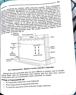
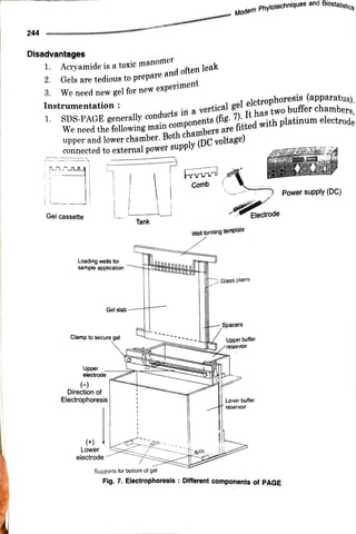
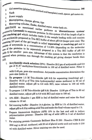
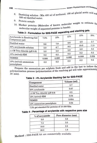
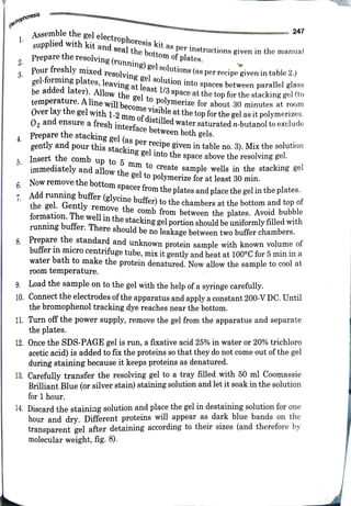
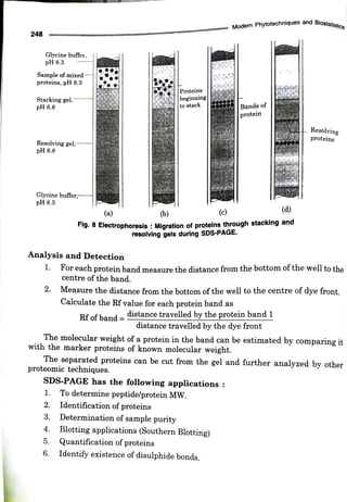
diamine
(TEMED)
a
catalyst
along
with
free
radical
is-acrylamide
(cross
linker).
This
process
can
be
make
easy
and
fast
by
addng
formed
as
a
result
of
polymerization
of
acrylamide
(monomer)
and
N,N'
methylene
The
gels
used
f
o
r
SDS-PAGE
ar
e
polyacrylamide
gels.
Polyacrylamide
gels
ar
e
Polyacryalamide
Gel
:
track
t
h
e
progress
of
t
h
e
protein
solution
through
t
h
e
gel
during
electrophoresis
process.
A
tracking
dye
such
as
bromophenol
blue
is
also
added
to
t
h
e
protein
solution
to
molecular
weight.
proteins
are
disturbed
into
linear
piece)
t
h
e
proteins
so
that
they
may
be
separated
strictly
by
their
some
proteins
fi
t
better
through
t
h
e
gel
matrix
than
others.
SDS
linearizes
(open
up
similar
molecular
weight
migrate
differently
due
to
differences
in
folding,
as
a
result
distance
of
migration
of
SDS-protein
complex.
Without
SDS,
different
proteins
wi
t
h
depends
largely
on
t
h
e
molecular
weight
(size)
of
t
h
e
protein
and
t
h
e
relative
with
both
SDS
and
DTT,
t
h
e
electrophoresis
separation
within
a
polyacrylamide
ge
l
roughly
constant
net
negative
charge
per
unit
mass.
When
protein
is
treated
overwhelms
any
charge
t
h
e
protein
may
have
and
form
anionic
complexes,
that
have
is
added
at
ratio
of
1.4g
SDS/lg
protein.
SDS
has
high
negative
charge
that
proteins.
SDS
has
strong
binding
capacity
to
t
h
e
individual
polypeptides.
When
SDS
B-mercaptoethanol
is
also
added
to
break
any
disulphide
bonds
present
in
t
h
e
(denaturation
of
proteins).
A
reducing
agent
like
DTT
(dithiothreitol)
or
disturbed
causing
their
separation
into
individual
polypetide
is
added
to
t
h
e
protein
sample,
as
a
result
al
l
non-covalent
interactions
in
t
h
e
native
An
ionic
detergent,
sodium
docecyl
sulphate
(CH,(CH,);6CH2
OS0,
Na*)
dodecyl
sulphate
(SDS).
SDS-PAGE.
This
technique
is
performed
in
polyacrylamide
gels
containing
sodium
For
this
purpose
a
new
variant
native
PAGE
has
been
developed
called
as
Principle
:
Various
polypeptides
can
not
be
separated
through
Native
PAGE.
purpose
a
new
variant
native
PAGE
has
been
developed
called
as
SDS-PAGE
But
various
polypeptides
can
not
be
separated
through
Native
PAGE.
For
this
basis
of
charge/mass
ratio.
three
dimentional
formn
protein
can
be
separated
through
Native
PAGE
on
t
h
e
charge,
net
negative
charge
or
no
charge
(isoelectric
point).
In
this
native
form
r
given
pH
of
t
h
e
buffer
protein
may
have
a
set
Each
protein
molecule
has
a
unique
mass
and
specific
charge
due
t
h
e
of
different
amino
acids.
At
a
set
pOsitive
presence
according
to
their
electrophoretic
mobility
and
on
t
h
e
basis
of
their
size.
used
in
biochemistry,
genetics
and
molecular
biology
to
separate
protein
(particulara
Sodium
dodecyl
sulphate
(SDS-PAGE)
gel
electrophoresis
technique
is
widely
Sodium
Dodecyl
Sulphate
Polyacrylamide
Ge
l
Electrophoresis
(SDS-PAGE)
Elecirophoresis
initiator,
ammonium per sulphate (APS), (Chemical method). These
components
induce
p
lvmerization. Modification can be made according to the
requirement of
slong
with acrylamide and appropriate buffer generate the free
radicals needed to
Dplication
bychangingthe amount of acrylamide (C,H;No). For larger pore size,
size,
the
ooncentration of monomer and/or cross linker can be
increased e.g. to
the
amountof acryamnide and/or cross linker can be decreased andfor smaller pore
esolve
(separate)small protein of 8,000 Dato 6000 Da,
small-pore-size gel required
r15-20%acrylamide) and for larger protein (15,000 Da-13000 Da),
larger pore size
gouldbe
required
(7.5-10% acrylamide). Thus poly acryamide gel
electrophores is
RCCOrdingtotheir size. Three dimensional mesh of poly
acryamide gel helps to 'catch'
Horksin a similar fashion as an agrose gel, by separating
protein
molecules
the
protein molecules asthey are transported bythe electric current. The
negatively
charged
protein molecules are pulled to the positive end but they
encounter by the
resistancefrom this polyacrylamide mesh. The smaller
molecules migrate faster
than
thelarger molecules (fig. 6).
Negative
Electrode
Stacking.
gel
Running
gel
2
Positive
Electrode
Lower buffer
reservoir
Load samples here
Buffer
big proteins
small proteins
Advantage of Polyacryamide gel
Fig. 6. Electrophoresis : Migration of protein molecules in SDS-PAGE,
Well
Although the poly acryamide gels are more difficult to prepare and to handle,
It ischemically stable and a cross linked gel
Glass
Plates
involving longer time for preparation than agarose gels but they have greater
resolving power and can accomnodate larger quantities of sample.
We get sharp bands ofseparated molecules
Spacer
lt is good for separation of lowmolecular weight molecules
243
244
Disadvantages
1. Acryamide is atoxic manomer
2. Gels are tedious to prepareand oftenleak
3. We need new gel for new
experiment
Instrumentation :
1 SDS-PAGE generallyconductsin a vertical gel
elctrophoresis(apparatus).
We need the following main
components(fig. 7). Ithastwobuffer
chambers,
upper and lower chamber. Bothchambersarefitted with platinum electrode
connected to external powersĂșpply(DCvoltage)
Gel cassette
Loading wells for
sample application
Clamp to secure gel
Upper
electrode
Gel slab
(
Direction of
Electrophoresis
(+)
Lower
electrode
Tank
Supports for bottom of get
Modern
Phhytotechniques and
Biostatistics
hrrn
Comb
Wellforming termplale
Glass plates
Spacers
Electrode
Upper buffer
reservoir
Lover buffer
reservoir
Fig. 7. Electrophoresis : Different components of PAGE
Power supply (DC)
Iohoresis
2.
4.
5
Gel cassettes
spacer, gel caster etc.
Tank
AGE.
ettes(platesboth notched and
baseplate)comb,
Power supply
Micropipettes,clamps, gloves, tips
SDS-PAGE
6. Measuring
cylinder, flasks, distilled water, water bath etc.
as mentioned earlier performs as
245
(onvented
by
Laemmli) to separate proteins. Inthis system1/3of the length of gel is
alled
stacking gel which includes (table 2) the sample loading wells and consists
4%acrylamide
preparedin Tris HCl buffer of about pH 6.8 (larger pore size). The
remaining2/3 part of the gel length is called'separating or resolving gel and it
consistsof
acryamide in a
concentration of 7.5-20% (depending on the molecular
SIzes of the proteins to be separated) prepared in a Tris HCI buffer of pH 8.8
(table3) for smaller pore size. Advantage of this system is that the resolution is
improved when proteins run through the stacking gel giving sharper bands than
discontinuous system
1. Acrylamide stock solution (30%): Dissolve 29.2 gm of acrlyamide and 0.ð
gm of N,N"-methylene-bisacrylamide in 100 ml of distilled water. Flter it
with 0.45 um. pore size membrane. Acrvamide concentration determines the
pore size (table 4)
2. To prepare 1.5 MTris-chloride (pH 8.8) for separating (resolving) gel :
Dissolve 18.15 g of Tris (tris hydroxymethyl amino methane) in 50 ml of
distilled water, adjust pH to 8.8 with HCl, raise yolume to 100 ml with
distilled water.
8. Toprepare 1.0 MTris-chloride (pH 6.8): Dissolve 12.08 gm ofTris in 60 ml
distilled water, adjust pH to 6.8 with HCl and raise to 100 ml.
4. 10% SDS: Dissolve 1 gm SDS in 5ml of distilled water and raise the final
volume to 10 ml.
5. Gel running buffer :Dissolve 14.4 glycine, lg. SDS in 1lt.of distilled water,
adjust pHto 8.3ĂŸy addingsolidTrisand make thefinal volume equal to1lt.
6. Prepare Ammonium Sulphate (APS) 10% in the last and add to induce
polymerization process : Dissolve 500 mg of solid APS in 5 ml of distilled
water.
I. For staining protein Coomassie Brilliant Blue R 250 :Dissolve CBBR-250
(600 mg) in 80ml. methanol, add 20 mlofglacial acetic acid and make it 200
ml with distilled water. Silver staining can also be used.
246
8.
9
Destining solution : Mix 400 ml of methanol, 100 ml glacial acetic
acid and
500 ml distilled water.
Protein sample
10. Marker proteins (Molecules of known molecular weight to estimate the
molecular weight of separated proteins inbands).
Acrylamide in Resolving Gel
|Component (ml)
Table 2: Formulationfor SDS-PAGE separating and stacking gels
Distilled water
TEMED
30% acrylamide solution
1.5 M Tris chloride (pH 8.8)
10% (wt/vol) SDS
10% (wt/vol) ammonium
persulphate
7.5%
|Component
Distilled water
9.6
5.0
0.008
TEMED
5.0
0.2
0.2
|30%acrylamide
1.0M Tris chloride (pH 6.8)
10% (wt/vol) SDS
5.0
7.5
10%
%ofacryamide
7.9
6.7
5.0
10.0
0.2
0.008
10% ammonium persulphate
20.0
0.2
Modern Phytotechniques and
Biostatistcs
12%
6.6
8.0
5.0
Table 3: 4% Acrylamide Stacking Gel for SDS-PAGE
0.2
0.008
0.2
repare the amnmonium per sulnhatefresh and add in the last to induce the
POyinenzation process (polymerization of the resolving gel willtake aPproximatel
30 min).
Volume (ml)
7.5% get standard for proteins of 10-300 Kda.
3.6
3.0
2.7
2.6
0.67
1.8
0.5
0.04
0.004
Pore diameter (nm)
0.04
Table 4:Percentage of acrylamidewith respective pore size
15%
4.6
10.0
Method : SDS-PAGE kitare commercially available
5.0
0.2
0.008
0.2
20%
2.7
11.9
5.0
0.2
0.008
0.2
gactrophoresis
1.
2
3.
4.
5.
Assemble the gel
electrophoresiskit as per
instructions given in the manual
supplied with kit and sealthe bottomof plates.
A
Preparethe
resolving (running) gel solutions(as per recipegivenin table 2.)
Pourfreshly mixed resolving gel solution into spaces between parallel glass
gel-forming plates,leaving at least 1/3space at thetopforthe stacking gel(to
be added
later). Allowthe gel to
polymerize for about 30 minutes at room
temperature. A
line will becomne visible at thetop for the gel asit polymerizes.
Over laythe gel with 1-2 mm of
distilled water saturated n-butanoltoexclude
O, andensure afresh interface between both gels.
247
Prepare the stacking gel(as per recipe given in table no. 3). Mix the solution
gently and pour this stacking gel intothe space abovethe resolving gel.
Insert the comb up to 5 mm to create samnle wells in the stacking 80
immediately and allowthe gel to polymerize for at least 30 min.
Now remove the bottom spacer from the plates and place the gel in the plates.
1 Add running buffer (glycine buffer) to the chambers at the bottom and top of
the gel. Gently remove the comb from between the plates. Avoid bubble
formation. The well in the stacking gel portion shouldbe uniformlyfilled with
running buffer. There should be no leakage between two buffer chambers.
8. Prepare the standard and unknown protein sample with known volume of
buffer in micro centrifuge tube, mix it gentlv and heat at 100°C for 5min in a
water bath to make the protein denatured. Now allow the sample tocool at
room temperature.
9. Load the sample on to the gel with the help of a syringe carefully.
10. Connectthe electrodes ofthe apparatus andapplyaconstant200-VDC. Until
the bromophenol tracking dye reaches near the bottom.
11. Turn off the power supply, remove the gel from the apparatus and separate
theplates.
12. Once the SDS-PAGE gel is run, a fixative acid 25% in water or 20% trichloro
aceticacid) isadded tofix the proteins so that they do not come out ofthe gel
during staining because it keeps proteins as denatured.
13. Carefully transfer the resolving gel to atray filled with 50 ml Coomassie
Brilliant Blue (or silver stain) staining solution and let it soak in the solution
for 1 hour.
14. Discard the staining solution and place the gel in destaining solution for one
hour and dry. Different proteins willappear as dark blue bands on the
transparent gel after detaining according to their sizes (and therefore by
molecular weight, fig. 8).
248
Glycine buffer,
pH 8.3
Sample of mixed
proteins, pH 8.3
Stacking gel.
pH 6.8
Resolving gel,
pH 8.8
Glycine buffer;
pH 8.3
Analysis and Detection
(a)
1.
2.
3.
4.
(b)
1. For each protein band measure the distance from the bottom of the well to the
centre of the band.
5.
2. Measure the distance from the bottom of the well to the centre of dye front.
6.
Proteins
beginning
tostack
Rf of band =
SDS-PAGE has the following applications
Fig. 8 Electrophoresis :Migration of proteins through stacking and
resolving gels during SDS-PAGE.
Calculate the Rf value for each protein band as
The molecular weight of a protein in the band can be estimated by comnparing it
with the marker proteins of known molecular weight.
The separated proteins can be cut from the gel and further analyzed by other
proteomic techniques.
(c)
distance travelled by the protein band 1
distance travelled bythedye front
Identification of proteins
Modern Phytotechniques and
Biostatistics
To determine peptidelprotein MW.
Bands of
protein
Determination of sample purity
(d)
Blotting applications (Southern Blotting)
Quantification of proteins
Identify existence of disulphide bonds.
Resolving
proteins

SDS PAGE (Electrophoresis) Book Notes