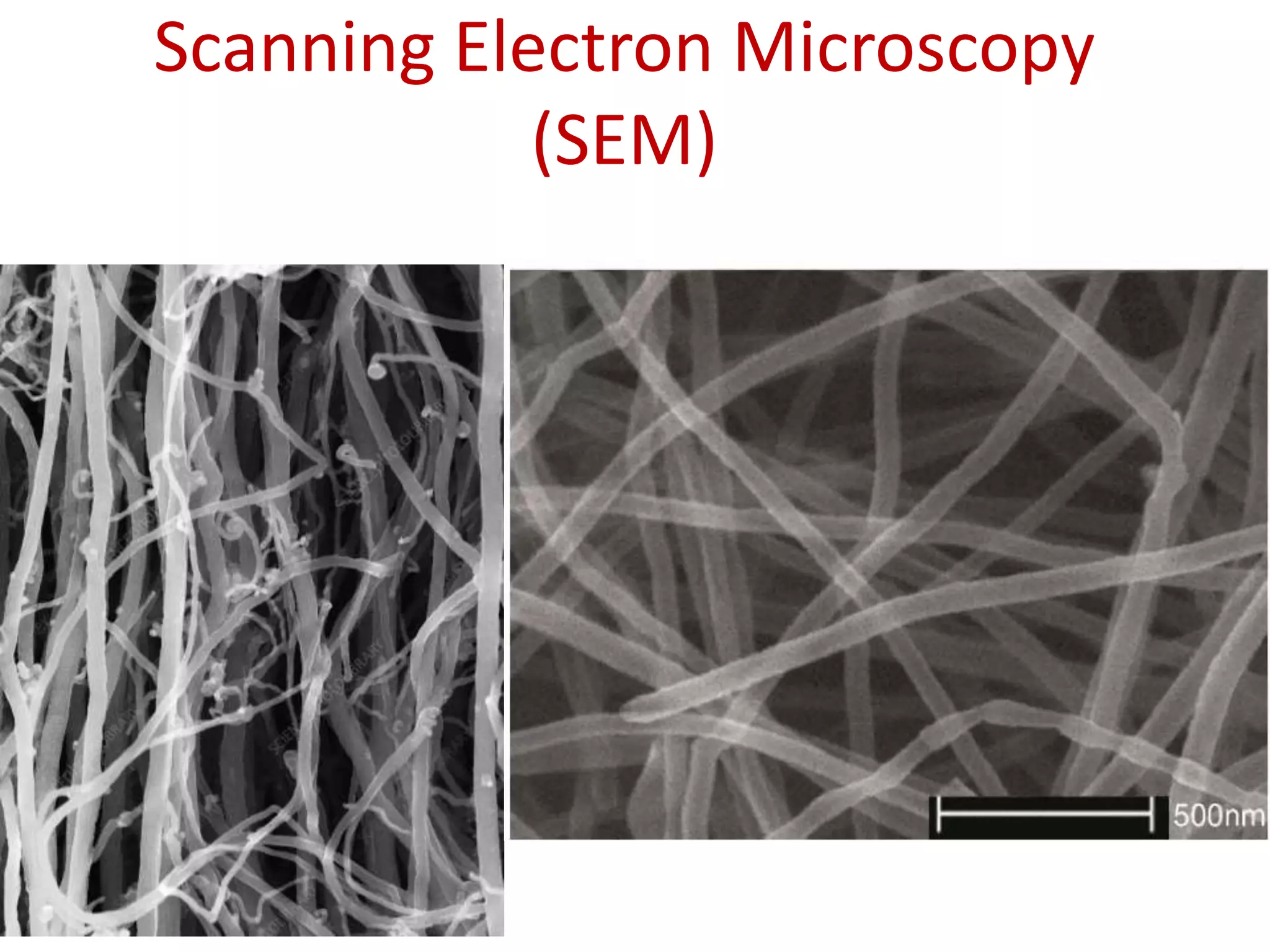
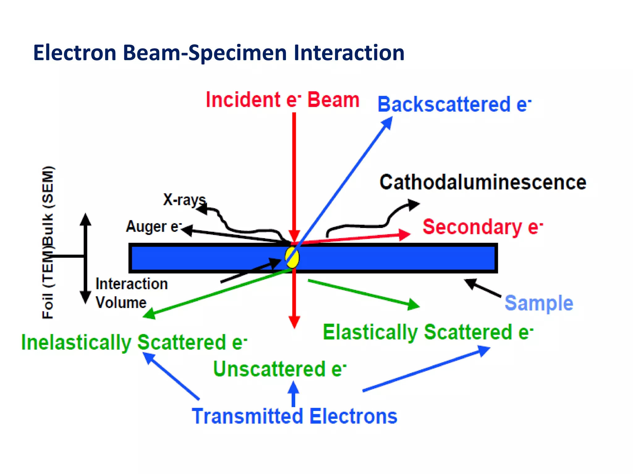
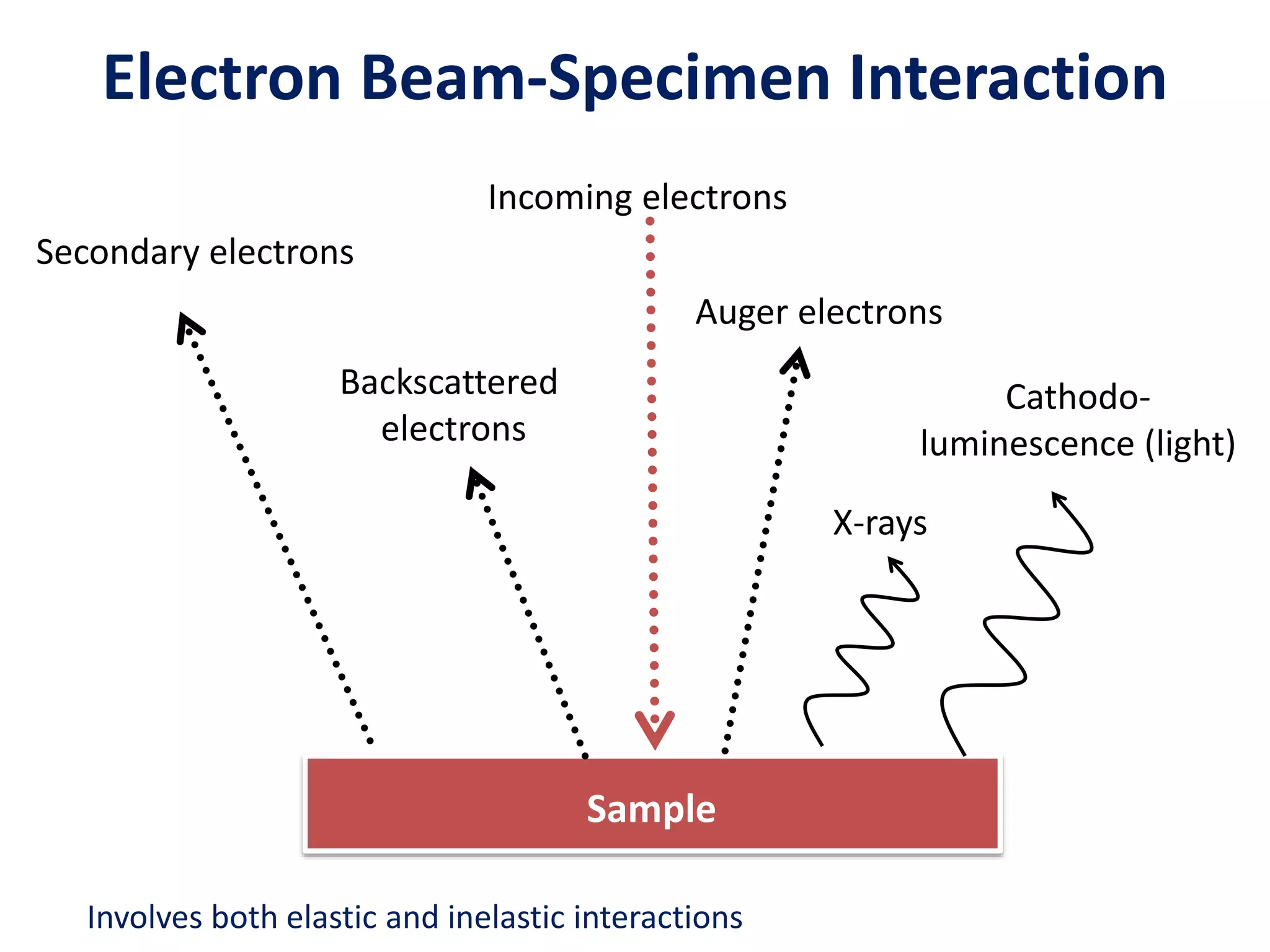
SlideShare a Scribd company logo
Scanning Electron Microscopy
(SEM)
A Short History
• V. K. Zworykin et al. 1942, first SEM for bulk samples
• 1965 first commercial SEM by Cambridge Scientific
Instruments
• Resolution at that time ~ 50 nm <-> Today ~ 10 nm
• Morphology only at that time <-> Today analytical
instrument
• Why e microscope ?
Electron Beam-Specimen Interaction
Incoming electrons
Secondary electrons
Backscattered
electrons
Auger electrons
X-rays
Cathodo-
luminescence (light)
Sample
Involves both elastic and inelastic interactions
Interaction Volume: I
The incident electrons do not go along a
straight line in the specimen, but a zig-zag
path instead.
Monte Carlo simulations of 100 electron trajectories
e-
Interaction Volume: II
The penetration or,
more precisely, the
interaction volume
depends on the
acceleration voltage
(energy of electron)
and the atomic
number of the
specimen.
Escape Volume of Various Signals
• The incident electrons interact with specimen
atoms along their path in the specimen and
generate various signals.
• Owing to the difference in energy of these
signals, their ‘penetration depths’ are
different
• Therefore different signal observable on the
specimen surface comes from different parts
of the interaction volume
• The volume responsible for the respective
signal is called the escape volume of that
signal.
If the diameter of primary
electron beam is ~5nm
- Dimensions of escape
zone of
Escape Volumes of Various Signals
•Secondary electron:
diameter~10nm; depth~10nm
•Backscattered electron:
diameter~1m; depth~1m
•X-ray: from the whole
interaction volume, i.e., ~5m
in diameter and depth
Where does the signals come from?
• Diameter of the interaction
volume is larger than the electron
spot
 resolution is poorer than the
size of the electron spot
Secondary Electrons (SE)
• Generated from the collision
between the incoming electrons and
the loosely bonded outer electrons
• Low energy electrons (~10-50 eV)
• Only SE generated close to surface
escape (topographic information is
obtained)
• Number of SE is greater than the
number of incoming electrons
• We differentiate between SE1 and
SE2
SE1
• The secondary electrons that are generated by
the incoming electron beam as they enter the
surface
• High resolution signal with a resolution which
is only limited by the electron beam diameter
SE2
• The secondary electrons that are generated by the backscattered
electrons that have returned to the surface after several inelastic
scattering events
• SE2 come from a surface area that is bigger than the spot from the
incoming electrons  resolution is poorer than for SE1 exclusively
Sample surface
Incoming electrons
SE2
Factors that affect SE emission
1. Work function of the surface
2. Beam energy and beam current
• Electron yield goes through a maximum at low acc.
voltage, then decreases with increasing acc. Voltage.
Incident electron energy / kV
Secondary
electron
yield
Factors that affect SE emission
3. Atomic number (Z)
• More SE2 are created with
increasing Z
• The Z-dependence is more
pronounced at lower beam
energies
4. The local curvature of the surface
(the most important factor)
By detecting SE1 electrons; a resolution of 1-2 nm can be achieved
Backscattered electrons (BSE)
High energy electrons (elastic scattering)
Fewer BSE than SE
We differentiate between BSE1 and BSE2
Sample surface
Incoming electrons
BSE2
• Most BSE are of BSE2 type
BSE as a function of atomic number
Signal intensity of the BSE increases with Z.
For phases containing more than one element, it is the average atomic number
that determines the backscatter coefficient h
Factors that affect BSE emission
• Direction of the surface w.r.t. electron beam
• Average atomic number
• When you want to study differences in atomic
numbers the sample should be as levelled as
possible (sample preparation is an issue!)
X-rays
• Photons not electrons
• Each element has a
fingerprint X-ray signal
• Poorer spatial resolution than
BSE and SE
• Relatively few X-ray signals
are emitted and the detector
is inefficient
 relatively long signal
collecting times are needed
X-rays
• Most common spectrometer: EDS (energy-
dispersive spectrometer)
• Signal overlap can be a problem
• We can analyze our sample in different modes
– spot analysis
– line scan
– chemical concentration map (elemental mapping)
Principles of SEM
Scanning Electron Microscope
– a Totally Different Imaging Concept
Instead of using the full-field image, a point-to-point
measurement strategy is used.
High energy electron beam is used to excite the specimen and
the signals are collected and analyzed so that an image can be
constructed.
The signals carry topological, chemical and crystallographic
information, respectively, of the samples surface.
Main Applications
• Topography
The surface features of an object and its texture
(hardness, reflectivity… etc.)
• Morphology
The shape and size of the particles making up the
object (strength, defects in IC and chips...etc.)
• Composition
The elements and compounds that the object is
composed of and the relative amounts of them
(melting point, reactivity, hardness...etc.)
• Crystallographic Information
How the grains are arranged in the object
(conductivity, electrical properties, strength...etc.)
SEM components
Source of Electrons
T: ~1500oC
Thermionic Gun
W and LaB6 Cold- and thermal FEG
Electron Gun Properties
Source Brightness Stability(%) Size Energy spread Vacuum
W 3X10⁵ ~1 50m 3.0(eV) 10-5 (Torr)
LaB6 3x10⁶ ~2 5m 1.5 10-6
C-FEG 109 ~5 5nm 0.3 10-10
T-FEG 109 <1 20nm 0.7 10-9
(5-50m)
E: >10MV/cm
(5nm)
Filament
W
Brightness – beam current density per unit solid angle
Magnetic Lenses
• Condenser lens – focusing
determines the beam current
which impinges on the sample.
• Objective lens – final probe
forming
determines the final spot size of
the electron beam, i.e., the
resolution of a SEM.
The objective lens
The objective lens aperture
Aperture in SEM: either to limit the amount of electrons or enhance contrast
Depth of Field
Resolution
Resolution
Can increase the resolution by:
How these changes affect
the depth of field?
Detector and Sample Stage
A detector placed within the column is known as an “in-lens” detector and
produces a very different image compared to a conventionally located detector
Secondary Electron Detector
Side Mounted In-Lens
Secondary Electron Detector
Side Mounted In-Lens
Image Magnification
Example of a series of increasing magnification (spherical lead particles imaged
in SE mode)
Topographical Contrast
Topographic contrast
arises because SE
generation depend on
the angle of incidence
between the beam and
sample.
Backscattered Electrons (BSE)
BSE are produced by elastic interactions of beam electrons with
nuclei of atoms in the specimen and they have high energy and
large escape depth.
BSE yield: h=nBS/nB ~ function of atomic number, Z
BSE images show characteristics of atomic number contrast,
i.e., high average Z appear brighter than those of low average Z.
h increases with tilt.
Primary
BSE image from flat surface of an Al (Z=13)
and Cu (Z=29) alloy
Scanning Electron Microscopy (SEM 2013).pptx

More Related Content

What's hot

Scanning probe microscope
Scanning probe microscopeScanning probe microscope
Scanning probe microscope
erfan.zaker@gmail.com
 
ELECTRON MICROSCOPY (TEM & SEM)
ELECTRON MICROSCOPY (TEM & SEM)ELECTRON MICROSCOPY (TEM & SEM)
ELECTRON MICROSCOPY (TEM & SEM)
ADITYA ARYA
 
Defects in crystalline materials
Defects in crystalline materialsDefects in crystalline materials
Defects in crystalline materials
onlinemetallurgy.com
 
Semiconductors
SemiconductorsSemiconductors
Semiconductors
Self-employed
 
Atomic Force Microscope
Atomic Force MicroscopeAtomic Force Microscope
Atomic Force Microscope
Ghalia Nawal
 
Radioactivity
RadioactivityRadioactivity
Radioactivity
Steve Bishop
 
para magnetism And Di magnetism
para magnetism And Di magnetism para magnetism And Di magnetism
para magnetism And Di magnetism
Nikhil Joshi
 
Semiconductor
SemiconductorSemiconductor
Semiconductor
Seti Saiman
 
TRANSMISSION ELECTRON MICROSCOPY
TRANSMISSION ELECTRON MICROSCOPYTRANSMISSION ELECTRON MICROSCOPY
TRANSMISSION ELECTRON MICROSCOPY
Sutapa Saha
 
X ray photoelecton spectroscopy
X ray photoelecton spectroscopy X ray photoelecton spectroscopy
X ray photoelecton spectroscopy
Gandhi Yellapu
 
BAND THEORY OF SOLIDS
BAND THEORY OF SOLIDSBAND THEORY OF SOLIDS
BAND THEORY OF SOLIDS
Mahi Shinde
 
Phy 4240 lec (7)
Phy 4240 lec (7)Phy 4240 lec (7)
Phy 4240 lec (7)
Dr. Abeer Kamal
 
Electron Microscope Shanthakumar
Electron Microscope ShanthakumarElectron Microscope Shanthakumar
Electron Microscope Shanthakumar
guestcd4662
 
Difference between plane waves and laser
Difference between plane waves and laserDifference between plane waves and laser
Difference between plane waves and laser
Lahiru Da Silva
 
FREE ELECTRON THEORY.pptx
FREE ELECTRON THEORY.pptxFREE ELECTRON THEORY.pptx
FREE ELECTRON THEORY.pptx
THANUSHS22ECR214
 
auger electron spectroscopy (AES)
auger electron spectroscopy  (AES)auger electron spectroscopy  (AES)
auger electron spectroscopy (AES)
Kamal Asadi Pakdel
 
Xps
XpsXps
Xps
cynon
 
Auger Electron Spectroscopy
Auger Electron SpectroscopyAuger Electron Spectroscopy
Auger Electron Spectroscopy
hasanjamal13
 
Basic Introduction of XANES and EXAFS
Basic Introduction of XANES and EXAFSBasic Introduction of XANES and EXAFS
Basic Introduction of XANES and EXAFS
Amlan Roy
 
X-ray spectroscopy
X-ray spectroscopyX-ray spectroscopy
X-ray spectroscopy
Vinit Gohel
 

What's hot (20)

Scanning probe microscope
Scanning probe microscopeScanning probe microscope
Scanning probe microscope
 
ELECTRON MICROSCOPY (TEM & SEM)
ELECTRON MICROSCOPY (TEM & SEM)ELECTRON MICROSCOPY (TEM & SEM)
ELECTRON MICROSCOPY (TEM & SEM)
 
Defects in crystalline materials
Defects in crystalline materialsDefects in crystalline materials
Defects in crystalline materials
 
Semiconductors
SemiconductorsSemiconductors
Semiconductors
 
Atomic Force Microscope
Atomic Force MicroscopeAtomic Force Microscope
Atomic Force Microscope
 
Radioactivity
RadioactivityRadioactivity
Radioactivity
 
para magnetism And Di magnetism
para magnetism And Di magnetism para magnetism And Di magnetism
para magnetism And Di magnetism
 
Semiconductor
SemiconductorSemiconductor
Semiconductor
 
TRANSMISSION ELECTRON MICROSCOPY
TRANSMISSION ELECTRON MICROSCOPYTRANSMISSION ELECTRON MICROSCOPY
TRANSMISSION ELECTRON MICROSCOPY
 
X ray photoelecton spectroscopy
X ray photoelecton spectroscopy X ray photoelecton spectroscopy
X ray photoelecton spectroscopy
 
BAND THEORY OF SOLIDS
BAND THEORY OF SOLIDSBAND THEORY OF SOLIDS
BAND THEORY OF SOLIDS
 
Phy 4240 lec (7)
Phy 4240 lec (7)Phy 4240 lec (7)
Phy 4240 lec (7)
 
Electron Microscope Shanthakumar
Electron Microscope ShanthakumarElectron Microscope Shanthakumar
Electron Microscope Shanthakumar
 
Difference between plane waves and laser
Difference between plane waves and laserDifference between plane waves and laser
Difference between plane waves and laser
 
FREE ELECTRON THEORY.pptx
FREE ELECTRON THEORY.pptxFREE ELECTRON THEORY.pptx
FREE ELECTRON THEORY.pptx
 
auger electron spectroscopy (AES)
auger electron spectroscopy  (AES)auger electron spectroscopy  (AES)
auger electron spectroscopy (AES)
 
Xps
XpsXps
Xps
 
Auger Electron Spectroscopy
Auger Electron SpectroscopyAuger Electron Spectroscopy
Auger Electron Spectroscopy
 
Basic Introduction of XANES and EXAFS
Basic Introduction of XANES and EXAFSBasic Introduction of XANES and EXAFS
Basic Introduction of XANES and EXAFS
 
X-ray spectroscopy
X-ray spectroscopyX-ray spectroscopy
X-ray spectroscopy
 

Similar to Scanning Electron Microscopy (SEM 2013).pptx

Scanning Electron Microscopy (SEM) Analysis.pdf
Scanning Electron Microscopy (SEM) Analysis.pdfScanning Electron Microscopy (SEM) Analysis.pdf
Scanning Electron Microscopy (SEM) Analysis.pdf
SanDeepSharma926061
 
SEM-for MS.pptx
SEM-for MS.pptxSEM-for MS.pptx
SEM-for MS.pptx
usamachaudhry14
 
Scanning electron microscopy
Scanning electron microscopyScanning electron microscopy
Scanning electron microscopy
Ganesh Shinde
 
Introduction to Electron Microscopyy.pdf
Introduction to Electron Microscopyy.pdfIntroduction to Electron Microscopyy.pdf
Introduction to Electron Microscopyy.pdf
Lapsik1
 
Sem mahfooz
Sem   mahfoozSem   mahfooz
Sem mahfooz
Mahfooz Alam
 
Electron microscopy
Electron microscopyElectron microscopy
Sem mahfooz
Sem   mahfoozSem   mahfooz
Sem mahfooz
Mahfooz Alam
 
SCANNING ELECTRON MICROSCOPE MITHILESH CHOUDHARY
SCANNING ELECTRON MICROSCOPE MITHILESH CHOUDHARYSCANNING ELECTRON MICROSCOPE MITHILESH CHOUDHARY
SCANNING ELECTRON MICROSCOPE MITHILESH CHOUDHARY
Anjan Anant
 
SEM,TEM & AFM
SEM,TEM & AFMSEM,TEM & AFM
SEM,TEM & AFM
Anamika Banerjee
 
2018 HM-scanning electron microscope
2018 HM-scanning electron microscope2018 HM-scanning electron microscope
2018 HM-scanning electron microscope
Harsh Mohan
 
Sem mahfooz
Sem mahfoozSem mahfooz
Sem mahfooz
Mahfooz Alam
 
overview on scanning electron microscope
overview on scanning electron microscopeoverview on scanning electron microscope
overview on scanning electron microscope
Bharathi59577
 
Scanning Electron Microscopy
Scanning Electron MicroscopyScanning Electron Microscopy
Scanning Electron Microscopy
Sejal9787
 
E mintro
E mintroE mintro
Advanced Characterization Technique - SEM
Advanced Characterization Technique - SEMAdvanced Characterization Technique - SEM
Advanced Characterization Technique - SEM
Ilyas Hussain
 
Scanning Electron Microscope
Scanning Electron MicroscopeScanning Electron Microscope
Scanning Electron Microscope
MallikarjunRaoPanaba
 
Scanning Electron microscopy
Scanning Electron microscopy Scanning Electron microscopy
Scanning Electron microscopy
sagar sen sen
 
Sem
SemSem
Scanning Electron Microscope.pptx
Scanning Electron Microscope.pptxScanning Electron Microscope.pptx
Scanning Electron Microscope.pptx
RABEYABASORI
 
Scanning electron microscope (sem)
Scanning electron microscope (sem)Scanning electron microscope (sem)
Scanning electron microscope (sem)
Mary Theresa
 

Similar to Scanning Electron Microscopy (SEM 2013).pptx (20)

Scanning Electron Microscopy (SEM) Analysis.pdf
Scanning Electron Microscopy (SEM) Analysis.pdfScanning Electron Microscopy (SEM) Analysis.pdf
Scanning Electron Microscopy (SEM) Analysis.pdf
 
SEM-for MS.pptx
SEM-for MS.pptxSEM-for MS.pptx
SEM-for MS.pptx
 
Scanning electron microscopy
Scanning electron microscopyScanning electron microscopy
Scanning electron microscopy
 
Introduction to Electron Microscopyy.pdf
Introduction to Electron Microscopyy.pdfIntroduction to Electron Microscopyy.pdf
Introduction to Electron Microscopyy.pdf
 
Sem mahfooz
Sem   mahfoozSem   mahfooz
Sem mahfooz
 
Electron microscopy
Electron microscopyElectron microscopy
Electron microscopy
 
Sem mahfooz
Sem   mahfoozSem   mahfooz
Sem mahfooz
 
SCANNING ELECTRON MICROSCOPE MITHILESH CHOUDHARY
SCANNING ELECTRON MICROSCOPE MITHILESH CHOUDHARYSCANNING ELECTRON MICROSCOPE MITHILESH CHOUDHARY
SCANNING ELECTRON MICROSCOPE MITHILESH CHOUDHARY
 
SEM,TEM & AFM
SEM,TEM & AFMSEM,TEM & AFM
SEM,TEM & AFM
 
2018 HM-scanning electron microscope
2018 HM-scanning electron microscope2018 HM-scanning electron microscope
2018 HM-scanning electron microscope
 
Sem mahfooz
Sem mahfoozSem mahfooz
Sem mahfooz
 
overview on scanning electron microscope
overview on scanning electron microscopeoverview on scanning electron microscope
overview on scanning electron microscope
 
Scanning Electron Microscopy
Scanning Electron MicroscopyScanning Electron Microscopy
Scanning Electron Microscopy
 
E mintro
E mintroE mintro
E mintro
 
Advanced Characterization Technique - SEM
Advanced Characterization Technique - SEMAdvanced Characterization Technique - SEM
Advanced Characterization Technique - SEM
 
Scanning Electron Microscope
Scanning Electron MicroscopeScanning Electron Microscope
Scanning Electron Microscope
 
Scanning Electron microscopy
Scanning Electron microscopy Scanning Electron microscopy
Scanning Electron microscopy
 
Sem
SemSem
Sem
 
Scanning Electron Microscope.pptx
Scanning Electron Microscope.pptxScanning Electron Microscope.pptx
Scanning Electron Microscope.pptx
 
Scanning electron microscope (sem)
Scanning electron microscope (sem)Scanning electron microscope (sem)
Scanning electron microscope (sem)
 

Recently uploaded

Engineering Drawings Lecture Detail Drawings 2014.pdf
Engineering Drawings Lecture Detail Drawings 2014.pdfEngineering Drawings Lecture Detail Drawings 2014.pdf
Engineering Drawings Lecture Detail Drawings 2014.pdf
abbyasa1014
 
Optimizing Gradle Builds - Gradle DPE Tour Berlin 2024
Optimizing Gradle Builds - Gradle DPE Tour Berlin 2024Optimizing Gradle Builds - Gradle DPE Tour Berlin 2024
Optimizing Gradle Builds - Gradle DPE Tour Berlin 2024
Sinan KOZAK
 
International Conference on NLP, Artificial Intelligence, Machine Learning an...
International Conference on NLP, Artificial Intelligence, Machine Learning an...International Conference on NLP, Artificial Intelligence, Machine Learning an...
International Conference on NLP, Artificial Intelligence, Machine Learning an...
gerogepatton
 
Certificates - Mahmoud Mohamed Moursi Ahmed
Certificates - Mahmoud Mohamed Moursi AhmedCertificates - Mahmoud Mohamed Moursi Ahmed
Certificates - Mahmoud Mohamed Moursi Ahmed
Mahmoud Morsy
 
Literature Review Basics and Understanding Reference Management.pptx
Literature Review Basics and Understanding Reference Management.pptxLiterature Review Basics and Understanding Reference Management.pptx
Literature Review Basics and Understanding Reference Management.pptx
Dr Ramhari Poudyal
 
Manufacturing Process of molasses based distillery ppt.pptx
Manufacturing Process of molasses based distillery ppt.pptxManufacturing Process of molasses based distillery ppt.pptx
Manufacturing Process of molasses based distillery ppt.pptx
Madan Karki
 
CHINA’S GEO-ECONOMIC OUTREACH IN CENTRAL ASIAN COUNTRIES AND FUTURE PROSPECT
CHINA’S GEO-ECONOMIC OUTREACH IN CENTRAL ASIAN COUNTRIES AND FUTURE PROSPECTCHINA’S GEO-ECONOMIC OUTREACH IN CENTRAL ASIAN COUNTRIES AND FUTURE PROSPECT
CHINA’S GEO-ECONOMIC OUTREACH IN CENTRAL ASIAN COUNTRIES AND FUTURE PROSPECT
jpsjournal1
 
john krisinger-the science and history of the alcoholic beverage.pptx
john krisinger-the science and history of the alcoholic beverage.pptxjohn krisinger-the science and history of the alcoholic beverage.pptx
john krisinger-the science and history of the alcoholic beverage.pptx
Madan Karki
 
IEEE Aerospace and Electronic Systems Society as a Graduate Student Member
IEEE Aerospace and Electronic Systems Society as a Graduate Student MemberIEEE Aerospace and Electronic Systems Society as a Graduate Student Member
IEEE Aerospace and Electronic Systems Society as a Graduate Student Member
VICTOR MAESTRE RAMIREZ
 
gray level transformation unit 3(image processing))
gray level transformation unit 3(image processing))gray level transformation unit 3(image processing))
gray level transformation unit 3(image processing))
shivani5543
 
学校原版美国波士顿大学毕业证学历学位证书原版一模一样
学校原版美国波士顿大学毕业证学历学位证书原版一模一样学校原版美国波士顿大学毕业证学历学位证书原版一模一样
学校原版美国波士顿大学毕业证学历学位证书原版一模一样
171ticu
 
Engine Lubrication performance System.pdf
Engine Lubrication performance System.pdfEngine Lubrication performance System.pdf
Engine Lubrication performance System.pdf
mamamaam477
 
ML Based Model for NIDS MSc Updated Presentation.v2.pptx
ML Based Model for NIDS MSc Updated Presentation.v2.pptxML Based Model for NIDS MSc Updated Presentation.v2.pptx
ML Based Model for NIDS MSc Updated Presentation.v2.pptx
JamalHussainArman
 
Transformers design and coooling methods
Transformers design and coooling methodsTransformers design and coooling methods
Transformers design and coooling methods
Roger Rozario
 
DEEP LEARNING FOR SMART GRID INTRUSION DETECTION: A HYBRID CNN-LSTM-BASED MODEL
DEEP LEARNING FOR SMART GRID INTRUSION DETECTION: A HYBRID CNN-LSTM-BASED MODELDEEP LEARNING FOR SMART GRID INTRUSION DETECTION: A HYBRID CNN-LSTM-BASED MODEL
DEEP LEARNING FOR SMART GRID INTRUSION DETECTION: A HYBRID CNN-LSTM-BASED MODEL
gerogepatton
 
Introduction to AI Safety (public presentation).pptx
Introduction to AI Safety (public presentation).pptxIntroduction to AI Safety (public presentation).pptx
Introduction to AI Safety (public presentation).pptx
MiscAnnoy1
 
Redefining brain tumor segmentation: a cutting-edge convolutional neural netw...
Redefining brain tumor segmentation: a cutting-edge convolutional neural netw...Redefining brain tumor segmentation: a cutting-edge convolutional neural netw...
Redefining brain tumor segmentation: a cutting-edge convolutional neural netw...
IJECEIAES
 
Generative AI leverages algorithms to create various forms of content
Generative AI leverages algorithms to create various forms of contentGenerative AI leverages algorithms to create various forms of content
Generative AI leverages algorithms to create various forms of content
Hitesh Mohapatra
 
Advanced control scheme of doubly fed induction generator for wind turbine us...
Advanced control scheme of doubly fed induction generator for wind turbine us...Advanced control scheme of doubly fed induction generator for wind turbine us...
Advanced control scheme of doubly fed induction generator for wind turbine us...
IJECEIAES
 
Curve Fitting in Numerical Methods Regression
Curve Fitting in Numerical Methods RegressionCurve Fitting in Numerical Methods Regression
Curve Fitting in Numerical Methods Regression
Nada Hikmah
 

Recently uploaded (20)

Engineering Drawings Lecture Detail Drawings 2014.pdf
Engineering Drawings Lecture Detail Drawings 2014.pdfEngineering Drawings Lecture Detail Drawings 2014.pdf
Engineering Drawings Lecture Detail Drawings 2014.pdf
 
Optimizing Gradle Builds - Gradle DPE Tour Berlin 2024
Optimizing Gradle Builds - Gradle DPE Tour Berlin 2024Optimizing Gradle Builds - Gradle DPE Tour Berlin 2024
Optimizing Gradle Builds - Gradle DPE Tour Berlin 2024
 
International Conference on NLP, Artificial Intelligence, Machine Learning an...
International Conference on NLP, Artificial Intelligence, Machine Learning an...International Conference on NLP, Artificial Intelligence, Machine Learning an...
International Conference on NLP, Artificial Intelligence, Machine Learning an...
 
Certificates - Mahmoud Mohamed Moursi Ahmed
Certificates - Mahmoud Mohamed Moursi AhmedCertificates - Mahmoud Mohamed Moursi Ahmed
Certificates - Mahmoud Mohamed Moursi Ahmed
 
Literature Review Basics and Understanding Reference Management.pptx
Literature Review Basics and Understanding Reference Management.pptxLiterature Review Basics and Understanding Reference Management.pptx
Literature Review Basics and Understanding Reference Management.pptx
 
Manufacturing Process of molasses based distillery ppt.pptx
Manufacturing Process of molasses based distillery ppt.pptxManufacturing Process of molasses based distillery ppt.pptx
Manufacturing Process of molasses based distillery ppt.pptx
 
CHINA’S GEO-ECONOMIC OUTREACH IN CENTRAL ASIAN COUNTRIES AND FUTURE PROSPECT
CHINA’S GEO-ECONOMIC OUTREACH IN CENTRAL ASIAN COUNTRIES AND FUTURE PROSPECTCHINA’S GEO-ECONOMIC OUTREACH IN CENTRAL ASIAN COUNTRIES AND FUTURE PROSPECT
CHINA’S GEO-ECONOMIC OUTREACH IN CENTRAL ASIAN COUNTRIES AND FUTURE PROSPECT
 
john krisinger-the science and history of the alcoholic beverage.pptx
john krisinger-the science and history of the alcoholic beverage.pptxjohn krisinger-the science and history of the alcoholic beverage.pptx
john krisinger-the science and history of the alcoholic beverage.pptx
 
IEEE Aerospace and Electronic Systems Society as a Graduate Student Member
IEEE Aerospace and Electronic Systems Society as a Graduate Student MemberIEEE Aerospace and Electronic Systems Society as a Graduate Student Member
IEEE Aerospace and Electronic Systems Society as a Graduate Student Member
 
gray level transformation unit 3(image processing))
gray level transformation unit 3(image processing))gray level transformation unit 3(image processing))
gray level transformation unit 3(image processing))
 
学校原版美国波士顿大学毕业证学历学位证书原版一模一样
学校原版美国波士顿大学毕业证学历学位证书原版一模一样学校原版美国波士顿大学毕业证学历学位证书原版一模一样
学校原版美国波士顿大学毕业证学历学位证书原版一模一样
 
Engine Lubrication performance System.pdf
Engine Lubrication performance System.pdfEngine Lubrication performance System.pdf
Engine Lubrication performance System.pdf
 
ML Based Model for NIDS MSc Updated Presentation.v2.pptx
ML Based Model for NIDS MSc Updated Presentation.v2.pptxML Based Model for NIDS MSc Updated Presentation.v2.pptx
ML Based Model for NIDS MSc Updated Presentation.v2.pptx
 
Transformers design and coooling methods
Transformers design and coooling methodsTransformers design and coooling methods
Transformers design and coooling methods
 
DEEP LEARNING FOR SMART GRID INTRUSION DETECTION: A HYBRID CNN-LSTM-BASED MODEL
DEEP LEARNING FOR SMART GRID INTRUSION DETECTION: A HYBRID CNN-LSTM-BASED MODELDEEP LEARNING FOR SMART GRID INTRUSION DETECTION: A HYBRID CNN-LSTM-BASED MODEL
DEEP LEARNING FOR SMART GRID INTRUSION DETECTION: A HYBRID CNN-LSTM-BASED MODEL
 
Introduction to AI Safety (public presentation).pptx
Introduction to AI Safety (public presentation).pptxIntroduction to AI Safety (public presentation).pptx
Introduction to AI Safety (public presentation).pptx
 
Redefining brain tumor segmentation: a cutting-edge convolutional neural netw...
Redefining brain tumor segmentation: a cutting-edge convolutional neural netw...Redefining brain tumor segmentation: a cutting-edge convolutional neural netw...
Redefining brain tumor segmentation: a cutting-edge convolutional neural netw...
 
Generative AI leverages algorithms to create various forms of content
Generative AI leverages algorithms to create various forms of contentGenerative AI leverages algorithms to create various forms of content
Generative AI leverages algorithms to create various forms of content
 
Advanced control scheme of doubly fed induction generator for wind turbine us...
Advanced control scheme of doubly fed induction generator for wind turbine us...Advanced control scheme of doubly fed induction generator for wind turbine us...
Advanced control scheme of doubly fed induction generator for wind turbine us...
 
Curve Fitting in Numerical Methods Regression
Curve Fitting in Numerical Methods RegressionCurve Fitting in Numerical Methods Regression
Curve Fitting in Numerical Methods Regression
 

Scanning Electron Microscopy (SEM 2013).pptx

  • 2.
  • 3. A Short History • V. K. Zworykin et al. 1942, first SEM for bulk samples • 1965 first commercial SEM by Cambridge Scientific Instruments • Resolution at that time ~ 50 nm <-> Today ~ 10 nm • Morphology only at that time <-> Today analytical instrument • Why e microscope ?
  • 5. Electron Beam-Specimen Interaction Incoming electrons Secondary electrons Backscattered electrons Auger electrons X-rays Cathodo- luminescence (light) Sample Involves both elastic and inelastic interactions
  • 6. Interaction Volume: I The incident electrons do not go along a straight line in the specimen, but a zig-zag path instead. Monte Carlo simulations of 100 electron trajectories e-
  • 7. Interaction Volume: II The penetration or, more precisely, the interaction volume depends on the acceleration voltage (energy of electron) and the atomic number of the specimen.
  • 8. Escape Volume of Various Signals • The incident electrons interact with specimen atoms along their path in the specimen and generate various signals. • Owing to the difference in energy of these signals, their ‘penetration depths’ are different • Therefore different signal observable on the specimen surface comes from different parts of the interaction volume • The volume responsible for the respective signal is called the escape volume of that signal.
  • 9. If the diameter of primary electron beam is ~5nm - Dimensions of escape zone of Escape Volumes of Various Signals •Secondary electron: diameter~10nm; depth~10nm •Backscattered electron: diameter~1m; depth~1m •X-ray: from the whole interaction volume, i.e., ~5m in diameter and depth
  • 10. Where does the signals come from? • Diameter of the interaction volume is larger than the electron spot  resolution is poorer than the size of the electron spot
  • 11. Secondary Electrons (SE) • Generated from the collision between the incoming electrons and the loosely bonded outer electrons • Low energy electrons (~10-50 eV) • Only SE generated close to surface escape (topographic information is obtained) • Number of SE is greater than the number of incoming electrons • We differentiate between SE1 and SE2
  • 12. SE1 • The secondary electrons that are generated by the incoming electron beam as they enter the surface • High resolution signal with a resolution which is only limited by the electron beam diameter
  • 13. SE2 • The secondary electrons that are generated by the backscattered electrons that have returned to the surface after several inelastic scattering events • SE2 come from a surface area that is bigger than the spot from the incoming electrons  resolution is poorer than for SE1 exclusively Sample surface Incoming electrons SE2
  • 14. Factors that affect SE emission 1. Work function of the surface 2. Beam energy and beam current • Electron yield goes through a maximum at low acc. voltage, then decreases with increasing acc. Voltage. Incident electron energy / kV Secondary electron yield
  • 15. Factors that affect SE emission 3. Atomic number (Z) • More SE2 are created with increasing Z • The Z-dependence is more pronounced at lower beam energies 4. The local curvature of the surface (the most important factor) By detecting SE1 electrons; a resolution of 1-2 nm can be achieved
  • 16. Backscattered electrons (BSE) High energy electrons (elastic scattering) Fewer BSE than SE We differentiate between BSE1 and BSE2
  • 17. Sample surface Incoming electrons BSE2 • Most BSE are of BSE2 type
  • 18. BSE as a function of atomic number Signal intensity of the BSE increases with Z. For phases containing more than one element, it is the average atomic number that determines the backscatter coefficient h
  • 19. Factors that affect BSE emission • Direction of the surface w.r.t. electron beam • Average atomic number • When you want to study differences in atomic numbers the sample should be as levelled as possible (sample preparation is an issue!)
  • 20. X-rays • Photons not electrons • Each element has a fingerprint X-ray signal • Poorer spatial resolution than BSE and SE • Relatively few X-ray signals are emitted and the detector is inefficient  relatively long signal collecting times are needed
  • 21. X-rays • Most common spectrometer: EDS (energy- dispersive spectrometer) • Signal overlap can be a problem • We can analyze our sample in different modes – spot analysis – line scan – chemical concentration map (elemental mapping)
  • 23. Scanning Electron Microscope – a Totally Different Imaging Concept Instead of using the full-field image, a point-to-point measurement strategy is used. High energy electron beam is used to excite the specimen and the signals are collected and analyzed so that an image can be constructed. The signals carry topological, chemical and crystallographic information, respectively, of the samples surface.
  • 24. Main Applications • Topography The surface features of an object and its texture (hardness, reflectivity… etc.) • Morphology The shape and size of the particles making up the object (strength, defects in IC and chips...etc.) • Composition The elements and compounds that the object is composed of and the relative amounts of them (melting point, reactivity, hardness...etc.) • Crystallographic Information How the grains are arranged in the object (conductivity, electrical properties, strength...etc.)
  • 25.
  • 27. Source of Electrons T: ~1500oC Thermionic Gun W and LaB6 Cold- and thermal FEG Electron Gun Properties Source Brightness Stability(%) Size Energy spread Vacuum W 3X10⁵ ~1 50m 3.0(eV) 10-5 (Torr) LaB6 3x10⁶ ~2 5m 1.5 10-6 C-FEG 109 ~5 5nm 0.3 10-10 T-FEG 109 <1 20nm 0.7 10-9 (5-50m) E: >10MV/cm (5nm) Filament W Brightness – beam current density per unit solid angle
  • 28.
  • 29.
  • 30. Magnetic Lenses • Condenser lens – focusing determines the beam current which impinges on the sample. • Objective lens – final probe forming determines the final spot size of the electron beam, i.e., the resolution of a SEM.
  • 32. The objective lens aperture Aperture in SEM: either to limit the amount of electrons or enhance contrast
  • 35. Resolution Can increase the resolution by: How these changes affect the depth of field?
  • 36. Detector and Sample Stage A detector placed within the column is known as an “in-lens” detector and produces a very different image compared to a conventionally located detector
  • 39. Image Magnification Example of a series of increasing magnification (spherical lead particles imaged in SE mode)
  • 40. Topographical Contrast Topographic contrast arises because SE generation depend on the angle of incidence between the beam and sample.
  • 41. Backscattered Electrons (BSE) BSE are produced by elastic interactions of beam electrons with nuclei of atoms in the specimen and they have high energy and large escape depth. BSE yield: h=nBS/nB ~ function of atomic number, Z BSE images show characteristics of atomic number contrast, i.e., high average Z appear brighter than those of low average Z. h increases with tilt. Primary BSE image from flat surface of an Al (Z=13) and Cu (Z=29) alloy