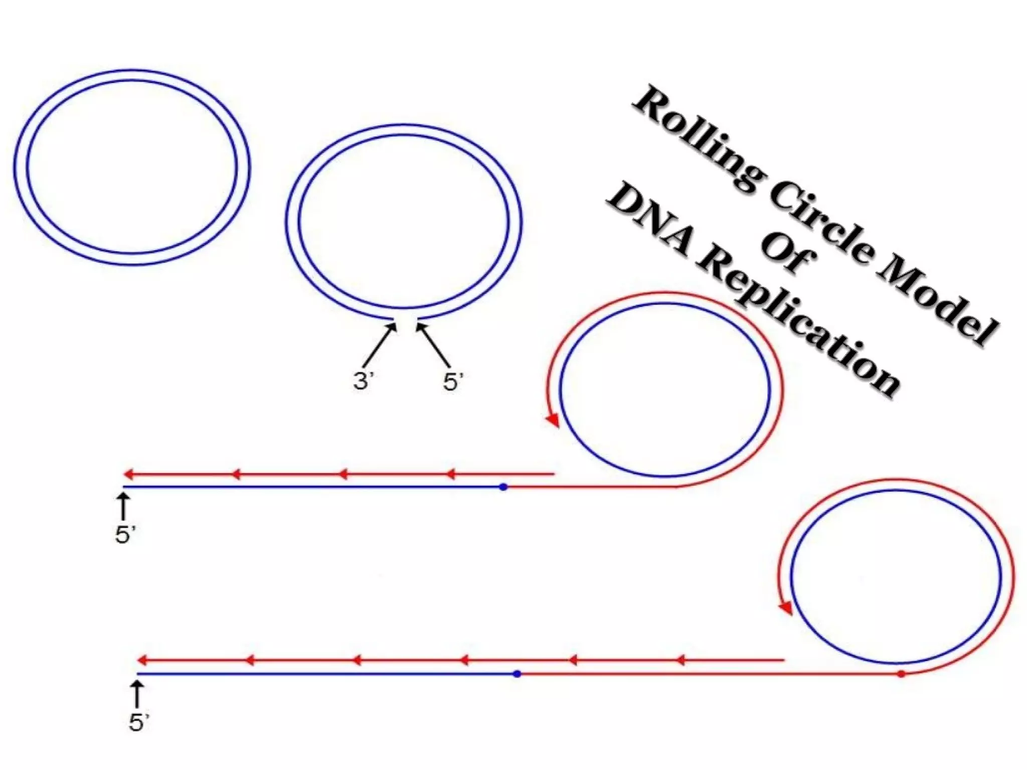
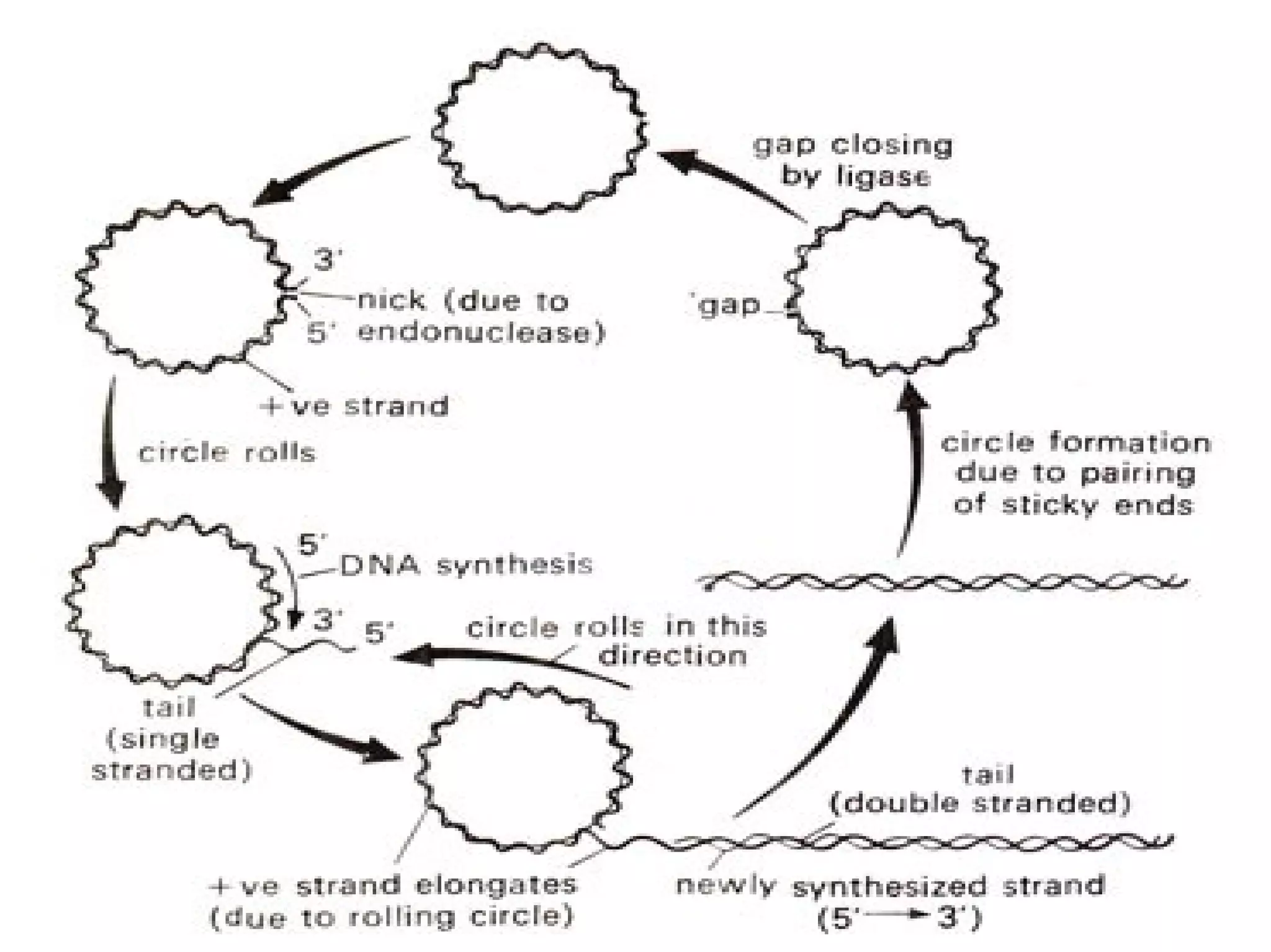
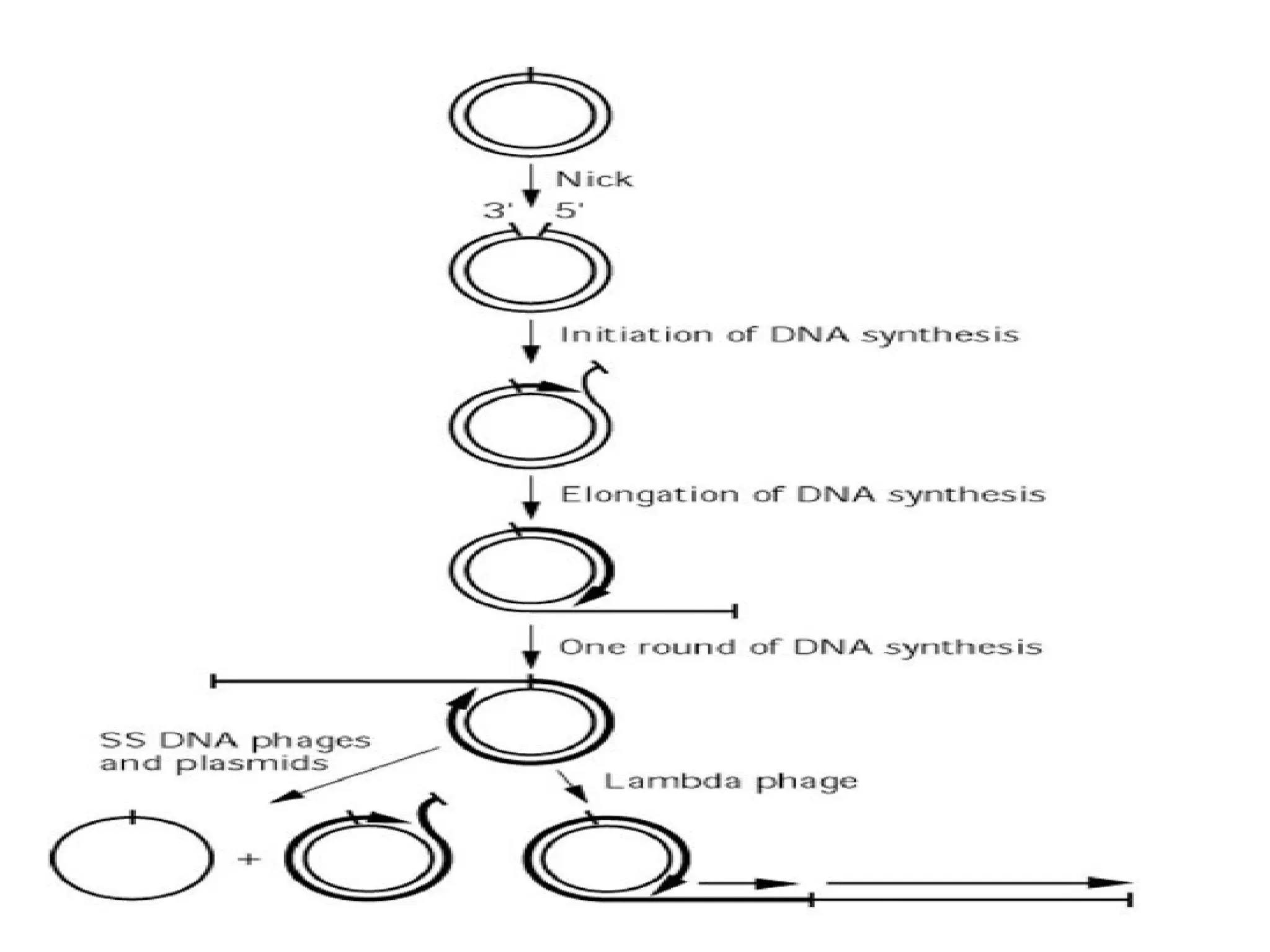
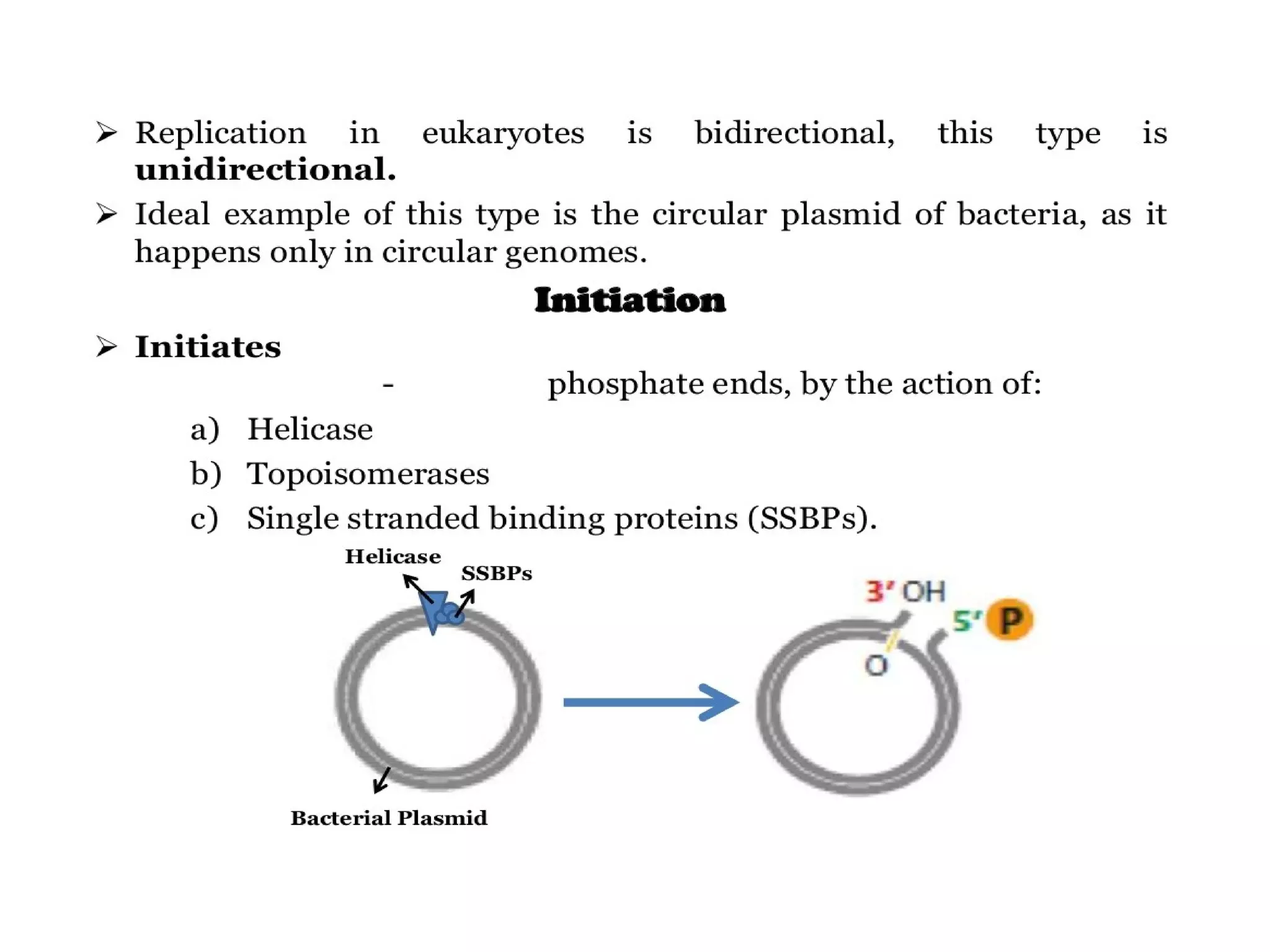
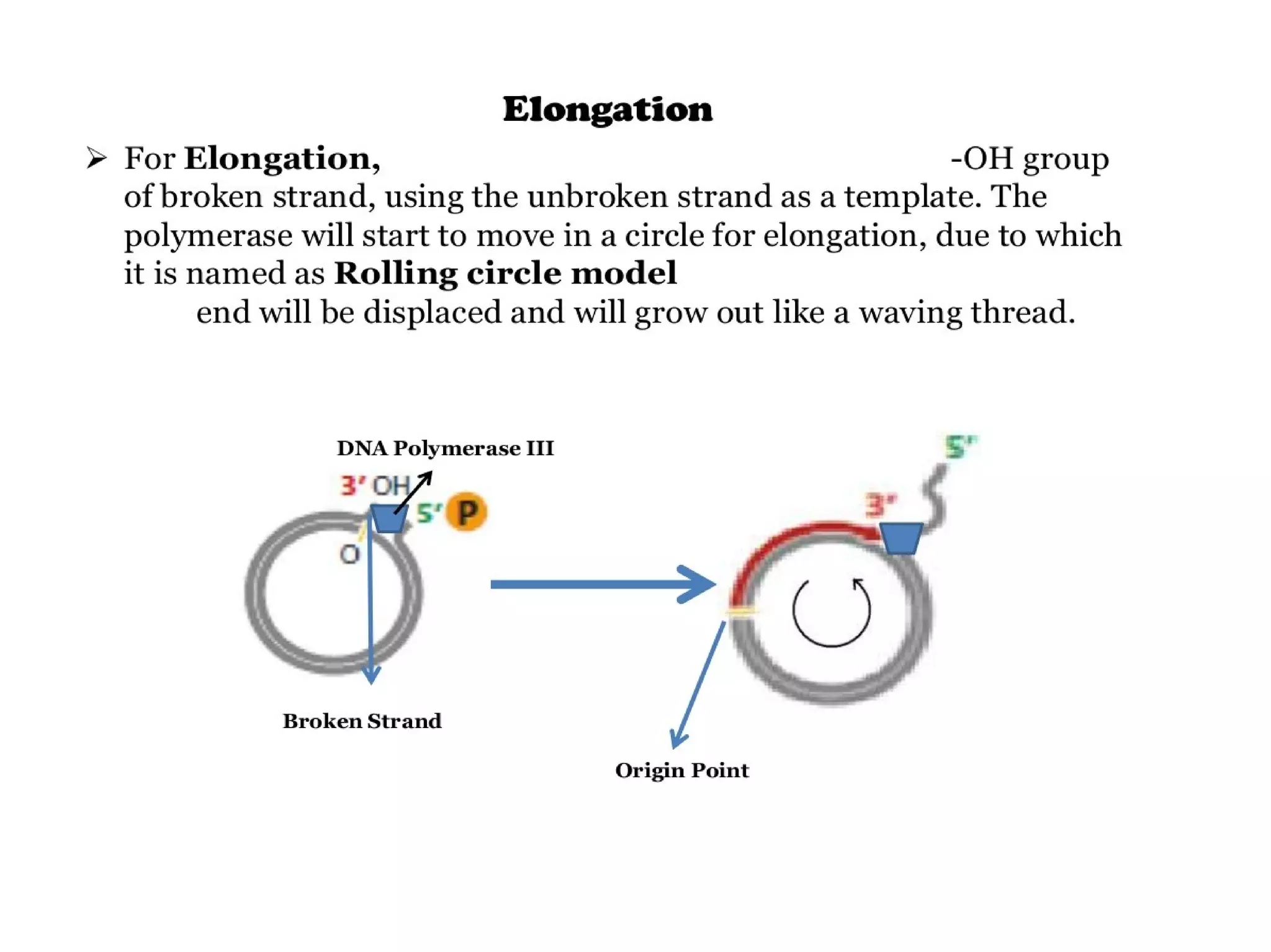
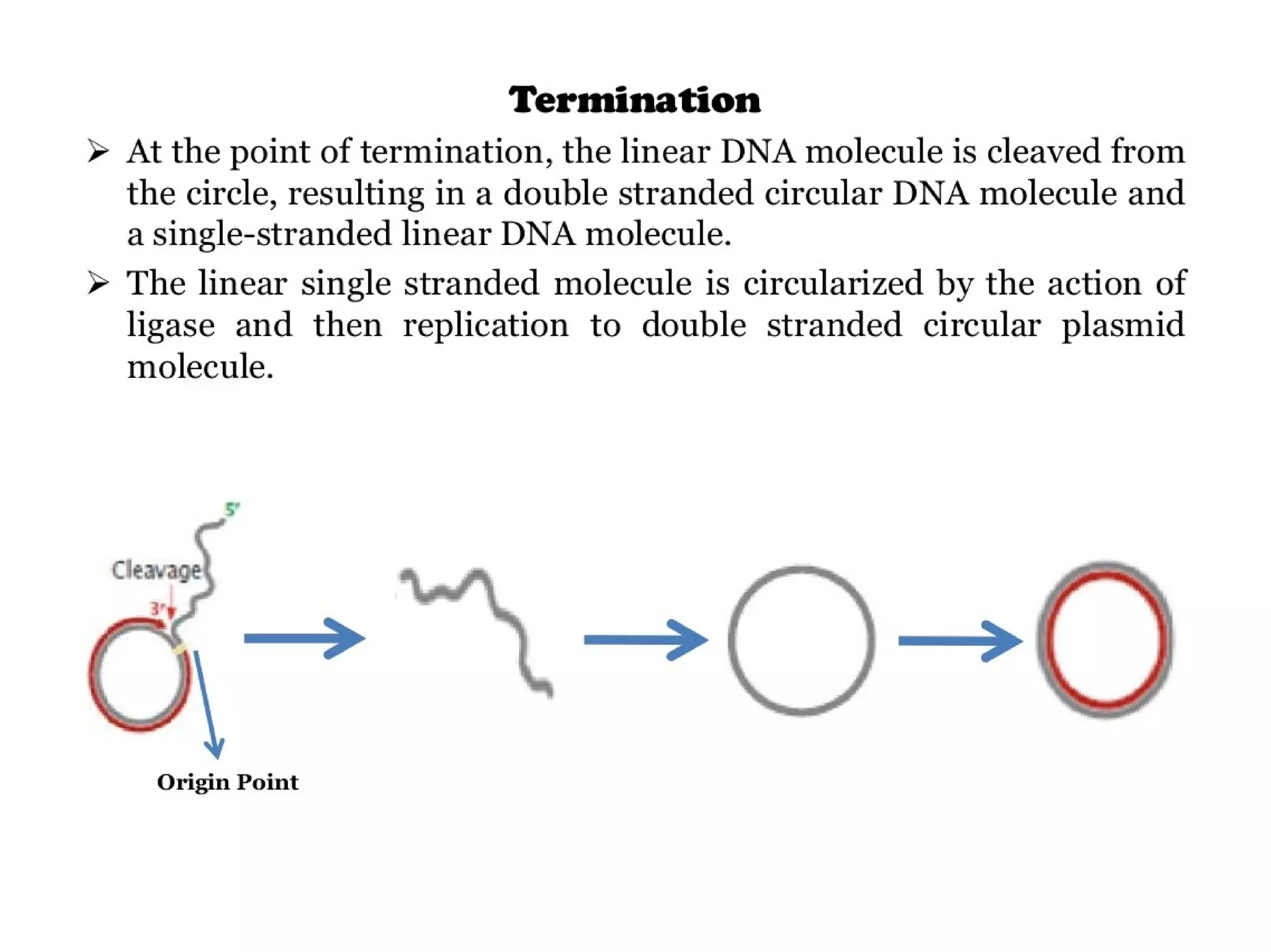
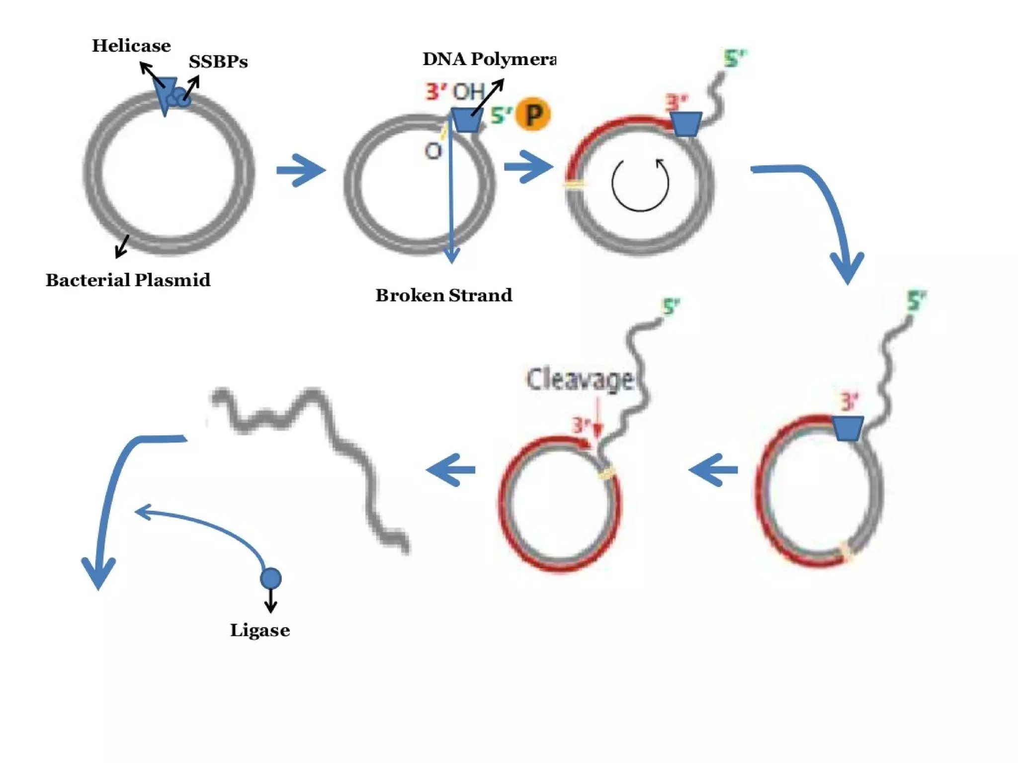
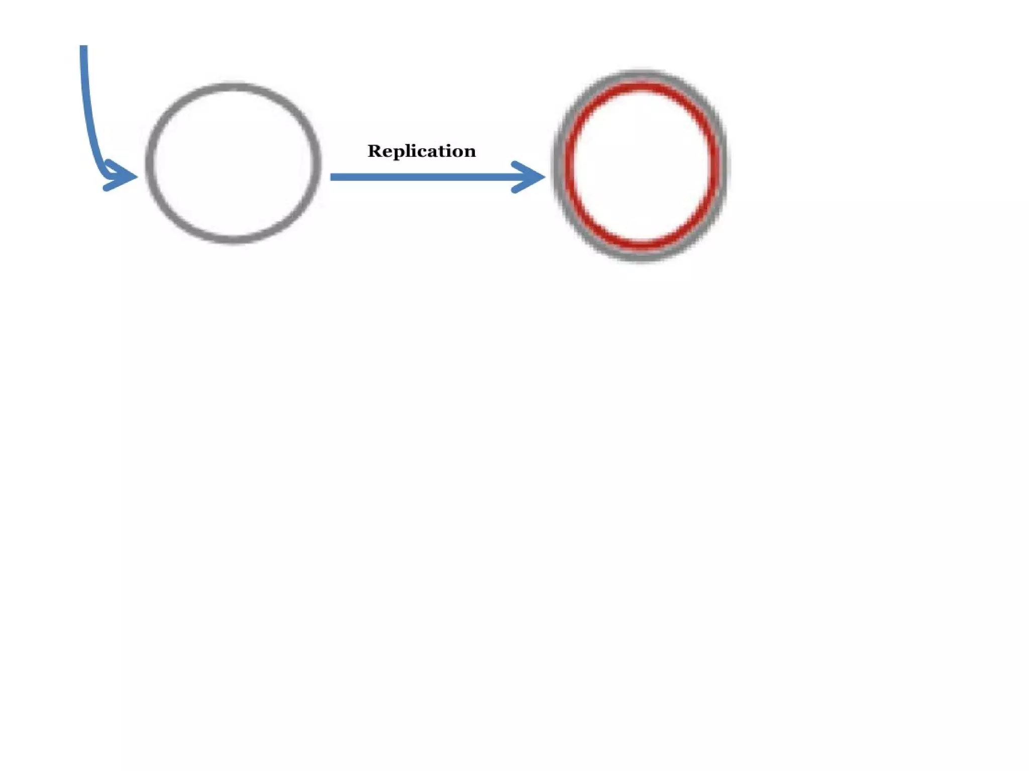
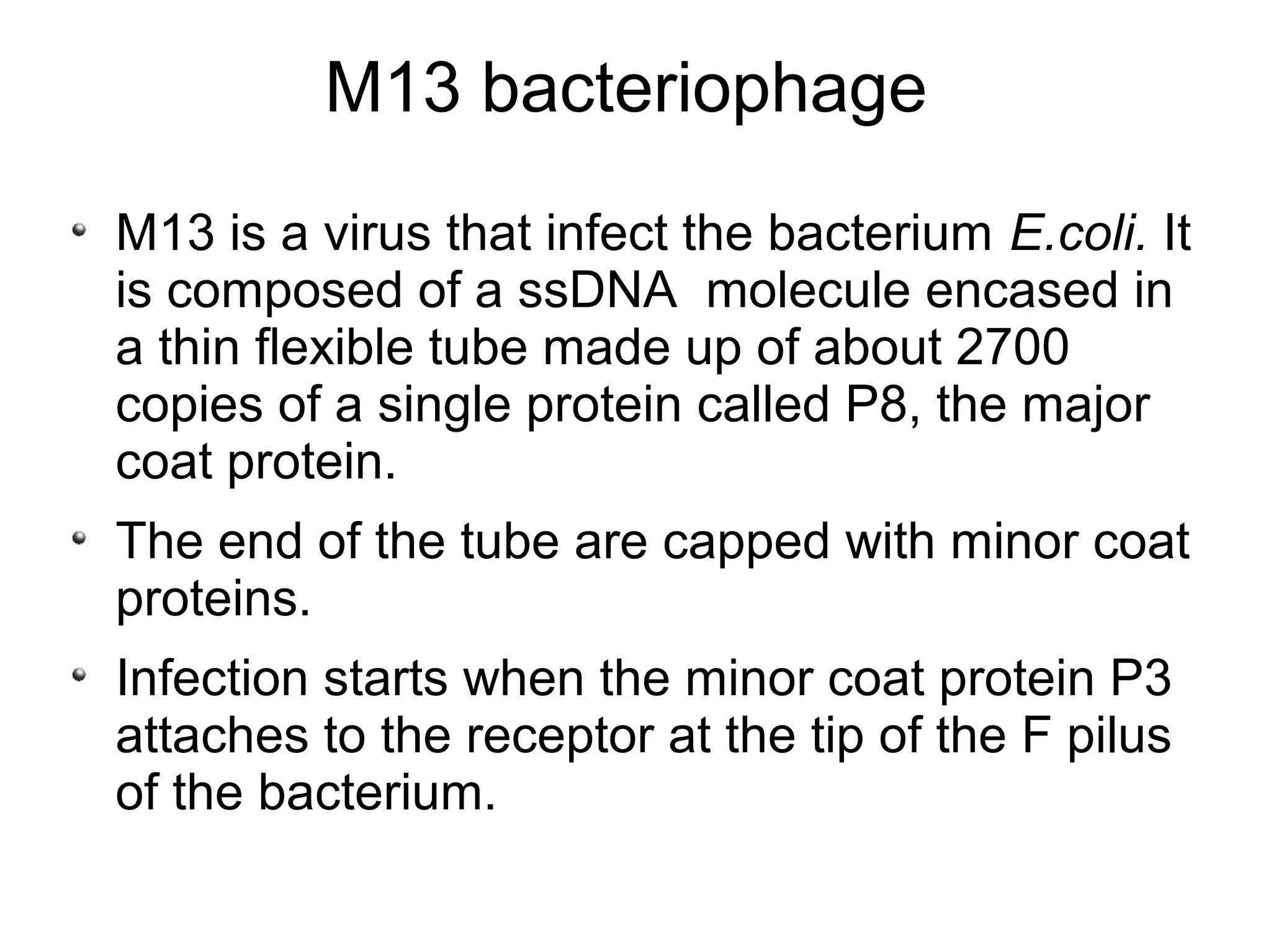
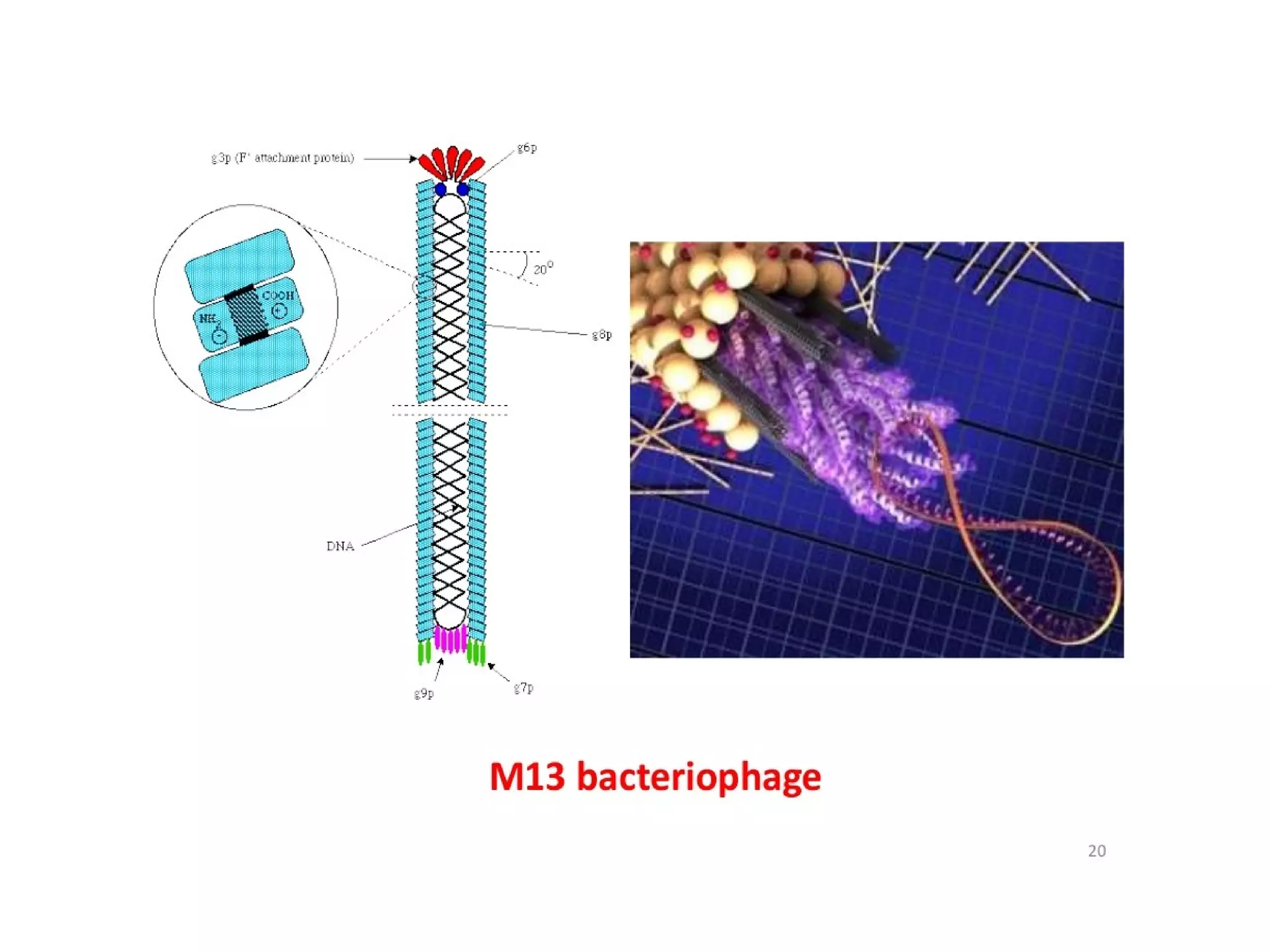
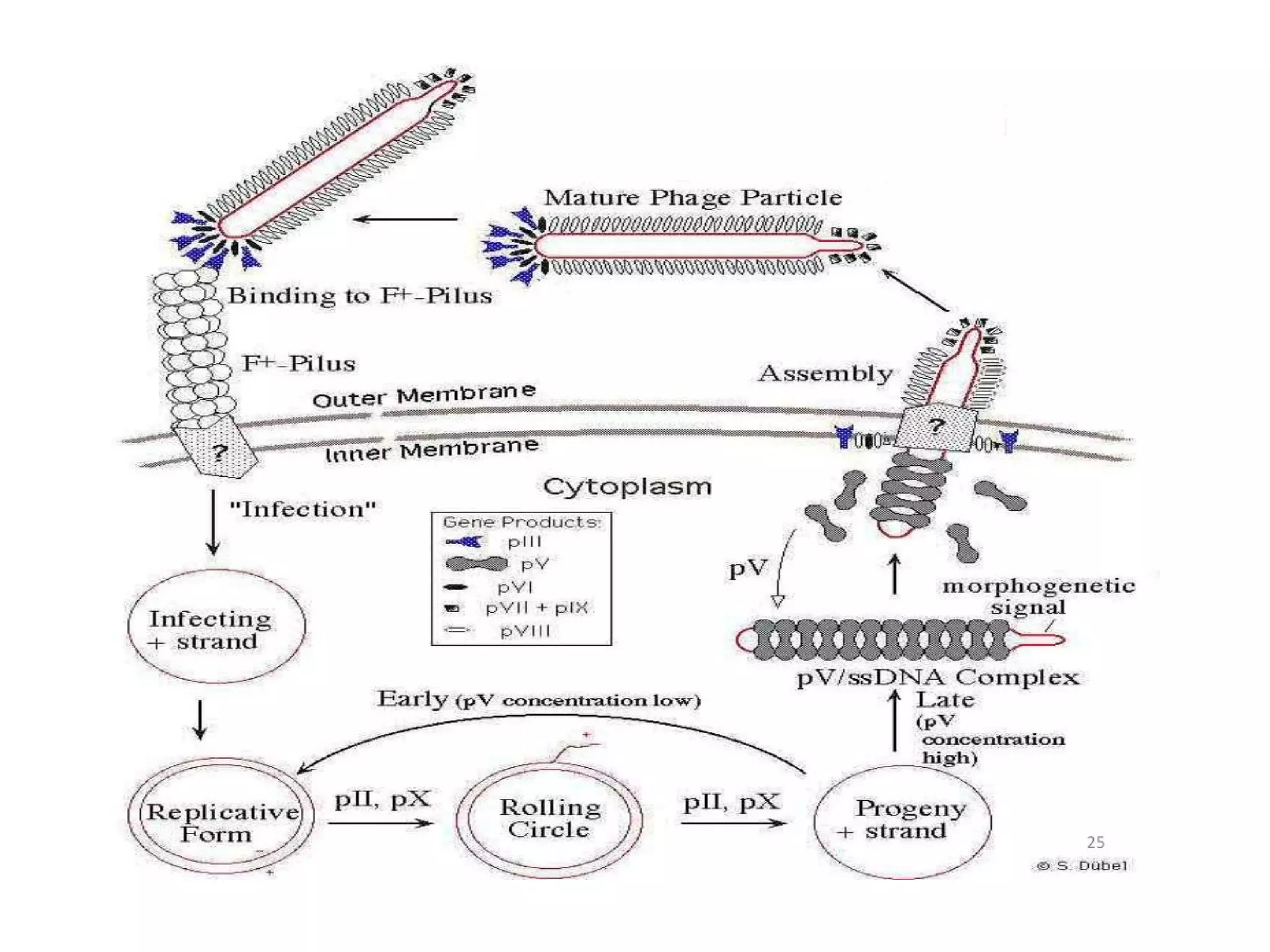
ROLLING CIRCLE MODEL AND
M13 BACTERIOPHAGE
REPLICATION
BY
Vivek kumar
M.SC.MICROBIOLOGY
Bangalore University
ROLLING CIRCLE REPLICATION
Rolling circle replication describes a process of
unidirectional nucleic acid replication that can
rapidly synthesize multiple copies of circular
molecules of DNA or RNA
Rolling circle model is initiated by an intiator
protein encoded by the plasmid or
bacteriophage DNA,
Which nicks one strand of the DS, circular DNA
molecule at a site called the DSO.
The initiator protein remainsbound to the 5'
phosphate end of the nicked strand, and the
free 3' hydroxyl end is released to serve as a
primer for DNA synthesis by DNA polymerase-
3.
Using the un nicked strand as a template,
replication proceeds around the circular DNA
molecule, displacing the nicked strand as SS is
carried out by a host encoded helicase called
PcrA.in the presence of the plasmid replication
initiation protein.
Continued DNA synthesis can produce multiple
SS linear copies of the original DNA in a
continuous head to-tail series – concatemer.
These linear copies can converted to DS
circular molecules , through the following
process:
First the initiator protein makes another nick in
the DNA to terminate synthesis of the
first(leading) strand.
RNA polymerase and DNA ploymerase3 then
replicate the SS origin DNA to make another
double stranded circle.
DNA polymerase1 removes the primer,replacing
it with DNA, and DNA ligase joints the ends to
make another molecule of DS circular DNA.
A typical DNA rolling circle replication has 5
steps
Circular dsDNA will be nicked.
The 3' end is elongated using unnicked DNA as
leading strand(template):5' end is displaced.
Displaced DNA is a lagging strand and is made
doubl stranded via a series of okazaki
fragments.
Replication of both unnicked and displaced
ssDNA.
Displaced DNA circularizes.
REPLICATION OF M13
BACTERIOPHAGE
M13 bacteriophage
M13 is a virus that infect the bacterium E.coli. It
is composed of a ssDNA molecule encased in
a thin flexible tube made up of about 2700
copies of a single protein called P8, the major
coat protein.
The end of the tube are capped with minor coat
proteins.
Infection starts when the minor coat protein P3
attaches to the receptor at the tip of the F pilus
of the bacterium.
Replication of M13 bacteriophage
Viral(+) strand DNA enters cytoplasm.
Complementary(-) strand is synthesized by
bacterial enzymes.
DNA gyrase, a type 2 topoisomerase, acts on
dsDNA and catalyses formation of negative
supercoils in dsDNA.
Final product is parental replicative
form(RF)DNA
A phage protein, P2 nicks the (+) strand in the
RF
3' hydroxyl acts as a primer in the creation of
new viral strand.
P2 circularizes displaced viral(+) strand DNA
Pool of progeny ds RF molecule produced
Negative strand of RF is template of
transcription
MRNAs are translated into the phage proteins
Phage proteins in the cytoplasm are P2, pX,
and pV, they are part of the replication process
of DNA.
PV dimers bind newly synthesized ssDNA and
prevent conversion to RF DNA
RF DNA synthesis continues and amount of pV
reaches critical concentration
DNA replication switches to synthesis of ss(+)
viral DNA
PV DNA structures from about 800nm long and
8nm in diameter
PV DNA complex is substrate in phage
assembly rection
REPLICATION OF M13
BACTERIOPHAGE
Thankyou

Rolling circle model and m13 bacteriophage replication

  • 1.
    ROLLING CIRCLE MODELAND M13 BACTERIOPHAGE REPLICATION BY Vivek kumar M.SC.MICROBIOLOGY Bangalore University
  • 3.
    ROLLING CIRCLE REPLICATION Rollingcircle replication describes a process of unidirectional nucleic acid replication that can rapidly synthesize multiple copies of circular molecules of DNA or RNA Rolling circle model is initiated by an intiator protein encoded by the plasmid or bacteriophage DNA, Which nicks one strand of the DS, circular DNA molecule at a site called the DSO.
  • 4.
    The initiator proteinremainsbound to the 5' phosphate end of the nicked strand, and the free 3' hydroxyl end is released to serve as a primer for DNA synthesis by DNA polymerase- 3. Using the un nicked strand as a template, replication proceeds around the circular DNA molecule, displacing the nicked strand as SS is carried out by a host encoded helicase called PcrA.in the presence of the plasmid replication initiation protein. Continued DNA synthesis can produce multiple SS linear copies of the original DNA in a continuous head to-tail series – concatemer.
  • 5.
    These linear copiescan converted to DS circular molecules , through the following process: First the initiator protein makes another nick in the DNA to terminate synthesis of the first(leading) strand. RNA polymerase and DNA ploymerase3 then replicate the SS origin DNA to make another double stranded circle. DNA polymerase1 removes the primer,replacing it with DNA, and DNA ligase joints the ends to make another molecule of DS circular DNA.
  • 6.
    A typical DNArolling circle replication has 5 steps Circular dsDNA will be nicked. The 3' end is elongated using unnicked DNA as leading strand(template):5' end is displaced. Displaced DNA is a lagging strand and is made doubl stranded via a series of okazaki fragments. Replication of both unnicked and displaced ssDNA. Displaced DNA circularizes.
  • 15.
  • 16.
    M13 bacteriophage M13 isa virus that infect the bacterium E.coli. It is composed of a ssDNA molecule encased in a thin flexible tube made up of about 2700 copies of a single protein called P8, the major coat protein. The end of the tube are capped with minor coat proteins. Infection starts when the minor coat protein P3 attaches to the receptor at the tip of the F pilus of the bacterium.
  • 17.
    Replication of M13bacteriophage Viral(+) strand DNA enters cytoplasm. Complementary(-) strand is synthesized by bacterial enzymes. DNA gyrase, a type 2 topoisomerase, acts on dsDNA and catalyses formation of negative supercoils in dsDNA. Final product is parental replicative form(RF)DNA A phage protein, P2 nicks the (+) strand in the RF
  • 18.
    3' hydroxyl actsas a primer in the creation of new viral strand. P2 circularizes displaced viral(+) strand DNA Pool of progeny ds RF molecule produced Negative strand of RF is template of transcription MRNAs are translated into the phage proteins Phage proteins in the cytoplasm are P2, pX, and pV, they are part of the replication process of DNA.
  • 19.
    PV dimers bindnewly synthesized ssDNA and prevent conversion to RF DNA RF DNA synthesis continues and amount of pV reaches critical concentration DNA replication switches to synthesis of ss(+) viral DNA PV DNA structures from about 800nm long and 8nm in diameter PV DNA complex is substrate in phage assembly rection
  • 22.
  • 24.