Notes by Prof. Sanjeev Puri, UIET, Panjab University on M13 Phage.
Large libraries can be prepared by cloning a mixture of cDNAs from a particular tissue or, less easily, by cloning genomic DNA fragments.
The library consists of phages displaying a range of different proteins and is used to identify those that interact with a test protein.
A phage display library made up of many recombinant phages, each displaying a different protein.
Oligonucleotide Directed Mutgenesis
uracil N-glycosylase
NEWLETTER FRANCE HELICES/ SDS SURFACE DRIVES - MAY 2024
Lectures PPT of Prof. Sanjeev Puri, on M13 phage
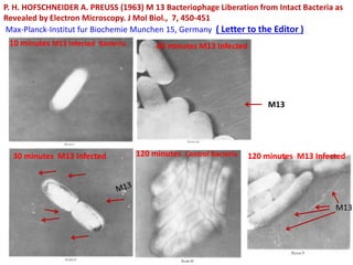
1. P. H. HOFSCHNEIDER A. PREUSS (1963) M 13 Bacteriophage Liberation from Intact Bacteria as
Revealed by Electron Microscopy. J Mol Biol., 7, 450-451
Max-Planck-Institut fur Biochemie Munchen 15, Germany ( Letter to the Editor )
10 minutes M13 Infected Bacteria
30 minutes M13 Infected
60 minutes M13 Infected
120 minutes Control Bacteria 120 minutes M13 Infected
M13
M13
2. M13 Phage is a filamentous Phage having (Sense Strand Serves template
for mRNA,) Covalently Closed Circle showing genes I to X coding for
about 1 5different proteins .
Double lines in fig. for ease of showing location of genes
Ori as origin of replication
IS intervening sequence (507 nucleotide long can be removed for insertion of GOI)
Genes I, II, IV-VII, IX & X codes for structural components and phage specific replication
enzymes
Gene V (codes for 2700 tubular protein
encasing genome & gene III codes
protein (5-8) at the end of
filamentous phage
M13 Phage
Endonuclease
3. This molecule is not inserted
into the bacterial genome, but
instead replicates until over 100
copies are present in the cell.
About 1000 new phages being
produced during each
generation of an infected cell
4. For bacterial infection it enters through “sex pilus”, once in host gets
converted to Double stranded Replicative forms (RF) used for making
copies of M13 positive single stranded form, via rolling circle model
of replication, and also for proteins
This RF behaves as Plasmid, can be selected, manipulated and
inserted in bacteria host
No size constraints for packaging DNA Protein V, a single strand
binding protein take it to Bacterial wall
There Protein VIII major coat protein bind for the release
Cloned genes in M13 Vector are obtained as Single
stranded DNA for multiple applications Viz. DNA
sequencing and in vitro mutagenesis, phage display
(Phagemid) methodologies
5. 1000
nm
Replicative
Form
v
P
P
OH
+ve strand
Rolling Circle Model
Host DNA Pol III
Bacteria
Genomic DNA
M13 Single stranded Circular DNA
Phage VIII Proteins (Structural)
Phage III Proteins
Nick
5nm
M13 Phages
Released
No Lysis of
Infected
Bacteria
(Plaques
are turbid)
continue to
grow and
divide
albeit slow
Sex pilus
~1000 Progeny phages
released/hr. post infection
6. Clear Plaques
Bacteria
Lysed contain phge
DNA
Turbid Plaques
Lysogenic in lambda
and productive
infection in M13
Clear Plaques Recombinant
Blue Plaques Non-Recombinant
Lawn of
Bacteria
7. M13 as Vector development steps
M13 ------- Wild Type Phage
M13 mp1 ------- Introduced lac Z’ gene
M13 mp2 ------- Mutate GGATTC near lacZ’
to GAATTC
To introduced EcoR1
Restriction endonuclease
site
M13 mp7 -------- Introduce Poly-Cloning Site
(PCS)within EcoR1
5’-AATTCCCC GGATCC GTCGAC CTGCAG GTCGAC GGATCC GGG G
3’-GGGG CCTAGG CAGCTG GACGTC CAGCTG CCTAGG CCC CTTAA-5’
EcoliR1 BamH1 Sal1 Pst1 Sal1 BamH1 EcoR1
PCS
LacZ’ and Ligase
Mutate
EcoR1, PCS, Ligase
Engineered PCS
with 4
Restriction
Endonucleases
MutateM13 Phage
8. M13 mp7 ------- 4 RE‘s Poly-Cloning Site
(PCS) within EcoR1
M13 mp8 ------- Remover mp7 PCS and
inserted 6-RE, PCS as
below within lacZ’
EcR1-----HindIII
M13 mp9 ------- Reverted Sites as
Hind III-----EcoRI
5’-AATTCCCGGGGATCC GTCGAC CTGCAG CCA
3’-GGGCCCCTAGG CAGCTG GACGTC GGTTCGA-5’
Ecoli RI Sma I Bam HI Sal I Pst I Hind III
PCS with 6- RE’S sites having different
terminal RE within LacZ’, Ligase
Eco R1 Hind III
Engineered PCS with 6
Restriction Endonucleases
M13 as Vector development steps
Hind III Eco R1
Revert the PCS
9. Phagemid:
A Construct or Vector generated by combining part of the DNA sequence
of Phage with the part of the DNA sequence of Plasmid DNA
Phagemids are named so because these are replicated and packaged by
M13 proteins. Upon further tranfection with M13 helper phage which
provides the necessary gene products to replicate and package phagemids as
ssDNA phages
Constraints of M13 phage vector removed one can clone a single stranded
gene upto 10 kb
Example is pEMBL8 ( plasmid European Molecular biology Laboratory )
1300 bp of M13 Phage + pUC vector sequence = M13 Phagemid
Plasmid DNA
Segment
Phage DNA
Segmment
Phagemid
13. Phage Display libraries ( For Protein- Protein interactions)
• Achieved by cloning the gene for
the protein in a special type of M13
vector.
• Results is cloned gene becoming
fused with a gene for a phage coat
protein.
• Transfect E. coli, this gene fusion
directs synthesis of a hybrid
protein, made up partly of the
coat protein and partly of the
product of the cloned gene.
• With luck this hybrid protein will
be inserted into the phage coat so
that the product of the cloned
gene is now located on the surface
of the phage particle
14. • A phage display library made up of many recombinant
phages, each displaying a different protein.
• Large libraries can be prepared by cloning a mixture of
cDNAs from a particular tissue or, less easily, by
cloning genomic DNA fragments.
• The library consists of phages displaying a range of
different proteins and is used to identify those that
interact with a test protein.