SlideShare a Scribd company logo
1 of 45
Right Heart Catheterization
Chairperson: Dr. Jahanara Arzu
Professor
Department of Cardiology, UCC, BSMMU
Presenter : Dr. Al-Amin
Resident(Phase B)
Werner Forssman Dr. Swan Dr Ganz
In 1929 Werner Forssmann from Germany first performed right heart catheter on himself
Dr Swan added a balloon tip the catheter and Dr. Ganz added thermistor at the tip
Introduction
Promoting catheters in the right heart chambers
and branches of the pulmonary artery in order to:
1.Record pressures and their waveforms
2.Measure cardiac output
3.Evaluate O2 saturation and content – oximetry run.
4. Evaluation of intracardiac shunt and other congenital heart
disease
Diagnostic indication
 Differentiation of various etiologies of shock and pulmonary
edema
 Diagnosis of intracardiac shunts
 hemodynamic assessment of shunt operability
 postoperative assessment
 Evaluation of pulmonary hypertension
 Quantification of pulmonary pressure
 Calculation of PVR
 Response to vasodilators
Therapeutic indication
Guide to fluid management and
Hemodynamic monitoring of patients after
 surgery,
 complicated myocardial infarction,
 shock & heart failure
Contraindications
Absolute
 Endocarditis(right cavities)
 Clot / mass in the right cavities
 Mechanical valves in the tricuspid / pulmonary
Relative
 Hemorrhagic diathesis (INR> 2, platelet count < 50,000 )
 Ventricular Arrhythmia
 Poor life expectancy
 Poor cooperation / patient
 Bioprosthetic valves
 Recent installation PM / ICD
 Contralateral pneumothorax
Complications
• Arrhythmia (Atrial and ventricular)
• Pulmonary Infarction
• Pulmonary artery rupture
• Air embolism
• Endocarditis
• Venous thrombosis
• Bleeding At the vascular access site
• Arterial puncture
• Pneumothorax
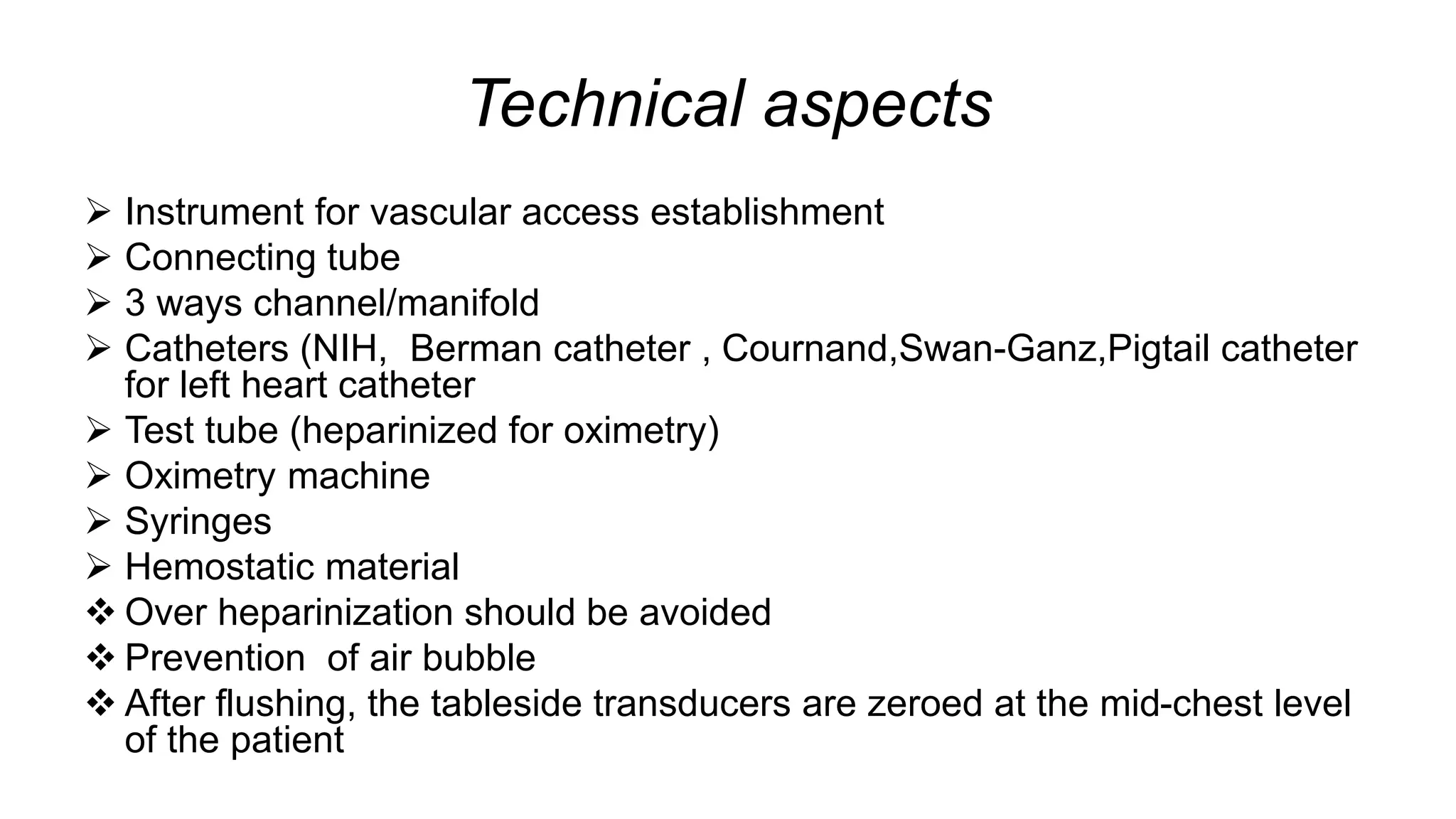
Technical aspects
 Instrument for vascular access establishment
 Connecting tube
 3 ways channel/manifold
 Catheters (NIH, Berman catheter , Cournand,Swan-Ganz,Pigtail catheter
for left heart catheter
 Test tube (heparinized for oximetry)
 Oximetry machine
 Syringes
 Hemostatic material
 Over heparinization should be avoided
 Prevention of air bubble
 After flushing, the tableside transducers are zeroed at the mid-chest level
of the patient
Swan Ganz catheter
venous access sites
 Internal jugular vein
 Subclavian vein
 Basilic vein (Right)
 Basilic vein (Left)
 Femoral vein
Procedure
• Establishment of vascular access
• Location of the catheter can be
determined by pressure recorded in
different chamber
• Advance the catheter into IVCRA
counterclock wise rotation to enter
SVC
• Record O2 saturation in SVC (high &
low)
• Record O2 saturation in RA (high,mid
& low)
• Record phasic & mean pressure of RA
Procedure
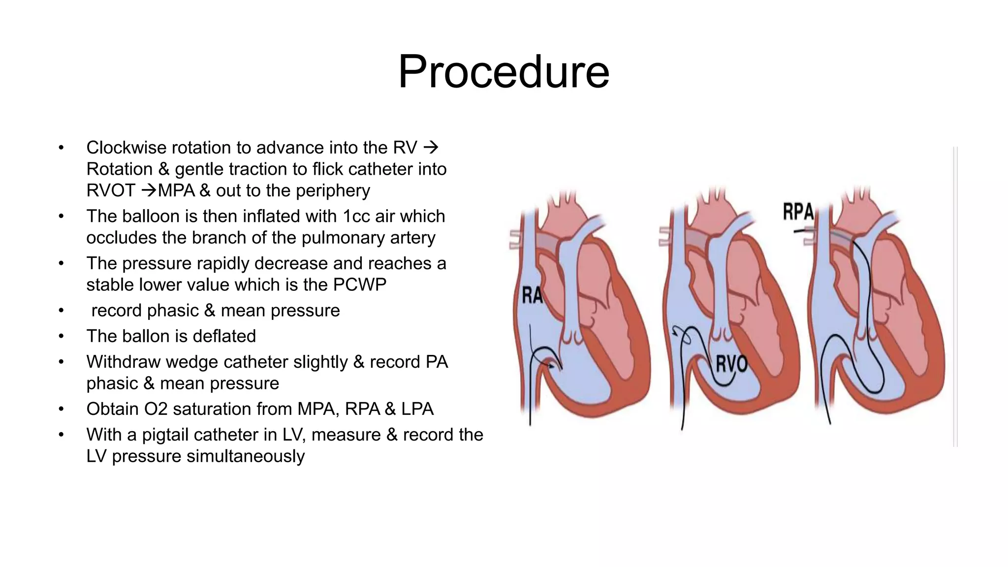
• Clockwise rotation to advance into the RV 
Rotation & gentle traction to flick catheter into
RVOT MPA & out to the periphery
• The balloon is then inflated with 1cc air which
occludes the branch of the pulmonary artery
• The pressure rapidly decrease and reaches a
stable lower value which is the PCWP
• record phasic & mean pressure
• The ballon is deflated
• Withdraw wedge catheter slightly & record PA
phasic & mean pressure
• Obtain O2 saturation from MPA, RPA & LPA
• With a pigtail catheter in LV, measure & record the
LV pressure simultaneously
Procedure
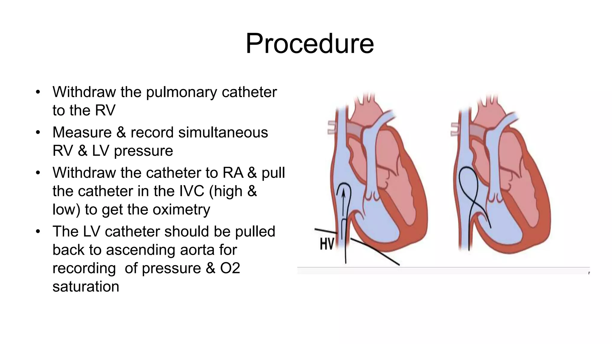
• Withdraw the pulmonary catheter
to the RV
• Measure & record simultaneous
RV & LV pressure
• Withdraw the catheter to RA & pull
the catheter in the IVC (high &
low) to get the oximetry
• The LV catheter should be pulled
back to ascending aorta for
recording of pressure & O2
saturation
Pressure recordings
Always record pressure at end expiration
Pressures will be lower in inspiration due to decrease in
intrathoracic pressure
Before any pressure measurements are taken, it is
imperative to perform zeroing and referencing of the
system
Normal pressure at different chambers of heart
Saturation run
• For shunts, a full saturation run should
be performed by obtaining 2 ml sample
from each of the location
– Left and right pulmonary artery
– Main pulmonary artery
– Right ventricle outflow tract, mid and apex
– Right atrium low, mid and high
– Superior vena cava low, high
– Inferior vena cava high, low
– Left ventricle
– Aorta
• Saturation syringe are heparinized and
new sample is taken after discarding few
ml of blood to avoid contamination
The normal pressure waves in the cardiac chambers during right heart catheterization with normal values
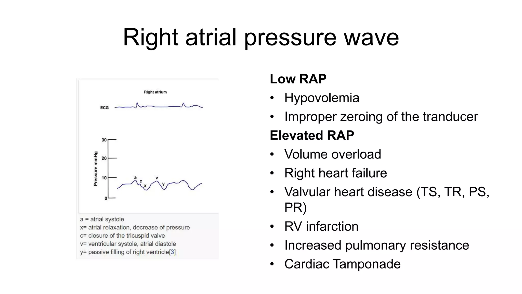
Right atrial pressure wave
Low RAP
• Hypovolemia
• Improper zeroing of the tranducer
Elevated RAP
• Volume overload
• Right heart failure
• Valvular heart disease (TS, TR, PS,
PR)
• RV infarction
• Increased pulmonary resistance
• Cardiac Tamponade
Right atrial pressure wave in different condition
 Elevated a wave:
tricuspid stenosis
decreased ventricular compliance due to ventricular
failure
pulmonic valve stenosis
 Elevated v wave:
tricuspid regurgitation (prominent y descent)
RV failure
 Absent a wave:
atrial fibrillation
 Equalization of a and v wave
ASD
 Prominent x descent and absent or reduced y
descent
Cardiac tamponade
 Prominent y descent
 Constrictive pericarditis
 Kussmaul sign
constrictive pericarditis,
RV infarct
Right ventricle pressure waves and abnormality
 Elevated Systolic pressure
 Pulmonary Hypertension
 Pulmonary stenosis
 Systolic pressure reduced:
 hypovolemia,
 cardiogenic shock,
 tamponade
 Elevated end diastolic pressure:
 cardiomyopathy,
 RV ischemia and infarction,
 RV failure
 tamponade
 Constrictive pericarditis
 Decreased end diastolic pressure:
tricuspid stenos
Pulmonary arterial pressure wave
 Elevated systolic pressure:
 idiopathic pulmonary hypertension,
 pulmonary disease like COPD,ILD
 mitral stenosis or regurgitation,
 significant cardiac left to right shunt,
pulmonary embolism
 Restrictive cardiomyopathy
 Decreased systolic pressure
 Hypotension
 Pulmonary stenosis
 Tricuspid valve stenosis
 Tricuspid valve atresia
 Ebstein’s anomaly
Pulmonary capillary wedge pressure
1. Low mean pressure:
– hypovolemia
2. Elevated mean pressure:
– intravascular overload,
– LV failure caused by myocardial disease,
– systemic hypertension
– valvular disease (MS, AoS, AoR)
– pericardial effusion with tamponade
3. Elevated a wave:
– mitral stenosis,
– diastolic dysfunction,
– LV volume overload
4. Cannon a wave
– atrial-ventricular asynchrony(3rd degree AVB, VT, V-pacer)
5. Elevated v wave:
– mitral regurgitation,
– LV failure,
– VSD
• Equal a and v waves
– Cardiac Tamponade
– Constrictive pericarditis
Pulmonary capillary wedge pressure
 PCW tracing “approximates”
actual LA tracing but is slightly
delayed since pressure wave is
transmitted retrograde through
pulmonary veins
 Normal LA pressure slightly higher
 Normal PCWP pressure range = 4-12 mmHg
(mean 9)
 When there is no obstruction between left
atrium and left ventricle: PCWP = LA
pressure = LVEDP. = PA diastolic pressure
 PCWP higher than LVEDP in
 MS,MR
 Pulmoary embolism
 A true wedge pressure can be measured
only in the absence of flow”, meaning with a
vessel closed by a balloon.
Pulmonary capillary wedge pressure
importance of measurement
It is important to measure PCWP to diagnose
 left ventricular failure
Quantify degree of mitral stenosis
Titration of I/V fluid or diuretics
Evaluation of pulmonary hypertension
Pulmonary Hypertension in Adult Congenital Heart Disease
Definition
Haemodynamic
characteristicsa Clinical settings
Pulmonary Hypertension (PH) Mean PAP >20 mmHg All
Pre-capillary PH (PAH)
•Mean PAP >20 mmHg
•PAWP ≤15 mmHg
•PVR ≥3 WU
•Shunt lesions prior to and after
repair (including Eisenmenger
syndrome)
•Complex CHD (including UVH,
segmental PAH)
Definition Haemodynamic
characteristicsa Clinical settings
isolated post-capillary PH
•Mean PAP >20 mmHg
•PAWP >15 mmHg
•PVR <3 WU
•Systemic ventricular dysfunction
•Systemic AV valve dysfunction
•Pulmonary vein obstruction
•Cor triatriatum
Combined pre- and post-capillary
PH
•Mean PAP >20 mmHg
•PAWP >15 mmHg
•PVR ≥3 WU
•Settings listed under isolated
post-capillary PH
•Settings listed under isolated
post-capillary PH in combination
with shunt lesions/complex CHD
Vasoreactivity test
• to detect residual properties of vasodilatation of small pulmonary arteries and
arterioles
• recommended in pt with group 1 PAH
• done by administration of short acting vasodilator drug then recording
hemodynamic response
• commonly used drugs for vasoreactivity testing-
– Inhaled nitric oxide
– Adenosine
– Epoprostenol
– Inhaled 100% oxygen
• Positive response is defined as reduction of mean PAP>10 mmhg to reach a
mean PAP < 40 mmhg with a increased or unchanged cardiac output
Cardiac output measurement
Fick-Oxygen Method
Thermodilution technique
Fick O2 method
 CO = O2 consumption/ arteriovenous 02 concentration difference
 O2 consumption roughly (3xwt in kg) ml/min
 LVO2 content= (1.36 x Hb x LVO2 sat x 10)
 Pulmonary arterial O2 content = (1.36 x Hb x PAO2 sat x 10)
 Arteriovenous 02 concentration difference = ( LVO2 content - Pulmonary arterial O2
content)
The value 1.36 is derived from the fact that 1 gram of hemoglobin, when 100%
saturated, combines with 1.36 ml of oxygen
Thermodilution indicator method
 Principle:
 injection of a given quantity of cold N / S to the
central circulation changes the temperature of
blood distally.
 Technique:
Injecting 10ml N /S at room temperature within 2
seconds from the proximal channel of the
Swan-Ganz record temperature -time curve
Catheter
 Complete mixing of the indicator causes a
decrease in blood tempdetected by distal
thermistor  CO computer calculates the
change in indicator concentration (temp over
time) to determine CO
Oximetry run
• Normally, pulmonary blood flow and systemic blood flow are equal
• When there is an abnormal communication between intracardiac
chambers or great vessels, blood flow is shunted either
– from the systemic circulation to the pulmonary circulation (left-to-right
shunt)
– from the pulmonary circulation to the systemic circulation (right-to-left
shunt)
– in both directions (bidirectional shunt)
Oximetry run
• Blood sampling from various cardiac chambers for oxygen
saturation determination
• A left-to-right shunt is detected when there is a significant
increase in blood oxygen saturation between two right-
sided vessels or chambers (step up)
• Despite its lack of sensitivity, clinically significant shunts
are generally detected by this technique
Step up for left to right shunt
Oxygen saturation values for shunt
detection
Level of shunt Significant step up
of O2
saturation(differen
ce between distal
and proximal
chamber
Possible causes of step up
Atrial(SVC/IVC
to rt atria)
>7 ASD,PAPVD
Ventricular(RA
to RV)
>5 VSD,Primum ASD
Great vessel(RV
to PA)
>5 PDA,Aortopulmonary widow
O2 step up in ASD and VSD
VSD
RA
RV
LV
ASD Secondum
RA LV
RV
• The flow ratio PBF/SBF (or QP/QS) is used clinically to
determine the significance of the shunt
• A flow ratio of less than 1.0 indicates a net right-to-left shunt
• A ratio of 1-1.5 indicates a small left-to-right shunt
• A ratio of 1.5- 2.0 or more indicates a large left-to-right shunt and
generally requires repair in order to prevent future pulmonary
and/or right ventricular complications
Shunt calculation
 Pulmonary flow(Qp)= O2 consumption ml/min
( pulmonary venous- pulmonary arterial) O2 content
 Systemic flow(Qs) = O2 consumption ml/min
( arterial- mixed venous )O2 content
 EPBF = O2 consumption ml/min
( pulmonary venous- mixed venous) O2 content
Mixed venous O2 content = 3 SVC+ 1 IVC
4
Effective pulmonary blood flow = systemic flow + shunt flow( left to right shunt)
(when shunt present) = systemic flow– shunt flow (right to left shunt)
PVR = (mean PAP-PCWP)X 80/CO
O2 consumption roughly (3xwt in kg) ml/min
O2 content= (1.36 x Hb x O2 sat x 10)
Example for Left to Right ASD shunt
calculation
Step 1- compute O2 content:
Arterial O2 content- 1.36x14.1x.98x10=188
Mixed venous O2content- 1.36x14.1x.71x10=136
Pulmonary artery O2 content- 1.36x14.1x.81x10=155
Pulmonary vein O2 content- 1.36x14.1x.98x10=188
Step 2- compute Qs
O2 Consumption/ (arterial- mixed venous) O2 content
= 225/(188-136)
=225/52
=4.3 L/min
Step 3- compute Qp
O2 Consumption/ (pulmonary venous-pulmonary artery) O2 content
=225/(188-155)
=225/33
=6.8L/min
Step 4- compute Qp/Qs
Qp/Qs= 6.8/4.3=1.6
The left to rt shunt is (6.8-4.3)=2.5 L/min
Rt to left shunt/bi-directional calculation
1. Compute O2 content
2. Compute Qs8.5
3. Compute Qp6.7
4. Qp/Qs= 6.7/8.5=0.79
< 1
(Right to Left shunt)
Right heart cathterization AL-AMIN.pptx

More Related Content

Similar to Right heart cathterization AL-AMIN.pptx

central venous pressure and intra-arterial blood pressure monitoring. invasiv...
central venous pressure and intra-arterial blood pressure monitoring. invasiv...central venous pressure and intra-arterial blood pressure monitoring. invasiv...
central venous pressure and intra-arterial blood pressure monitoring. invasiv...prateek gupta
 
PA cathrater ashish.pptx
PA cathrater ashish.pptxPA cathrater ashish.pptx
PA cathrater ashish.pptxdrashish05
 
Pa catheter ctvac
Pa catheter ctvacPa catheter ctvac
Pa catheter ctvacchainapaul
 
Pericardial compressive syndromes
Pericardial compressive syndromesPericardial compressive syndromes
Pericardial compressive syndromesSLNursesAssociation
 
Hemodynamics.kiran rai
Hemodynamics.kiran raiHemodynamics.kiran rai
Hemodynamics.kiran raiKiran Sotang
 
cardiacoutput2-131218012010-phpapp02.pptx
cardiacoutput2-131218012010-phpapp02.pptxcardiacoutput2-131218012010-phpapp02.pptx
cardiacoutput2-131218012010-phpapp02.pptxMSrujanaDevi
 
CVP Pulmonary artery wedge pressure monitoring: Physiology
CVP Pulmonary artery wedge pressure monitoring: PhysiologyCVP Pulmonary artery wedge pressure monitoring: Physiology
CVP Pulmonary artery wedge pressure monitoring: PhysiologySaneesh P J
 
EVALUATION OF PULMONARY HEMODYNAMICS AND ALTERATION IN DISEASE STATES
EVALUATION OF PULMONARY HEMODYNAMICS AND ALTERATION IN DISEASE STATESEVALUATION OF PULMONARY HEMODYNAMICS AND ALTERATION IN DISEASE STATES
EVALUATION OF PULMONARY HEMODYNAMICS AND ALTERATION IN DISEASE STATESsoumenprasad
 
pulmonaryarterycatheter-151008070656-lva1-app6892 (1) (1).pptx
pulmonaryarterycatheter-151008070656-lva1-app6892 (1) (1).pptxpulmonaryarterycatheter-151008070656-lva1-app6892 (1) (1).pptx
pulmonaryarterycatheter-151008070656-lva1-app6892 (1) (1).pptxdrashish05
 
Pulmonary artery catheter
Pulmonary artery catheterPulmonary artery catheter
Pulmonary artery catheterrajkumarsrihari
 
pulmonaryarterycatheter-151008070656-lva1-app6892.pdf
pulmonaryarterycatheter-151008070656-lva1-app6892.pdfpulmonaryarterycatheter-151008070656-lva1-app6892.pdf
pulmonaryarterycatheter-151008070656-lva1-app6892.pdfAshishSharma907946
 

Similar to Right heart cathterization AL-AMIN.pptx (20)

central venous pressure and intra-arterial blood pressure monitoring. invasiv...
central venous pressure and intra-arterial blood pressure monitoring. invasiv...central venous pressure and intra-arterial blood pressure monitoring. invasiv...
central venous pressure and intra-arterial blood pressure monitoring. invasiv...
 
Hemodynamics
HemodynamicsHemodynamics
Hemodynamics
 
Hemodynamics
HemodynamicsHemodynamics
Hemodynamics
 
PA cathrater ashish.pptx
PA cathrater ashish.pptxPA cathrater ashish.pptx
PA cathrater ashish.pptx
 
cath Lab Hemoduhynamic
cath Lab Hemoduhynamiccath Lab Hemoduhynamic
cath Lab Hemoduhynamic
 
Pa catheter ctvac
Pa catheter ctvacPa catheter ctvac
Pa catheter ctvac
 
Pericardial compressive syndromes
Pericardial compressive syndromesPericardial compressive syndromes
Pericardial compressive syndromes
 
Cardiac output 2
Cardiac output 2Cardiac output 2
Cardiac output 2
 
Hemodynamics.kiran rai
Hemodynamics.kiran raiHemodynamics.kiran rai
Hemodynamics.kiran rai
 
Nir Hus Absite review q6
Nir Hus Absite review q6Nir Hus Absite review q6
Nir Hus Absite review q6
 
Bedside invasive procedures in ccu
Bedside invasive procedures in ccuBedside invasive procedures in ccu
Bedside invasive procedures in ccu
 
cardiacoutput2-131218012010-phpapp02.pptx
cardiacoutput2-131218012010-phpapp02.pptxcardiacoutput2-131218012010-phpapp02.pptx
cardiacoutput2-131218012010-phpapp02.pptx
 
cvs physiology part1.pptx
cvs physiology part1.pptxcvs physiology part1.pptx
cvs physiology part1.pptx
 
CVP Pulmonary artery wedge pressure monitoring: Physiology
CVP Pulmonary artery wedge pressure monitoring: PhysiologyCVP Pulmonary artery wedge pressure monitoring: Physiology
CVP Pulmonary artery wedge pressure monitoring: Physiology
 
EVALUATION OF PULMONARY HEMODYNAMICS AND ALTERATION IN DISEASE STATES
EVALUATION OF PULMONARY HEMODYNAMICS AND ALTERATION IN DISEASE STATESEVALUATION OF PULMONARY HEMODYNAMICS AND ALTERATION IN DISEASE STATES
EVALUATION OF PULMONARY HEMODYNAMICS AND ALTERATION IN DISEASE STATES
 
pulmonaryarterycatheter-151008070656-lva1-app6892 (1) (1).pptx
pulmonaryarterycatheter-151008070656-lva1-app6892 (1) (1).pptxpulmonaryarterycatheter-151008070656-lva1-app6892 (1) (1).pptx
pulmonaryarterycatheter-151008070656-lva1-app6892 (1) (1).pptx
 
Monitoring in ICU
Monitoring in ICUMonitoring in ICU
Monitoring in ICU
 
Pulmonary artery catheter
Pulmonary artery catheterPulmonary artery catheter
Pulmonary artery catheter
 
pulmonaryarterycatheter-151008070656-lva1-app6892.pdf
pulmonaryarterycatheter-151008070656-lva1-app6892.pdfpulmonaryarterycatheter-151008070656-lva1-app6892.pdf
pulmonaryarterycatheter-151008070656-lva1-app6892.pdf
 
Rush protocol yaya
Rush protocol yayaRush protocol yaya
Rush protocol yaya
 

Recently uploaded

Paris 2024 Olympic Geographies - an activity
Paris 2024 Olympic Geographies - an activityParis 2024 Olympic Geographies - an activity
Paris 2024 Olympic Geographies - an activityGeoBlogs
 
Science 7 - LAND and SEA BREEZE and its Characteristics
Science 7 - LAND and SEA BREEZE and its CharacteristicsScience 7 - LAND and SEA BREEZE and its Characteristics
Science 7 - LAND and SEA BREEZE and its CharacteristicsKarinaGenton
 
Enzyme, Pharmaceutical Aids, Miscellaneous Last Part of Chapter no 5th.pdf
Enzyme, Pharmaceutical Aids, Miscellaneous Last Part of Chapter no 5th.pdfEnzyme, Pharmaceutical Aids, Miscellaneous Last Part of Chapter no 5th.pdf
Enzyme, Pharmaceutical Aids, Miscellaneous Last Part of Chapter no 5th.pdfSumit Tiwari
 
Computed Fields and api Depends in the Odoo 17
Computed Fields and api Depends in the Odoo 17Computed Fields and api Depends in the Odoo 17
Computed Fields and api Depends in the Odoo 17Celine George
 
Proudly South Africa powerpoint Thorisha.pptx
Proudly South Africa powerpoint Thorisha.pptxProudly South Africa powerpoint Thorisha.pptx
Proudly South Africa powerpoint Thorisha.pptxthorishapillay1
 
Introduction to ArtificiaI Intelligence in Higher Education
Introduction to ArtificiaI Intelligence in Higher EducationIntroduction to ArtificiaI Intelligence in Higher Education
Introduction to ArtificiaI Intelligence in Higher Educationpboyjonauth
 
A Critique of the Proposed National Education Policy Reform
A Critique of the Proposed National Education Policy ReformA Critique of the Proposed National Education Policy Reform
A Critique of the Proposed National Education Policy ReformChameera Dedduwage
 
Biting mechanism of poisonous snakes.pdf
Biting mechanism of poisonous snakes.pdfBiting mechanism of poisonous snakes.pdf
Biting mechanism of poisonous snakes.pdfadityarao40181
 
Science lesson Moon for 4th quarter lesson
Science lesson Moon for 4th quarter lessonScience lesson Moon for 4th quarter lesson
Science lesson Moon for 4th quarter lessonJericReyAuditor
 
The Most Excellent Way | 1 Corinthians 13
The Most Excellent Way | 1 Corinthians 13The Most Excellent Way | 1 Corinthians 13
The Most Excellent Way | 1 Corinthians 13Steve Thomason
 
call girls in Kamla Market (DELHI) 🔝 >༒9953330565🔝 genuine Escort Service 🔝✔️✔️
call girls in Kamla Market (DELHI) 🔝 >༒9953330565🔝 genuine Escort Service 🔝✔️✔️call girls in Kamla Market (DELHI) 🔝 >༒9953330565🔝 genuine Escort Service 🔝✔️✔️
call girls in Kamla Market (DELHI) 🔝 >༒9953330565🔝 genuine Escort Service 🔝✔️✔️9953056974 Low Rate Call Girls In Saket, Delhi NCR
 
Employee wellbeing at the workplace.pptx
Employee wellbeing at the workplace.pptxEmployee wellbeing at the workplace.pptx
Employee wellbeing at the workplace.pptxNirmalaLoungPoorunde1
 
Introduction to AI in Higher Education_draft.pptx
Introduction to AI in Higher Education_draft.pptxIntroduction to AI in Higher Education_draft.pptx
Introduction to AI in Higher Education_draft.pptxpboyjonauth
 
Class 11 Legal Studies Ch-1 Concept of State .pdf
Class 11 Legal Studies Ch-1 Concept of State .pdfClass 11 Legal Studies Ch-1 Concept of State .pdf
Class 11 Legal Studies Ch-1 Concept of State .pdfakmcokerachita
 
Painted Grey Ware.pptx, PGW Culture of India
Painted Grey Ware.pptx, PGW Culture of IndiaPainted Grey Ware.pptx, PGW Culture of India
Painted Grey Ware.pptx, PGW Culture of IndiaVirag Sontakke
 
ENGLISH5 QUARTER4 MODULE1 WEEK1-3 How Visual and Multimedia Elements.pptx
ENGLISH5 QUARTER4 MODULE1 WEEK1-3 How Visual and Multimedia Elements.pptxENGLISH5 QUARTER4 MODULE1 WEEK1-3 How Visual and Multimedia Elements.pptx
ENGLISH5 QUARTER4 MODULE1 WEEK1-3 How Visual and Multimedia Elements.pptxAnaBeatriceAblay2
 
Software Engineering Methodologies (overview)
Software Engineering Methodologies (overview)Software Engineering Methodologies (overview)
Software Engineering Methodologies (overview)eniolaolutunde
 
ECONOMIC CONTEXT - LONG FORM TV DRAMA - PPT
ECONOMIC CONTEXT - LONG FORM TV DRAMA - PPTECONOMIC CONTEXT - LONG FORM TV DRAMA - PPT
ECONOMIC CONTEXT - LONG FORM TV DRAMA - PPTiammrhaywood
 
Blooming Together_ Growing a Community Garden Worksheet.docx
Blooming Together_ Growing a Community Garden Worksheet.docxBlooming Together_ Growing a Community Garden Worksheet.docx
Blooming Together_ Growing a Community Garden Worksheet.docxUnboundStockton
 

Recently uploaded (20)

Paris 2024 Olympic Geographies - an activity
Paris 2024 Olympic Geographies - an activityParis 2024 Olympic Geographies - an activity
Paris 2024 Olympic Geographies - an activity
 
Science 7 - LAND and SEA BREEZE and its Characteristics
Science 7 - LAND and SEA BREEZE and its CharacteristicsScience 7 - LAND and SEA BREEZE and its Characteristics
Science 7 - LAND and SEA BREEZE and its Characteristics
 
Enzyme, Pharmaceutical Aids, Miscellaneous Last Part of Chapter no 5th.pdf
Enzyme, Pharmaceutical Aids, Miscellaneous Last Part of Chapter no 5th.pdfEnzyme, Pharmaceutical Aids, Miscellaneous Last Part of Chapter no 5th.pdf
Enzyme, Pharmaceutical Aids, Miscellaneous Last Part of Chapter no 5th.pdf
 
Computed Fields and api Depends in the Odoo 17
Computed Fields and api Depends in the Odoo 17Computed Fields and api Depends in the Odoo 17
Computed Fields and api Depends in the Odoo 17
 
Proudly South Africa powerpoint Thorisha.pptx
Proudly South Africa powerpoint Thorisha.pptxProudly South Africa powerpoint Thorisha.pptx
Proudly South Africa powerpoint Thorisha.pptx
 
Introduction to ArtificiaI Intelligence in Higher Education
Introduction to ArtificiaI Intelligence in Higher EducationIntroduction to ArtificiaI Intelligence in Higher Education
Introduction to ArtificiaI Intelligence in Higher Education
 
A Critique of the Proposed National Education Policy Reform
A Critique of the Proposed National Education Policy ReformA Critique of the Proposed National Education Policy Reform
A Critique of the Proposed National Education Policy Reform
 
TataKelola dan KamSiber Kecerdasan Buatan v022.pdf
TataKelola dan KamSiber Kecerdasan Buatan v022.pdfTataKelola dan KamSiber Kecerdasan Buatan v022.pdf
TataKelola dan KamSiber Kecerdasan Buatan v022.pdf
 
Biting mechanism of poisonous snakes.pdf
Biting mechanism of poisonous snakes.pdfBiting mechanism of poisonous snakes.pdf
Biting mechanism of poisonous snakes.pdf
 
Science lesson Moon for 4th quarter lesson
Science lesson Moon for 4th quarter lessonScience lesson Moon for 4th quarter lesson
Science lesson Moon for 4th quarter lesson
 
The Most Excellent Way | 1 Corinthians 13
The Most Excellent Way | 1 Corinthians 13The Most Excellent Way | 1 Corinthians 13
The Most Excellent Way | 1 Corinthians 13
 
call girls in Kamla Market (DELHI) 🔝 >༒9953330565🔝 genuine Escort Service 🔝✔️✔️
call girls in Kamla Market (DELHI) 🔝 >༒9953330565🔝 genuine Escort Service 🔝✔️✔️call girls in Kamla Market (DELHI) 🔝 >༒9953330565🔝 genuine Escort Service 🔝✔️✔️
call girls in Kamla Market (DELHI) 🔝 >༒9953330565🔝 genuine Escort Service 🔝✔️✔️
 
Employee wellbeing at the workplace.pptx
Employee wellbeing at the workplace.pptxEmployee wellbeing at the workplace.pptx
Employee wellbeing at the workplace.pptx
 
Introduction to AI in Higher Education_draft.pptx
Introduction to AI in Higher Education_draft.pptxIntroduction to AI in Higher Education_draft.pptx
Introduction to AI in Higher Education_draft.pptx
 
Class 11 Legal Studies Ch-1 Concept of State .pdf
Class 11 Legal Studies Ch-1 Concept of State .pdfClass 11 Legal Studies Ch-1 Concept of State .pdf
Class 11 Legal Studies Ch-1 Concept of State .pdf
 
Painted Grey Ware.pptx, PGW Culture of India
Painted Grey Ware.pptx, PGW Culture of IndiaPainted Grey Ware.pptx, PGW Culture of India
Painted Grey Ware.pptx, PGW Culture of India
 
ENGLISH5 QUARTER4 MODULE1 WEEK1-3 How Visual and Multimedia Elements.pptx
ENGLISH5 QUARTER4 MODULE1 WEEK1-3 How Visual and Multimedia Elements.pptxENGLISH5 QUARTER4 MODULE1 WEEK1-3 How Visual and Multimedia Elements.pptx
ENGLISH5 QUARTER4 MODULE1 WEEK1-3 How Visual and Multimedia Elements.pptx
 
Software Engineering Methodologies (overview)
Software Engineering Methodologies (overview)Software Engineering Methodologies (overview)
Software Engineering Methodologies (overview)
 
ECONOMIC CONTEXT - LONG FORM TV DRAMA - PPT
ECONOMIC CONTEXT - LONG FORM TV DRAMA - PPTECONOMIC CONTEXT - LONG FORM TV DRAMA - PPT
ECONOMIC CONTEXT - LONG FORM TV DRAMA - PPT
 
Blooming Together_ Growing a Community Garden Worksheet.docx
Blooming Together_ Growing a Community Garden Worksheet.docxBlooming Together_ Growing a Community Garden Worksheet.docx
Blooming Together_ Growing a Community Garden Worksheet.docx
 

Right heart cathterization AL-AMIN.pptx

  • 1. Right Heart Catheterization Chairperson: Dr. Jahanara Arzu Professor Department of Cardiology, UCC, BSMMU Presenter : Dr. Al-Amin Resident(Phase B)
  • 2. Werner Forssman Dr. Swan Dr Ganz In 1929 Werner Forssmann from Germany first performed right heart catheter on himself Dr Swan added a balloon tip the catheter and Dr. Ganz added thermistor at the tip
  • 3. Introduction Promoting catheters in the right heart chambers and branches of the pulmonary artery in order to: 1.Record pressures and their waveforms 2.Measure cardiac output 3.Evaluate O2 saturation and content – oximetry run. 4. Evaluation of intracardiac shunt and other congenital heart disease
  • 4. Diagnostic indication  Differentiation of various etiologies of shock and pulmonary edema  Diagnosis of intracardiac shunts  hemodynamic assessment of shunt operability  postoperative assessment  Evaluation of pulmonary hypertension  Quantification of pulmonary pressure  Calculation of PVR  Response to vasodilators
  • 5. Therapeutic indication Guide to fluid management and Hemodynamic monitoring of patients after  surgery,  complicated myocardial infarction,  shock & heart failure
  • 6. Contraindications Absolute  Endocarditis(right cavities)  Clot / mass in the right cavities  Mechanical valves in the tricuspid / pulmonary Relative  Hemorrhagic diathesis (INR> 2, platelet count < 50,000 )  Ventricular Arrhythmia  Poor life expectancy  Poor cooperation / patient  Bioprosthetic valves  Recent installation PM / ICD  Contralateral pneumothorax
  • 7. Complications • Arrhythmia (Atrial and ventricular) • Pulmonary Infarction • Pulmonary artery rupture • Air embolism • Endocarditis • Venous thrombosis • Bleeding At the vascular access site • Arterial puncture • Pneumothorax
  • 8. Technical aspects  Instrument for vascular access establishment  Connecting tube  3 ways channel/manifold  Catheters (NIH, Berman catheter , Cournand,Swan-Ganz,Pigtail catheter for left heart catheter  Test tube (heparinized for oximetry)  Oximetry machine  Syringes  Hemostatic material  Over heparinization should be avoided  Prevention of air bubble  After flushing, the tableside transducers are zeroed at the mid-chest level of the patient
  • 9.
  • 11. venous access sites  Internal jugular vein  Subclavian vein  Basilic vein (Right)  Basilic vein (Left)  Femoral vein
  • 12. Procedure • Establishment of vascular access • Location of the catheter can be determined by pressure recorded in different chamber • Advance the catheter into IVCRA counterclock wise rotation to enter SVC • Record O2 saturation in SVC (high & low) • Record O2 saturation in RA (high,mid & low) • Record phasic & mean pressure of RA
  • 13. Procedure • Clockwise rotation to advance into the RV  Rotation & gentle traction to flick catheter into RVOT MPA & out to the periphery • The balloon is then inflated with 1cc air which occludes the branch of the pulmonary artery • The pressure rapidly decrease and reaches a stable lower value which is the PCWP • record phasic & mean pressure • The ballon is deflated • Withdraw wedge catheter slightly & record PA phasic & mean pressure • Obtain O2 saturation from MPA, RPA & LPA • With a pigtail catheter in LV, measure & record the LV pressure simultaneously
  • 14. Procedure • Withdraw the pulmonary catheter to the RV • Measure & record simultaneous RV & LV pressure • Withdraw the catheter to RA & pull the catheter in the IVC (high & low) to get the oximetry • The LV catheter should be pulled back to ascending aorta for recording of pressure & O2 saturation
  • 15. Pressure recordings Always record pressure at end expiration Pressures will be lower in inspiration due to decrease in intrathoracic pressure Before any pressure measurements are taken, it is imperative to perform zeroing and referencing of the system
  • 16. Normal pressure at different chambers of heart
  • 17. Saturation run • For shunts, a full saturation run should be performed by obtaining 2 ml sample from each of the location – Left and right pulmonary artery – Main pulmonary artery – Right ventricle outflow tract, mid and apex – Right atrium low, mid and high – Superior vena cava low, high – Inferior vena cava high, low – Left ventricle – Aorta • Saturation syringe are heparinized and new sample is taken after discarding few ml of blood to avoid contamination
  • 18. The normal pressure waves in the cardiac chambers during right heart catheterization with normal values
  • 19. Right atrial pressure wave Low RAP • Hypovolemia • Improper zeroing of the tranducer Elevated RAP • Volume overload • Right heart failure • Valvular heart disease (TS, TR, PS, PR) • RV infarction • Increased pulmonary resistance • Cardiac Tamponade
  • 20. Right atrial pressure wave in different condition  Elevated a wave: tricuspid stenosis decreased ventricular compliance due to ventricular failure pulmonic valve stenosis  Elevated v wave: tricuspid regurgitation (prominent y descent) RV failure  Absent a wave: atrial fibrillation  Equalization of a and v wave ASD  Prominent x descent and absent or reduced y descent Cardiac tamponade  Prominent y descent  Constrictive pericarditis  Kussmaul sign constrictive pericarditis, RV infarct
  • 21. Right ventricle pressure waves and abnormality  Elevated Systolic pressure  Pulmonary Hypertension  Pulmonary stenosis  Systolic pressure reduced:  hypovolemia,  cardiogenic shock,  tamponade  Elevated end diastolic pressure:  cardiomyopathy,  RV ischemia and infarction,  RV failure  tamponade  Constrictive pericarditis  Decreased end diastolic pressure: tricuspid stenos
  • 22. Pulmonary arterial pressure wave  Elevated systolic pressure:  idiopathic pulmonary hypertension,  pulmonary disease like COPD,ILD  mitral stenosis or regurgitation,  significant cardiac left to right shunt, pulmonary embolism  Restrictive cardiomyopathy  Decreased systolic pressure  Hypotension  Pulmonary stenosis  Tricuspid valve stenosis  Tricuspid valve atresia  Ebstein’s anomaly
  • 23. Pulmonary capillary wedge pressure 1. Low mean pressure: – hypovolemia 2. Elevated mean pressure: – intravascular overload, – LV failure caused by myocardial disease, – systemic hypertension – valvular disease (MS, AoS, AoR) – pericardial effusion with tamponade 3. Elevated a wave: – mitral stenosis, – diastolic dysfunction, – LV volume overload 4. Cannon a wave – atrial-ventricular asynchrony(3rd degree AVB, VT, V-pacer) 5. Elevated v wave: – mitral regurgitation, – LV failure, – VSD • Equal a and v waves – Cardiac Tamponade – Constrictive pericarditis
  • 24. Pulmonary capillary wedge pressure  PCW tracing “approximates” actual LA tracing but is slightly delayed since pressure wave is transmitted retrograde through pulmonary veins  Normal LA pressure slightly higher  Normal PCWP pressure range = 4-12 mmHg (mean 9)  When there is no obstruction between left atrium and left ventricle: PCWP = LA pressure = LVEDP. = PA diastolic pressure  PCWP higher than LVEDP in  MS,MR  Pulmoary embolism  A true wedge pressure can be measured only in the absence of flow”, meaning with a vessel closed by a balloon.
  • 25. Pulmonary capillary wedge pressure importance of measurement It is important to measure PCWP to diagnose  left ventricular failure Quantify degree of mitral stenosis Titration of I/V fluid or diuretics Evaluation of pulmonary hypertension
  • 26. Pulmonary Hypertension in Adult Congenital Heart Disease Definition Haemodynamic characteristicsa Clinical settings Pulmonary Hypertension (PH) Mean PAP >20 mmHg All Pre-capillary PH (PAH) •Mean PAP >20 mmHg •PAWP ≤15 mmHg •PVR ≥3 WU •Shunt lesions prior to and after repair (including Eisenmenger syndrome) •Complex CHD (including UVH, segmental PAH)
  • 27. Definition Haemodynamic characteristicsa Clinical settings isolated post-capillary PH •Mean PAP >20 mmHg •PAWP >15 mmHg •PVR <3 WU •Systemic ventricular dysfunction •Systemic AV valve dysfunction •Pulmonary vein obstruction •Cor triatriatum Combined pre- and post-capillary PH •Mean PAP >20 mmHg •PAWP >15 mmHg •PVR ≥3 WU •Settings listed under isolated post-capillary PH •Settings listed under isolated post-capillary PH in combination with shunt lesions/complex CHD
  • 28. Vasoreactivity test • to detect residual properties of vasodilatation of small pulmonary arteries and arterioles • recommended in pt with group 1 PAH • done by administration of short acting vasodilator drug then recording hemodynamic response • commonly used drugs for vasoreactivity testing- – Inhaled nitric oxide – Adenosine – Epoprostenol – Inhaled 100% oxygen • Positive response is defined as reduction of mean PAP>10 mmhg to reach a mean PAP < 40 mmhg with a increased or unchanged cardiac output
  • 29.
  • 30.
  • 31. Cardiac output measurement Fick-Oxygen Method Thermodilution technique
  • 32. Fick O2 method  CO = O2 consumption/ arteriovenous 02 concentration difference  O2 consumption roughly (3xwt in kg) ml/min  LVO2 content= (1.36 x Hb x LVO2 sat x 10)  Pulmonary arterial O2 content = (1.36 x Hb x PAO2 sat x 10)  Arteriovenous 02 concentration difference = ( LVO2 content - Pulmonary arterial O2 content) The value 1.36 is derived from the fact that 1 gram of hemoglobin, when 100% saturated, combines with 1.36 ml of oxygen
  • 33. Thermodilution indicator method  Principle:  injection of a given quantity of cold N / S to the central circulation changes the temperature of blood distally.  Technique: Injecting 10ml N /S at room temperature within 2 seconds from the proximal channel of the Swan-Ganz record temperature -time curve Catheter  Complete mixing of the indicator causes a decrease in blood tempdetected by distal thermistor  CO computer calculates the change in indicator concentration (temp over time) to determine CO
  • 34. Oximetry run • Normally, pulmonary blood flow and systemic blood flow are equal • When there is an abnormal communication between intracardiac chambers or great vessels, blood flow is shunted either – from the systemic circulation to the pulmonary circulation (left-to-right shunt) – from the pulmonary circulation to the systemic circulation (right-to-left shunt) – in both directions (bidirectional shunt)
  • 35. Oximetry run • Blood sampling from various cardiac chambers for oxygen saturation determination • A left-to-right shunt is detected when there is a significant increase in blood oxygen saturation between two right- sided vessels or chambers (step up) • Despite its lack of sensitivity, clinically significant shunts are generally detected by this technique
  • 36. Step up for left to right shunt
  • 37. Oxygen saturation values for shunt detection Level of shunt Significant step up of O2 saturation(differen ce between distal and proximal chamber Possible causes of step up Atrial(SVC/IVC to rt atria) >7 ASD,PAPVD Ventricular(RA to RV) >5 VSD,Primum ASD Great vessel(RV to PA) >5 PDA,Aortopulmonary widow
  • 38. O2 step up in ASD and VSD VSD RA RV LV
  • 40. • The flow ratio PBF/SBF (or QP/QS) is used clinically to determine the significance of the shunt • A flow ratio of less than 1.0 indicates a net right-to-left shunt • A ratio of 1-1.5 indicates a small left-to-right shunt • A ratio of 1.5- 2.0 or more indicates a large left-to-right shunt and generally requires repair in order to prevent future pulmonary and/or right ventricular complications
  • 41. Shunt calculation  Pulmonary flow(Qp)= O2 consumption ml/min ( pulmonary venous- pulmonary arterial) O2 content  Systemic flow(Qs) = O2 consumption ml/min ( arterial- mixed venous )O2 content  EPBF = O2 consumption ml/min ( pulmonary venous- mixed venous) O2 content Mixed venous O2 content = 3 SVC+ 1 IVC 4 Effective pulmonary blood flow = systemic flow + shunt flow( left to right shunt) (when shunt present) = systemic flow– shunt flow (right to left shunt) PVR = (mean PAP-PCWP)X 80/CO O2 consumption roughly (3xwt in kg) ml/min O2 content= (1.36 x Hb x O2 sat x 10)
  • 42. Example for Left to Right ASD shunt calculation Step 1- compute O2 content: Arterial O2 content- 1.36x14.1x.98x10=188 Mixed venous O2content- 1.36x14.1x.71x10=136 Pulmonary artery O2 content- 1.36x14.1x.81x10=155 Pulmonary vein O2 content- 1.36x14.1x.98x10=188 Step 2- compute Qs O2 Consumption/ (arterial- mixed venous) O2 content = 225/(188-136) =225/52 =4.3 L/min
  • 43. Step 3- compute Qp O2 Consumption/ (pulmonary venous-pulmonary artery) O2 content =225/(188-155) =225/33 =6.8L/min Step 4- compute Qp/Qs Qp/Qs= 6.8/4.3=1.6 The left to rt shunt is (6.8-4.3)=2.5 L/min
  • 44. Rt to left shunt/bi-directional calculation 1. Compute O2 content 2. Compute Qs8.5 3. Compute Qp6.7 4. Qp/Qs= 6.7/8.5=0.79 < 1 (Right to Left shunt)