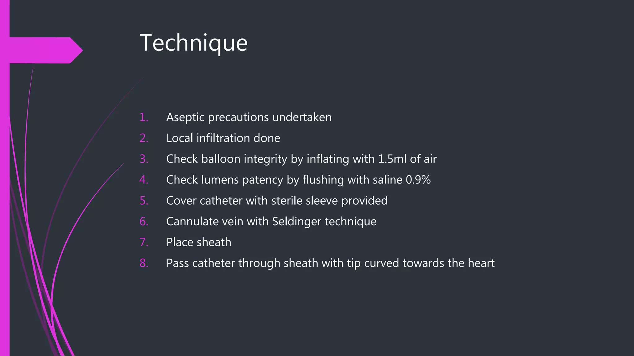
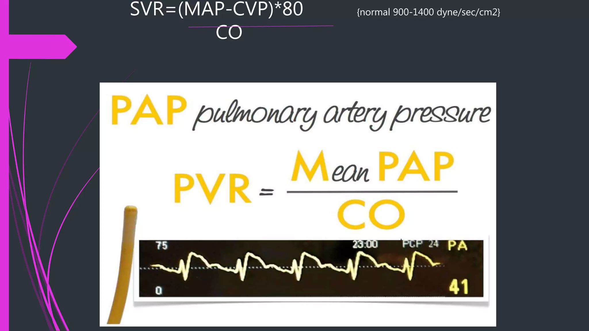
Pulmonary Artery Catheter
Speaker: Dr. Arun
Moderator Asst Prof. Dr. Madhavakrishna
Professor: Dr. Kumudha Lingaraj
 Introduction
 Indications
 Contraindications
 Preparation
 Technique
 Interpretation of physiological values and waveforms
 Complications
Indications
 Diagnostic:
 Differentiation among causes of shock
 Differentiation between mechanisms of pulmonary edema
 Evaluation of pulmonary hypertension
 Diagnosis of pericardial tamponade
 Diagnosis of right to left intracardiac shunts
 Unexplained dyspnea
 Therapeutic:
 Management of perioperative patients with unstable cardiac status
 Management of complicated myocardial infarction
 Management of patients following cardiac surgery/high risk surgery
 Management of severe preecclampsia
 Guide to pharmacologic therapy
 Burns/ Renal Failure/ Heart failure/Sepsis/ Decompensated cirrhosis
 Assess response to pulmonary hypertension specific therapy
Contraindication:
Absolute:
 Tricuspid and pulmonary valvular
stenosis
 Tetrology of fallot
 Infection at insertion site
 Presence of RV assist device
 Insertion during CPB
 Lack of consent
Relative:
 Coagulopathy
 Thrombocytopenia
 Electrolyte disturbances
(K/Mg/Na/Ca)
 Severe Pulmonary HTN
Preparation
 Patient has to be monitored with continuous ECG throughout the procedure, in
supine position regardless of the approach
 Aseptic precautions must be employed
 Cautions should be taken while cannulating via IJV/ Subclavian vein
 Equipments:
 2% chlorhexidine skin preparation solution
 Sterile gown, gloves, face shield and cap
 Sterile gauze pads
 1% lidocaine -5 cc
 Seeker needle 23G
 Introducer needle  18G
 J-tip guidewire
 Transduction tubing
 Sterile catheter flush solution
 Sheath
 Pulonary catheter
 Sterile sleeve for catheter
 2-0 silk suture
 Sterile dressing
Catheters:
The standard PAC has 7.0,7.5 or 8.0 Fr
circumference and in 110cm in length with distances
marked at 10cm intervals.
DOUBLE LUMEN- for ballon inflation and PA
pressure
TRIPLE LUMEN- above 2 + RA pressure
QUADRUPLE LUMEN- above 3 + THERMISTOR for
thermodilution {CO measurement}
FIVE LUMEN –additional lumen for drug infusion
Technique
1. Aseptic precautions undertaken
2. Local infiltration done
3. Check balloon integrity by inflating with 1.5ml of air
4. Check lumens patency by flushing with saline 0.9%
5. Cover catheter with sterile sleeve provided
6. Cannulate vein with Seldinger technique
7. Place sheath
8. Pass catheter through sheath with tip curved towards the heart
9. Once tip of catheter passed through introducer sheath inflate balloon at level of
right ventricle
10. The progress of the catheter through right atrium and ventricle into pulmonary
artery and wedge position can be monitored by changes in pressure trace
11. After acquiring wedge pressure  deflate balloon
Interpretations of wave forms
Physiological Measurements:
Direct measurements of the following can be
obtained from an accurately placed pulmonary artery
catheter(PAC)
Central Venous Pressure(CVP)
Right sided intracardiac pressures(RA/V)
Pulmonary artery pressure(Pap)
Pulmonary artery occlusion pressure (PAOP)
Cardiac Output
Mixed Venous Oxygen Saturation(SvO2)
Indirect measurements that are possible:
Systemic Vascular Resistance
Pulmonary Vascular Resistance
Cardiac Index
Stroke volume index
Oxygen delivery
Oxygen uptake
Calculation of Cardiac Output:
Thermodilution versus Ficks method
•Thermodilution: Add an indicator substance (5ml of dextrose or
saline) that is cooler than blood. Indicator in injected through the
proximal port of the PA catheter and mixes with the blood in the
RV. The mixing lowers the temperature of the flowing blood which
is carried to the distal thermistor port. The thermistor records the
temperature change and electronically displays a temperature/time
curve. The area under the curve is inversely proportional to the
flow rate in the pulmonary artery which equals the cardiac output in
absence of intracardiac shunt
-sources of error with thermodilution are seen with
tricuspid regurgitation and intracardiac shunts
Fick’s Method
 General principle: the release or uptake of a substance by
an organ equals the product of the bloodflow through
that organ times the difference of arteriovenous
concentrations of that substance.
 CO= O2 consumption (ml/min)
------------------------------------------------------------
---
arterial O2 content(PCWP)-mixed venous (PA) O2
content
 O2 consumption varies according to individual, by age
and sex. Usually estimated as being 250mL for a 70kg
male. Generally estimated at 130mL x BSA
 Blood O2 content=% saturation X Hb x 1.39 ml O2/gm
Hb
 Errors: assumptions of O2 consumption, wont work at all
with intracardiac shunts. But works better with TR
SVR=(MAP-CVP)*80 {normal 900-1400 dyne/sec/cm2}
CO
Pulmonary arterial occlusion pressure:
 Once catheter tip has reached PA, it should be advanced until
PAOP is identified by decrease in pressure and change in
waveform
The balloon should then be deflated and PA tracing should
reappear
If PCOP tracing persists catheter should be withdrawn with
definitive PA tracing obtained
 Final position of the catheter within PA must be such that PCOP tracing is
 obtained whenever 75-100% of 1.5ml maximum volume of balloon is
 insufflated.
 If < 1ml of air is injected and PAOP is seen then it is overwedged  needs to be
withdrawn
 If after maximal inflation fails to result in PCOP tracing or after 2-3 seconds delay 
too proximal – advanced with balloon inflated
 PCWP/PAOP  interprets Left atrial pressures
more importantly – LVEDP
PAEDP ~ PAOP ~ PVP ~ LAP ~ LVEDP
 Best measured in
 Supine position
 At end of expiration
 Zone 3 (most dependent region)
 Normal PCWP- 6-15 mmHg ; Mean :9mmHg
 Abnormal PAOP:
Increased LVEDP  Increased PAOP
LV systolic HF
LV Distolic HF
Mitral and Aortic valve disease
Hypertrophic cardiomyopathy
Hypervolemia
Large R-L shunts
Pericardial disease
 Decreased PCWP:
 Hypovolemia
 Obstructive shock due to large pulmonary embolus
 Abnormal waveforms
 Large a waves:
 MS
 LV systolic /diastolic function
 LV volume overload
 MI
 Large v waves - MR
Complications
 General:
 Immediate:
 Bleeding
 Arterial Puncture
 Air embolism
 Thoracic duct injury ( L side)
 Pneumothorax/hemothorax
 Delayed:
 Infections
 Thrombosis
 Related to insertion of PAC:
 Arrhythmias (most common- Ventricular/ RBBB)
 Misplacement
 Knotting
 Myocardial/valve/vessel rupture
 Related to maintenance and use of PAC:
 Pulmonary artery perforation
 Thromboembolism
 Infection
THANK YOU. . .

Pulmonary artery catheter

  • 1.
    Pulmonary Artery Catheter Speaker:Dr. Arun Moderator Asst Prof. Dr. Madhavakrishna Professor: Dr. Kumudha Lingaraj
  • 2.
     Introduction  Indications Contraindications  Preparation  Technique  Interpretation of physiological values and waveforms  Complications
  • 4.
    Indications  Diagnostic:  Differentiationamong causes of shock  Differentiation between mechanisms of pulmonary edema  Evaluation of pulmonary hypertension  Diagnosis of pericardial tamponade  Diagnosis of right to left intracardiac shunts  Unexplained dyspnea
  • 5.
     Therapeutic:  Managementof perioperative patients with unstable cardiac status  Management of complicated myocardial infarction  Management of patients following cardiac surgery/high risk surgery  Management of severe preecclampsia  Guide to pharmacologic therapy  Burns/ Renal Failure/ Heart failure/Sepsis/ Decompensated cirrhosis  Assess response to pulmonary hypertension specific therapy
  • 6.
    Contraindication: Absolute:  Tricuspid andpulmonary valvular stenosis  Tetrology of fallot  Infection at insertion site  Presence of RV assist device  Insertion during CPB  Lack of consent Relative:  Coagulopathy  Thrombocytopenia  Electrolyte disturbances (K/Mg/Na/Ca)  Severe Pulmonary HTN
  • 7.
    Preparation  Patient hasto be monitored with continuous ECG throughout the procedure, in supine position regardless of the approach  Aseptic precautions must be employed  Cautions should be taken while cannulating via IJV/ Subclavian vein
  • 8.
     Equipments:  2%chlorhexidine skin preparation solution  Sterile gown, gloves, face shield and cap  Sterile gauze pads  1% lidocaine -5 cc  Seeker needle 23G  Introducer needle  18G  J-tip guidewire  Transduction tubing  Sterile catheter flush solution  Sheath  Pulonary catheter  Sterile sleeve for catheter  2-0 silk suture  Sterile dressing
  • 10.
    Catheters: The standard PAChas 7.0,7.5 or 8.0 Fr circumference and in 110cm in length with distances marked at 10cm intervals. DOUBLE LUMEN- for ballon inflation and PA pressure TRIPLE LUMEN- above 2 + RA pressure QUADRUPLE LUMEN- above 3 + THERMISTOR for thermodilution {CO measurement} FIVE LUMEN –additional lumen for drug infusion
  • 12.
    Technique 1. Aseptic precautionsundertaken 2. Local infiltration done 3. Check balloon integrity by inflating with 1.5ml of air 4. Check lumens patency by flushing with saline 0.9% 5. Cover catheter with sterile sleeve provided 6. Cannulate vein with Seldinger technique 7. Place sheath 8. Pass catheter through sheath with tip curved towards the heart
  • 13.
    9. Once tipof catheter passed through introducer sheath inflate balloon at level of right ventricle 10. The progress of the catheter through right atrium and ventricle into pulmonary artery and wedge position can be monitored by changes in pressure trace 11. After acquiring wedge pressure  deflate balloon
  • 14.
  • 16.
    Physiological Measurements: Direct measurementsof the following can be obtained from an accurately placed pulmonary artery catheter(PAC) Central Venous Pressure(CVP) Right sided intracardiac pressures(RA/V) Pulmonary artery pressure(Pap) Pulmonary artery occlusion pressure (PAOP) Cardiac Output Mixed Venous Oxygen Saturation(SvO2)
  • 17.
    Indirect measurements thatare possible: Systemic Vascular Resistance Pulmonary Vascular Resistance Cardiac Index Stroke volume index Oxygen delivery Oxygen uptake
  • 18.
    Calculation of CardiacOutput: Thermodilution versus Ficks method •Thermodilution: Add an indicator substance (5ml of dextrose or saline) that is cooler than blood. Indicator in injected through the proximal port of the PA catheter and mixes with the blood in the RV. The mixing lowers the temperature of the flowing blood which is carried to the distal thermistor port. The thermistor records the temperature change and electronically displays a temperature/time curve. The area under the curve is inversely proportional to the flow rate in the pulmonary artery which equals the cardiac output in absence of intracardiac shunt -sources of error with thermodilution are seen with tricuspid regurgitation and intracardiac shunts
  • 19.
    Fick’s Method  Generalprinciple: the release or uptake of a substance by an organ equals the product of the bloodflow through that organ times the difference of arteriovenous concentrations of that substance.  CO= O2 consumption (ml/min) ------------------------------------------------------------ --- arterial O2 content(PCWP)-mixed venous (PA) O2 content  O2 consumption varies according to individual, by age and sex. Usually estimated as being 250mL for a 70kg male. Generally estimated at 130mL x BSA  Blood O2 content=% saturation X Hb x 1.39 ml O2/gm Hb  Errors: assumptions of O2 consumption, wont work at all with intracardiac shunts. But works better with TR
  • 22.
  • 23.
    Pulmonary arterial occlusionpressure:  Once catheter tip has reached PA, it should be advanced until PAOP is identified by decrease in pressure and change in waveform The balloon should then be deflated and PA tracing should reappear If PCOP tracing persists catheter should be withdrawn with definitive PA tracing obtained
  • 24.
     Final positionof the catheter within PA must be such that PCOP tracing is  obtained whenever 75-100% of 1.5ml maximum volume of balloon is  insufflated.  If < 1ml of air is injected and PAOP is seen then it is overwedged  needs to be withdrawn  If after maximal inflation fails to result in PCOP tracing or after 2-3 seconds delay  too proximal – advanced with balloon inflated
  • 25.
     PCWP/PAOP interprets Left atrial pressures more importantly – LVEDP PAEDP ~ PAOP ~ PVP ~ LAP ~ LVEDP  Best measured in  Supine position  At end of expiration  Zone 3 (most dependent region)  Normal PCWP- 6-15 mmHg ; Mean :9mmHg
  • 26.
     Abnormal PAOP: IncreasedLVEDP  Increased PAOP LV systolic HF LV Distolic HF Mitral and Aortic valve disease Hypertrophic cardiomyopathy Hypervolemia Large R-L shunts Pericardial disease
  • 27.
     Decreased PCWP: Hypovolemia  Obstructive shock due to large pulmonary embolus  Abnormal waveforms  Large a waves:  MS  LV systolic /diastolic function  LV volume overload  MI  Large v waves - MR
  • 28.
    Complications  General:  Immediate: Bleeding  Arterial Puncture  Air embolism  Thoracic duct injury ( L side)  Pneumothorax/hemothorax  Delayed:  Infections  Thrombosis
  • 29.
     Related toinsertion of PAC:  Arrhythmias (most common- Ventricular/ RBBB)  Misplacement  Knotting  Myocardial/valve/vessel rupture  Related to maintenance and use of PAC:  Pulmonary artery perforation  Thromboembolism  Infection
  • 31.