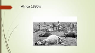
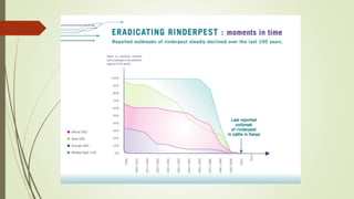
3. Introduction
Synonym : Cattle plaque
Acute, highly contagious viral disease of cattle characterized by high fever, necrotic
stomatitis, gastroenteritis, diarrhoea and high mortality caused by morbillivirus
The virus is antigenically closely related to the viruses of canine distemper, PPR
of sheep and goats and measles of humans
Globally eradicated in 2011
4. The need to combat rinderpest outbreaks was instrumental in the establishment
of the world’s first veterinary school in 1762 in Lyon, France, and the Office
International des Epizooties (OIE) in 1924, also in France
In 1994 the Global Rinderpest Eradication Program (GREP) was created to
undertake complete eradicationn of the disease
The principal vaccine used to control rinderpest was the tissue culture rinderpest
vaccine (TCRV) produced in calf kidney cells for cattle.
Plowright and others developed the vaccine in Kabete, Kenya, in 1957.
5. The last recorded outbreak of lineage 3 virus was in Pakistan in 2000 and of
lineages 1 and 2 in Africa in 2001
A joint meeting of the 79th General Session of the OIE and the 37th FAO
Conference adopted a resolution declaring the “Global Freedom from Rinderpest”
on June 28, 2011.
The virus still exists in research laboratories in the world.
On 14 June 2019 the largest stock of the rinderpest virus was destroyed at
the Pirbright Institute
6.
7. India has been free from clinical Rinderpest since June, 1995.
However, as per norms, India submitted the dossier on August, 2005 for obtaining freedom
from Rinderpest. The International Committee of World Organization for Animal Health,
OIE recognized India as free from Rinderpest infection on 25th May, 2006.
8.
9.
10. Susceptibility
Mainly cattle and buffaloes, but also reported in sheep,goat and pigs
Reported in deer, antelope, wild buffaloes, wild boars, bushbuck, warthogs and
giraffe
Mortality is 100 % in exotic breeds and 20-50 % mortality in indigenous breeds
11. Transmission
Incubation period is 2 to 3 days in experimental inoculation and in contact infection
is 6 to 9 days
Virus excreted in body secretions
Ingestion of contaminated feed and water
Inhalation
12. Pathogenesis
The virus is inhaled in infected droplets
It penetrates through the epithelium of upper respiratory tract and multiplies in the tonsils and
regional lymph nodes
From here it enters the blood in mononuclear cells which disseminate to other lymphoid organs,
lungs and epithelial cells of mucous membranes
Rinderpest virus has a high degree affinity for lymphoid tissue and mucous membrane of
alimentary tract
Pronounced destruction of lymphocytes in tissues and it is responsible for marked leucopoenia
Focal necrotic stomatitis and enteritis are the direct result of viral infection and replication in the
epithelial cells in the alimentary tract.
Death is usually from severe dehydration but in less acute cases, death may be from activated
latent parasitic or bacterial infections
These infections aggravate because the animal is immunosuppressed as a result of destruction of
lymphoid organs by the virus
13. Clinical signs
Fever (104-105 0F), restlessness, dryness of the muzzle and constipation
Other signs include photophobia, excessive thirst, starry coat, retarded rumination,
anorexia and excessive salivation
Rashes may develop in those parts of the body where the hair is fine in nature
Mucous membrane of lips, gums and tongue revealed small vesicles resembling
bran like deposits
'Shooting diarrhoea' with foetid odour
Dehydration
Marked leucopoenia with drop in lymphocyte count
14. Post mortem findings
Virus produce lesions in the oral mucosa after settling in the cells following a viremic
state
Fore stomach are free
Abomasum reveals necrotic foci and haemorrhagic streaks more seen at the pyloric
region.
Folds of abomasum are thick and oedematous
If the disease progress, abomasal mucosa shows irregular ulcers of different size.
The virus has got affinity for lymphoid tissue and in the intestine, peyer's patches are
swollen and ulcerated
Duodenum and ileum revealed streaks of haemorrhages and erosions
In the large intestine, ileo caecal valve may be markedly swollen
15. Linear haemorrhages on the folds of mucosa of rectum appear like 'Zebra
marking' which is pathognomonic in Rinder pest
Lesions are more severe in large intestine with ulceration and diphtheitic patches
Liver: Chronic passive congestion resulting from cardiac and pulmonary complications
Petechiae and erosions in the larynx
Tracheal haemorrhages
Alveolar and interstitial emphysema
Subepithelial and subendocardial haemorrhages
Petechiae and erosions may be seen in the bladder and vagina
Purulent conjunctivitis and ulceration of cornea may be noticed
In sheep and goat, mouth lesions are usually not seen
16. Histopathology
Epithelial surface reveals ulcers, haemorrhages, oedema, necrosis and leucocytic
infiltration along with multinucleated cells
Eosinophilic cytoplasmic inclusion bodies form in the mucosal epithelial cells and
giant cells. Intranuclear inclusion bodies are rare.
17. Samples
• Animals showing a pyrexia are probably viraemic and usually the best source of blood for
detecting or isolating virus; take blood from several pyrexic animals
o sterile whole blood preserved in heparin (10 IU/ml) or EDTA (0.5 mg/ml) and transferred to
laboratory on ice (but not frozen); do not use glycerol as preservative transport media as it
inactivates RPV
o blood for serum, although antibodies are unlikely to be detected until the mucosal phase
• Spleen, prescapular or mesenteric lymph nodes of dead animals chilled to sub-zero temperatures
for virus isolation
• Full set of tissue samples is advised in 10% neutral buffered formalin for histopathology and
immunohistochemistry
o base of the tongue, retropharyngeal lymph node and third eyelid are suitable tissues
• Ocular and nasal secretions of infected animals during either the prodromal or the erosive phase
18. Diagnosis
Symptoms and lesions
Complement fixation test (CFT)
Agar Gel Precipitation Test (AGPT)
Virus isolation and diagnosis on tissue cultures
Using specific cDNA probes, isolates of rinderpest and PPR viruses can be
differentiated presently
Polymerase chain Reaction (PCR)