This document provides an overview of rickets, including:
- It is a disorder of defective bone mineralization caused by vitamin D deficiency that disturbs calcium and phosphorus homeostasis.
- Those at risk include children with dark skin and those exclusively breastfed without vitamin D supplementation.
- Vitamin D is synthesized in the body from sun exposure and obtained through diet. A deficiency can be caused by inadequate intake or absorption.
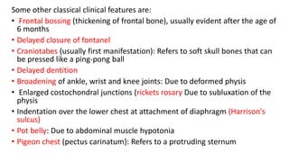
- Symptoms include bone deformities, muscle weakness, and fractures. Diagnosis involves blood tests showing low vitamin D and elevated alkaline phosphatase. Treatment is vitamin D supplementation.


![TYPES OF RICKETS & LAB FINDINGS
1) NUTRITIONAL RICKETS
• This is the most common variety in developing countries.
• The recommended daily intake of vitamin D is 400 IU for infants, 600 IU from 1
year to 70 years of age, and 800 IU for those over 70.
• Inability to take these recommended levels leads to dietary deficiency of
vitamin D, so active vitamin D [l,25(0H)2 D] levels are low.
• This leads to inability to absorb calcium and phosphorus.
• PTH is elevated in response to hypocalcemia, corrects the serum calcium, so
calcium levels are normal to low while phosphate levels may be low to normal.
Alkaline phosphatase (ALP) is elevated.
• Cut-off value for vitamin D deficiency: Most investigators have used different
cut-off levels to define vitamin D deficiency .
• Most commonly used cut-off value to define vitamin D deficiency is 25(0H)-
vitamin D less than 20 ng/mL, insufficiency as 20- 29 ng/mL and sufficiency as
more than or equal to 30 ng/ mL. Severe deficiency is defined as a level less
than 5 ng/mL.](https://image.slidesharecdn.com/rickets-230308173406-8c9b9846/85/RICKETS-pptx-16-320.jpg)
![2) Vitamin D-dependent Rickets (VDDR)
VDDR-1:
• It is an autosomal recessive disorder due to deficiency of 1-alpha-hydroxylase
renal enzyme.
• It is necessary for the formation of the active metabolite of vitamin D which is not
formed in adequate amount [ 1,25( OH- vitamin D levels low].
• This leads to inability to absorb calcium and phosphorus and levels of both
minerals are low in serum.
• PTH is elevated in response to hypocalcemia.
• ALP is also elevated.
• To differentiate it from nutritional rickets, levels of 25(0H)-vitamin D and
l,25(0H)2 -vitamin are measured. In nutritional variety, both will be low while in
vitamin D-dependent rickets (VDDR) type I, the levels of 25( OH)-vitamin D will be
increased while the active form [l,25(0H)2-vitamin D] will be markedly decreased.
The rachitic features appear early with renal tubular dysfunction.](https://image.slidesharecdn.com/rickets-230308173406-8c9b9846/85/RICKETS-pptx-17-320.jpg)