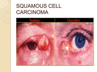
EYELID TUMOURS
-SOLANKI RIDDHI
2ND YEAR (B.OPTOM)
Almost all types of tumours arising
from the skin, connective tissue,
glandular tissue, blood vessels,
nerves & muscles can involve the lids.
A few common tumours are listed
here.
CLASSIFICATION
1. BENIGN TUMOURS
1. Papillomas
2. Xanthelasma
3. Haemangioma
4. Neurofibroma
2. MALIGNANT TUMOURS
1. Basal-cell Carcinoma
2. Squamous cell Carcinoma
3. Sebaceous Gland Carcinoma
4. Malignant Melanoma (Melanocarcinoma)
1. BENIGN TUMOURS
1. Papillomas:
These are the most common benign
tumours arising from the surface epithelium.
i. Squamous papillomas occur in adults, as
very slow growing or stationary, raspberry-
like growths or as a pedunculate lesion,
usually involving the lid margin.
ii. Seborrhoeic keratosis occurs in middle-
aged & older persons. Their surface is
friable, verrucous & slightly pigmented.
TREATMENT:
Simple excision.
SQUAMOUS PAPILLOMAS
SEBORRHOEIC KERATOSIS
2. Xanthelasma:
 These are creamy-yellow plaque-like
lesions which frequently involve the skin of
upper & lower lids near the inner canthus.
 Xanthelasma represents lipid deposits in
histiocytes in the dermis of the lid.
 These may be associated with diabetes
mellitus or high cholesterol levels.
TREATMENT:
Excision may be advised for cosmetic
reasons; but recurrences are common.
XANTHELASMA
3. Haemangioma:
Haemangiomas of the lids are common
tumours.
i. Capillary haemangioma:
 Is the most common variety which
occurs at or shortly after birth, often grows
rapidly & in many cases resolves
spontaneously by the age of 7 years.
 These may be superficial & bright red in
colour or deep & bluish or violet in colour.
 They consists of proliferating capillaries
& endothelial cells.
TREATMENT:
Excision, Intralesional steroid,
Superficial radiotherapy.
CAPILLARY HAEMANGIOMA
ii. Naevus flammeus (port wine stain):
 It may occur side-by-side or more
commonly as a part of Sturge-Weber
syndrome.
 It consists of dilated vascular channels &
does not grow or regress like the capillary
haemangioma.
iii. Cavernous haemangioma:
 Are developmental & usually occur after
first decade of life.
 It consists of large endothelium-lined
vascular channels & usually does not show
any regression.
NAEVUS FLAMMEUS
CAVERNOUS HAEMANGIOMA
4. Neurofibroma:
 Lids & orbits are commonly affected in
neurofibromatosis (von Recklinghausen’s
disease).
 The tumour is usually plexiform type.
NEUROFIBROMA
2. MALIGNANT TUMOURS
1. Basal-cell Carcinoma:
 It is the commonest malignant tumour of
the lids (90%) usually seen in elderly
people.
 It is locally malignant & involves most
commonly lower lid (50%) followed by
medial canthus (25%), upper lid (10-15%)
& outer canthus (5-10%).
TREATMENT:
Surgery, Radiotherapy & Cryotherapy.
CLINICAL FEATURES OF BASAL-
CELL CARCINOMA
It may present in four forms:
 Noduloulcerative basal cell carcinoma is
the most common presentation.
 It starts as a small nodule which
undergoes central ulceration with pearly
rolled margins.
 The tumour grows by burrowing &
destroying the tissues locally like a rodent &
hence the name rodent ulcer.
 Other rare presentations include: non-
ulcerated nodular form, sclerosing or
morphea type & pigmented basal cell
BASAL-CELL CARCINOMA
2. Squamous cell Carcinoma:
 It forms the second commonest
malignant tumour of the lid.
 Its incidence (5%) is much less than the
basal cell carcinoma.
 It commonly arises from the lid margin
(mucocutaneous junction) in elderly
patients.
 Affects upper & lower lids equally.
TREATMENT:
Treatment on the lines of basal cell
carcinoma.
CLINICAL FEATURES OF
SQUAMOUS CELL CARCINOMA
It may present in two forms:
 An ulcerated growth with elevated &
indurated margins is the common
presentation.
 The second form, fungating or
polypoid verrucous lesion without
ulceration, is a rare presentation.
 Metastasis: It metastatises in
preauricular & sub-mandibular lymph
nodes.
SQUAMOUS CELL
CARCINOMA
3. Sebaceous gland Carcinoma:
 It is a rare tumour arising from the
meibomian glands.
TREATMENT:
Surgical excision with reconstruction of
lids; recurrences are common.
CLINICAL FEATURES:
 It usually presents initially as a nodule
(which may be mistaken for a chalazion).
Which then grows to form a big growth.
 Rarely, a diffuse tumour along the lid
margin may be mistaken as chronic
blepharitis.
SEBACEOUS GLAND
CARCINOMA
4. Malignant Melanoma
(Melanocarcinoma):
 It is a rare tumour of the lid (less than
1% of all eyelid lesions).
 It may arise from a pre-existing naevus,
but usually arises from the beginning from
the melanocytes present in the skin.
TREATMENT:
Surgical excision with reconstruction of
lid.
CLINICAL FEATURES OF
MALIGNANT MELANOMA
(MELANOCARCINOMA):
 It often appears as a flat or slightly
elevated naevus which has variegated
pigmentation & irregular borders.
 It may ulcerate & bleed.
 Metastasis: The tumour spreads locally
as well as to distant sites by lymphatics &
blood stream.
MALIGNANT MELANOMA
Eyelid tumours

Eyelid tumours

  • 1.
  • 2.
    Almost all typesof tumours arising from the skin, connective tissue, glandular tissue, blood vessels, nerves & muscles can involve the lids. A few common tumours are listed here.
  • 3.
    CLASSIFICATION 1. BENIGN TUMOURS 1.Papillomas 2. Xanthelasma 3. Haemangioma 4. Neurofibroma 2. MALIGNANT TUMOURS 1. Basal-cell Carcinoma 2. Squamous cell Carcinoma 3. Sebaceous Gland Carcinoma 4. Malignant Melanoma (Melanocarcinoma)
  • 4.
    1. BENIGN TUMOURS 1.Papillomas: These are the most common benign tumours arising from the surface epithelium. i. Squamous papillomas occur in adults, as very slow growing or stationary, raspberry- like growths or as a pedunculate lesion, usually involving the lid margin. ii. Seborrhoeic keratosis occurs in middle- aged & older persons. Their surface is friable, verrucous & slightly pigmented. TREATMENT: Simple excision.
  • 5.
  • 6.
  • 7.
    2. Xanthelasma:  Theseare creamy-yellow plaque-like lesions which frequently involve the skin of upper & lower lids near the inner canthus.  Xanthelasma represents lipid deposits in histiocytes in the dermis of the lid.  These may be associated with diabetes mellitus or high cholesterol levels. TREATMENT: Excision may be advised for cosmetic reasons; but recurrences are common.
  • 8.
  • 9.
    3. Haemangioma: Haemangiomas ofthe lids are common tumours. i. Capillary haemangioma:  Is the most common variety which occurs at or shortly after birth, often grows rapidly & in many cases resolves spontaneously by the age of 7 years.  These may be superficial & bright red in colour or deep & bluish or violet in colour.  They consists of proliferating capillaries & endothelial cells. TREATMENT: Excision, Intralesional steroid, Superficial radiotherapy.
  • 10.
  • 11.
    ii. Naevus flammeus(port wine stain):  It may occur side-by-side or more commonly as a part of Sturge-Weber syndrome.  It consists of dilated vascular channels & does not grow or regress like the capillary haemangioma. iii. Cavernous haemangioma:  Are developmental & usually occur after first decade of life.  It consists of large endothelium-lined vascular channels & usually does not show any regression.
  • 12.
  • 13.
  • 14.
    4. Neurofibroma:  Lids& orbits are commonly affected in neurofibromatosis (von Recklinghausen’s disease).  The tumour is usually plexiform type.
  • 15.
  • 16.
    2. MALIGNANT TUMOURS 1.Basal-cell Carcinoma:  It is the commonest malignant tumour of the lids (90%) usually seen in elderly people.  It is locally malignant & involves most commonly lower lid (50%) followed by medial canthus (25%), upper lid (10-15%) & outer canthus (5-10%). TREATMENT: Surgery, Radiotherapy & Cryotherapy.
  • 17.
    CLINICAL FEATURES OFBASAL- CELL CARCINOMA It may present in four forms:  Noduloulcerative basal cell carcinoma is the most common presentation.  It starts as a small nodule which undergoes central ulceration with pearly rolled margins.  The tumour grows by burrowing & destroying the tissues locally like a rodent & hence the name rodent ulcer.  Other rare presentations include: non- ulcerated nodular form, sclerosing or morphea type & pigmented basal cell
  • 18.
  • 19.
    2. Squamous cellCarcinoma:  It forms the second commonest malignant tumour of the lid.  Its incidence (5%) is much less than the basal cell carcinoma.  It commonly arises from the lid margin (mucocutaneous junction) in elderly patients.  Affects upper & lower lids equally. TREATMENT: Treatment on the lines of basal cell carcinoma.
  • 20.
    CLINICAL FEATURES OF SQUAMOUSCELL CARCINOMA It may present in two forms:  An ulcerated growth with elevated & indurated margins is the common presentation.  The second form, fungating or polypoid verrucous lesion without ulceration, is a rare presentation.  Metastasis: It metastatises in preauricular & sub-mandibular lymph nodes.
  • 21.
  • 22.
    3. Sebaceous glandCarcinoma:  It is a rare tumour arising from the meibomian glands. TREATMENT: Surgical excision with reconstruction of lids; recurrences are common. CLINICAL FEATURES:  It usually presents initially as a nodule (which may be mistaken for a chalazion). Which then grows to form a big growth.  Rarely, a diffuse tumour along the lid margin may be mistaken as chronic blepharitis.
  • 23.
  • 24.
    4. Malignant Melanoma (Melanocarcinoma): It is a rare tumour of the lid (less than 1% of all eyelid lesions).  It may arise from a pre-existing naevus, but usually arises from the beginning from the melanocytes present in the skin. TREATMENT: Surgical excision with reconstruction of lid.
  • 25.
    CLINICAL FEATURES OF MALIGNANTMELANOMA (MELANOCARCINOMA):  It often appears as a flat or slightly elevated naevus which has variegated pigmentation & irregular borders.  It may ulcerate & bleed.  Metastasis: The tumour spreads locally as well as to distant sites by lymphatics & blood stream.
  • 26.