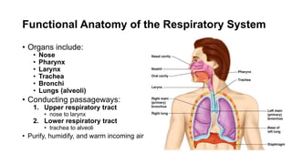
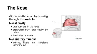
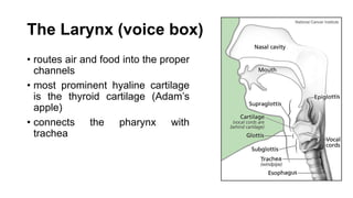
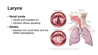
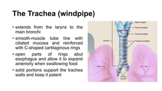
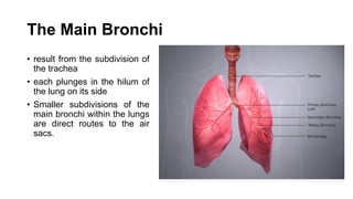
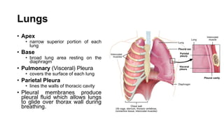
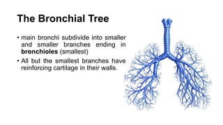
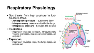
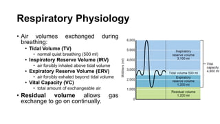
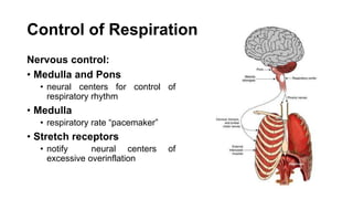
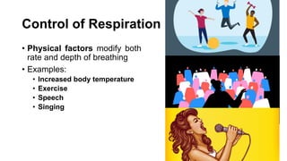
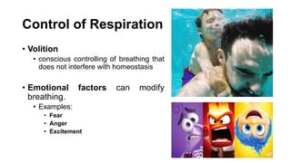
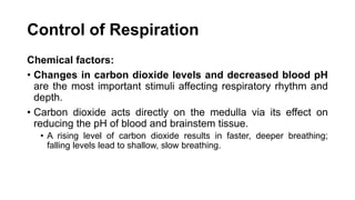
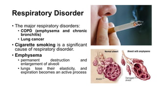
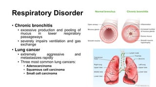
THE RESPIRATORY SYSTEM
Functional Anatomy of the Respiratory System
• Organs include:
• Nose
• Pharynx
• Larynx
• Trachea
• Bronchi
• Lungs (alveoli)
• Conducting passageways:
1. Upper respiratory tract
• nose to larynx
2. Lower respiratory tract
• trachea to alveoli
• Purify, humidify, and warm incoming air
The Nose
• Air enters the nose by passing
through the nostrils.
• Nasal cavity
• chamber within the nose
• separated from oral cavity by
palate
• lined with mucosa
• Respiratory mucosa
• warms, filters and moistens
incoming air
Nose
• Ciliated cells of nasal mucosa
create gentle current that moves
sheet of contaminated mucus
posteriorly toward the throat
• Conchae
• three mucosa-covered lobes in the
lateral walls of nasal cavity
• greatly increase the surface area
of the mucosa exposed to the air
• increase air turbulence
Cleft Palate
• genetic defect
• failure of the bones forming the
palate to fuse medially
• results in breathing difficulty
• problems with oral cavity
functions such as nursing and
speaking
Nose
• The nasal cavity is surrounded
by a ring of paranasal sinuses.
• Paranasal sinuses
• lighten the skull and act as
resonance chambers for speech
• produce mucus which drains
into the nasal cavities
• Nasolacrimal ducts
• drain tears from the eyes
• empty into the nasal cavities
Rhinitis
• caused by cold viruses and
various allergens
• inflammation of the nasal
mucosa
• excessive mucus produced
results in nasal congestion and
postnasal drip
• nasal cavity infections often
spread
Sinusitis
• sinus inflammation
• difficult to treat and can cause
marked changes in voice quality
• result is a partial vacuum and a
sinus headache localized over
the inflamed area
The Pharynx (throat)
• mucosa-lined, muscular tube
• passageway for food and air
• Three regions:
• Nasopharynx
• Oropharynx
• Laryngopharynx
• Air enters nasopharynx then
descends through oropharynx and
laryngopharynx.
• Food enters the mouth, travels
along with air through the
oropharynx and laryngopharynx.
Pharynx
• Pharyngotympanic tubes
• drain the middle ears and open into
nasopharynx
• Ear infections may follow a sore throat
• Tonsils
• clusters of lymphatic tissue
• play a role in protecting the body from
infection
Tonsilitis
• Pharyngeal tonsil becomes
inflamed and swollen during a
bacterial infection
• Obstruction in the nasopharynx
forces the person to breathe
through the mouth.
• Mouth breathing makes the air not
properly moistened, warmed, or
filtered.
The Larynx (voice box)
• routes air and food into the proper
channels
• most prominent hyaline cartilage
is the thyroid cartilage (Adam’s
apple)
• connects the pharynx with
trachea
Larynx
• Epiglottis
• protects the superior opening of
the larynx
• forming lid over larynx’s opening
when swallowing
• larynx is pulled upward and the
epiglottis tips when swallowing
• Cough reflex is triggered to
prevent substance from
continuing into the lungs.
Larynx
• Vocal cords
• vibrate with expelled air
• vibration allows speaking
• Glottis
• between the vocal folds and the
slitlike passageway
The Trachea (windpipe)
• extends from the larynx to the
main bronchi
• smooth-muscle tube line with
ciliated mucosa and reinforced
with C-shaped cartilaginous rings
• open parts of rings abut
esophagus and allow it to expand
anteriorly when swallowing food
• solid portions support the trachea
walls and keep it patent
Trachea
• Many people have
suffocated after choking
on a piece of food that
suddenly closed off the
trachea.
• Heimlich maneuver
• air in a person’s own lungs
is used to expel an
obstructing piece of food
• cracked ribs are a distinct
possibility when it is done
incorrectly
Trachea
• lined with ciliated mucosa
• cilia propel this mucus away from
the lungs to the throat
• Smoking inhibits and ultimately
destroys the cilia.
The Main Bronchi
• result from the subdivision of
the trachea
• each plunges in the hilum of
the lung on its side
• Smaller subdivisions of the
main bronchi within the lungs
are direct routes to the air
sacs.
Lungs
• Apex
• narrow superior portion of each
lung
• Base
• broad lung area resting on the
diaphragm
• Pulmonary (Visceral) Pleura
• covers the surface of each lung
• Parietal Pleura
• lines the walls of thoracic cavity
• Pleural membranes produce
pleural fluid which allows lungs
to glide over thorax wall during
breathing.
Pleurisy
• inflammation of the pleurae
• caused by insufficient secretion
of pleural fluid
• pleural surfaces become dry
and rough, resulting in friction
and stabbing pain with each
breath
• The pleurae may produce
excessive amounts of fluid
which exerts pressure on the
lungs.
The Bronchial Tree
• main bronchi subdivide into smaller
and smaller branches ending in
bronchioles (smallest)
• All but the smallest branches have
reinforcing cartilage in their walls.
Respiratory Zone Structure
• terminal bronchioles lead into
respiratory zone
• includes the respiratory
bronchioles, alveolar ducts,
alveolar sacs, and alveoli, is
the only site of gas exchange
• All other respiratory passages
are conducting zone
structures that serve as
conduits to and from the
respiratory zone.
Alveoli
• millions clustered inside the
lungs that resembles bunches
of grapes
• composed largely of a single,
thin layer of simple squamous
epithelial cells
• walls are very thin (thinner than
tissue paper)
• Alveolar pores
• connect neighboring air sacs and
provide alternative routes for air
Alveoli
• external surfaces are covered
with pulmonary capillaries
• alveolar and capillary walls
construct respiratory membrane
(air-blood barrier)
• Respiratory membrane has gas
(air) flowing past on one side and
blood flowing past on the other.
• Gas exchange occurs by simple
diffusion through the respiratory
membrane.
Alveoli
• Alveolar Macrophages
• also known as dust cells
• wander in and out of the
alveoli picking up bacteria,
carbon particles, and other
debris
• Surfactant-secreting Cells
• produce a lipid molecule
(surfactant) which coats the
gas-exposed alveolar
surfaces
Respiratory Physiology
Four distinct events of respiration:
1. Pulmonary ventilation
• air move into and out of the lungs (breathing)
2. External respiration
• gas exchanges are being made between the blood and the body exterior
3. Respiratory gas transport
• oxygen and carbon dioxide are transported to and from the lungs and tissue cells
4. Internal respiration
• gas exchange occurs between the blood and cells inside the body
Respiratory Physiology
• Gas travels from high pressure to low-
pressure areas.
• Atmospheric pressure – outside the body
• Intrapulmonary pressure – inside the lungs
• Intrapleural pressure – between the pleurae
• Inspiration
• inspiratory muscles contract, intrapulmonary
volume increases, its pressure decreases, air
rushes in
• Expiration
• inspiratory muscles relax, the lungs recoil, air
rushes out
Respiratory Physiology
• Air volumes exchanged during
breathing:
• Tidal Volume (TV)
• normal quiet breathing (500 ml)
• Inspiratory Reserve Volume (IRV)
• air forcibly inhaled above tidal volume
• Expiratory Reserve Volume (ERV)
• air forcibly exhaled beyond tidal volume
• Vital Capacity (VC)
• total amount of exchangeable air
• Residual volume allows gas
exchange to go on continually.
Respiratory Physiology
• Non-respiratory air movements
• voluntary or reflex activities that
move air into or out of the lungs
• Examples:
• Coughing
• Sneezing
• Laughing
• Crying
• Hiccupping
• Yawning
Control of Respiration
Nervous control:
• Medulla and Pons
• neural centers for control of
respiratory rhythm
• Medulla
• respiratory rate “pacemaker”
• Stretch receptors
• notify neural centers of
excessive overinflation
Control of Respiration
• Physical factors modify both
rate and depth of breathing
• Examples:
• Increased body temperature
• Exercise
• Speech
• Singing
Control of Respiration
• Volition
• conscious controlling of breathing that
does not interfere with homeostasis
• Emotional factors can modify
breathing.
• Examples:
• Fear
• Anger
• Excitement
Control of Respiration
Chemical factors:
• Changes in carbon dioxide levels and decreased blood pH
are the most important stimuli affecting respiratory rhythm and
depth.
• Carbon dioxide acts directly on the medulla via its effect on
reducing the pH of blood and brainstem tissue.
• A rising level of carbon dioxide results in faster, deeper breathing;
falling levels lead to shallow, slow breathing.
Respiratory Disorder
• The major respiratory disorders:
• COPD (emphysema and chronic
bronchitis)
• Lung cancer
• Cigarette smoking is a significant
cause of respiratory disorder.
• Emphysema
• permanent destruction and
enlargement of alveoli
• lungs lose their elasticity, and
expiration becomes an active process
Respiratory Disorder
• Chronic bronchitis
• excessive production and pooling of
mucus in lower respiratory
passageways
• severely impairs ventilation and gas
exchange
• Lung cancer
• extremely aggressive and
metastasizes rapidly
• Three most common lung cancers:
• Adenocarcinoma
• Squamous cell carcinoma
• Small cell carcinoma

Respiratory System.pdf

  • 1.
  • 2.
    Functional Anatomy ofthe Respiratory System • Organs include: • Nose • Pharynx • Larynx • Trachea • Bronchi • Lungs (alveoli) • Conducting passageways: 1. Upper respiratory tract • nose to larynx 2. Lower respiratory tract • trachea to alveoli • Purify, humidify, and warm incoming air
  • 3.
    The Nose • Airenters the nose by passing through the nostrils. • Nasal cavity • chamber within the nose • separated from oral cavity by palate • lined with mucosa • Respiratory mucosa • warms, filters and moistens incoming air
  • 4.
    Nose • Ciliated cellsof nasal mucosa create gentle current that moves sheet of contaminated mucus posteriorly toward the throat • Conchae • three mucosa-covered lobes in the lateral walls of nasal cavity • greatly increase the surface area of the mucosa exposed to the air • increase air turbulence
  • 5.
    Cleft Palate • geneticdefect • failure of the bones forming the palate to fuse medially • results in breathing difficulty • problems with oral cavity functions such as nursing and speaking
  • 6.
    Nose • The nasalcavity is surrounded by a ring of paranasal sinuses. • Paranasal sinuses • lighten the skull and act as resonance chambers for speech • produce mucus which drains into the nasal cavities • Nasolacrimal ducts • drain tears from the eyes • empty into the nasal cavities
  • 7.
    Rhinitis • caused bycold viruses and various allergens • inflammation of the nasal mucosa • excessive mucus produced results in nasal congestion and postnasal drip • nasal cavity infections often spread
  • 8.
    Sinusitis • sinus inflammation •difficult to treat and can cause marked changes in voice quality • result is a partial vacuum and a sinus headache localized over the inflamed area
  • 9.
    The Pharynx (throat) •mucosa-lined, muscular tube • passageway for food and air • Three regions: • Nasopharynx • Oropharynx • Laryngopharynx • Air enters nasopharynx then descends through oropharynx and laryngopharynx. • Food enters the mouth, travels along with air through the oropharynx and laryngopharynx.
  • 10.
    Pharynx • Pharyngotympanic tubes •drain the middle ears and open into nasopharynx • Ear infections may follow a sore throat • Tonsils • clusters of lymphatic tissue • play a role in protecting the body from infection
  • 11.
    Tonsilitis • Pharyngeal tonsilbecomes inflamed and swollen during a bacterial infection • Obstruction in the nasopharynx forces the person to breathe through the mouth. • Mouth breathing makes the air not properly moistened, warmed, or filtered.
  • 12.
    The Larynx (voicebox) • routes air and food into the proper channels • most prominent hyaline cartilage is the thyroid cartilage (Adam’s apple) • connects the pharynx with trachea
  • 13.
    Larynx • Epiglottis • protectsthe superior opening of the larynx • forming lid over larynx’s opening when swallowing • larynx is pulled upward and the epiglottis tips when swallowing • Cough reflex is triggered to prevent substance from continuing into the lungs.
  • 14.
    Larynx • Vocal cords •vibrate with expelled air • vibration allows speaking • Glottis • between the vocal folds and the slitlike passageway
  • 15.
    The Trachea (windpipe) •extends from the larynx to the main bronchi • smooth-muscle tube line with ciliated mucosa and reinforced with C-shaped cartilaginous rings • open parts of rings abut esophagus and allow it to expand anteriorly when swallowing food • solid portions support the trachea walls and keep it patent
  • 16.
    Trachea • Many peoplehave suffocated after choking on a piece of food that suddenly closed off the trachea. • Heimlich maneuver • air in a person’s own lungs is used to expel an obstructing piece of food • cracked ribs are a distinct possibility when it is done incorrectly
  • 17.
    Trachea • lined withciliated mucosa • cilia propel this mucus away from the lungs to the throat • Smoking inhibits and ultimately destroys the cilia.
  • 18.
    The Main Bronchi •result from the subdivision of the trachea • each plunges in the hilum of the lung on its side • Smaller subdivisions of the main bronchi within the lungs are direct routes to the air sacs.
  • 19.
    Lungs • Apex • narrowsuperior portion of each lung • Base • broad lung area resting on the diaphragm • Pulmonary (Visceral) Pleura • covers the surface of each lung • Parietal Pleura • lines the walls of thoracic cavity • Pleural membranes produce pleural fluid which allows lungs to glide over thorax wall during breathing.
  • 20.
    Pleurisy • inflammation ofthe pleurae • caused by insufficient secretion of pleural fluid • pleural surfaces become dry and rough, resulting in friction and stabbing pain with each breath • The pleurae may produce excessive amounts of fluid which exerts pressure on the lungs.
  • 21.
    The Bronchial Tree •main bronchi subdivide into smaller and smaller branches ending in bronchioles (smallest) • All but the smallest branches have reinforcing cartilage in their walls.
  • 22.
    Respiratory Zone Structure •terminal bronchioles lead into respiratory zone • includes the respiratory bronchioles, alveolar ducts, alveolar sacs, and alveoli, is the only site of gas exchange • All other respiratory passages are conducting zone structures that serve as conduits to and from the respiratory zone.
  • 23.
    Alveoli • millions clusteredinside the lungs that resembles bunches of grapes • composed largely of a single, thin layer of simple squamous epithelial cells • walls are very thin (thinner than tissue paper) • Alveolar pores • connect neighboring air sacs and provide alternative routes for air
  • 24.
    Alveoli • external surfacesare covered with pulmonary capillaries • alveolar and capillary walls construct respiratory membrane (air-blood barrier) • Respiratory membrane has gas (air) flowing past on one side and blood flowing past on the other. • Gas exchange occurs by simple diffusion through the respiratory membrane.
  • 25.
    Alveoli • Alveolar Macrophages •also known as dust cells • wander in and out of the alveoli picking up bacteria, carbon particles, and other debris • Surfactant-secreting Cells • produce a lipid molecule (surfactant) which coats the gas-exposed alveolar surfaces
  • 26.
    Respiratory Physiology Four distinctevents of respiration: 1. Pulmonary ventilation • air move into and out of the lungs (breathing) 2. External respiration • gas exchanges are being made between the blood and the body exterior 3. Respiratory gas transport • oxygen and carbon dioxide are transported to and from the lungs and tissue cells 4. Internal respiration • gas exchange occurs between the blood and cells inside the body
  • 27.
    Respiratory Physiology • Gastravels from high pressure to low- pressure areas. • Atmospheric pressure – outside the body • Intrapulmonary pressure – inside the lungs • Intrapleural pressure – between the pleurae • Inspiration • inspiratory muscles contract, intrapulmonary volume increases, its pressure decreases, air rushes in • Expiration • inspiratory muscles relax, the lungs recoil, air rushes out
  • 28.
    Respiratory Physiology • Airvolumes exchanged during breathing: • Tidal Volume (TV) • normal quiet breathing (500 ml) • Inspiratory Reserve Volume (IRV) • air forcibly inhaled above tidal volume • Expiratory Reserve Volume (ERV) • air forcibly exhaled beyond tidal volume • Vital Capacity (VC) • total amount of exchangeable air • Residual volume allows gas exchange to go on continually.
  • 29.
    Respiratory Physiology • Non-respiratoryair movements • voluntary or reflex activities that move air into or out of the lungs • Examples: • Coughing • Sneezing • Laughing • Crying • Hiccupping • Yawning
  • 30.
    Control of Respiration Nervouscontrol: • Medulla and Pons • neural centers for control of respiratory rhythm • Medulla • respiratory rate “pacemaker” • Stretch receptors • notify neural centers of excessive overinflation
  • 31.
    Control of Respiration •Physical factors modify both rate and depth of breathing • Examples: • Increased body temperature • Exercise • Speech • Singing
  • 32.
    Control of Respiration •Volition • conscious controlling of breathing that does not interfere with homeostasis • Emotional factors can modify breathing. • Examples: • Fear • Anger • Excitement
  • 33.
    Control of Respiration Chemicalfactors: • Changes in carbon dioxide levels and decreased blood pH are the most important stimuli affecting respiratory rhythm and depth. • Carbon dioxide acts directly on the medulla via its effect on reducing the pH of blood and brainstem tissue. • A rising level of carbon dioxide results in faster, deeper breathing; falling levels lead to shallow, slow breathing.
  • 34.
    Respiratory Disorder • Themajor respiratory disorders: • COPD (emphysema and chronic bronchitis) • Lung cancer • Cigarette smoking is a significant cause of respiratory disorder. • Emphysema • permanent destruction and enlargement of alveoli • lungs lose their elasticity, and expiration becomes an active process
  • 35.
    Respiratory Disorder • Chronicbronchitis • excessive production and pooling of mucus in lower respiratory passageways • severely impairs ventilation and gas exchange • Lung cancer • extremely aggressive and metastasizes rapidly • Three most common lung cancers: • Adenocarcinoma • Squamous cell carcinoma • Small cell carcinoma