
Recommended
More Related Content
What's hot
What's hot (20)
Similar to RESPIRATORY MEDICAL SURGICAL CONDITIONS
Similar to RESPIRATORY MEDICAL SURGICAL CONDITIONS (20)
Recently uploaded
Recently uploaded (20)
RESPIRATORY MEDICAL SURGICAL CONDITIONS
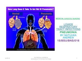
- 1. MEDICAL SURGICAL NURSING LOWER RESPIRATORY TRACT INFECTIONS PNEUMONIA BY NATIJUKA ANDREW 15/BSU/BNS/016 10/28/18 1 NATWIUKA ANDREW NEWSON:0775570748/0754755068
- 2. Review of Lung Anatomy RU L RM L RL L LUL LLL Lingula 10/28/18 2 NATWIUKA ANDREW NEWSON:0775570748/0754755068
- 4. Pneumonia • Pneumonia is an inflammation of the lung • parenchyma caused by various microorganisms • , including bacteria, mycobacteria, fungi, and viruses • Pneumonitis is a more general term that describes the inflammatory process in the lung tissue that may predispose and place the patient at risk for • microbial invasion. 10/28/18 4 NATWIUKA ANDREW NEWSON:0775570748/0754755068
- 5. Classification • Pneumonia is classified into four: community-acquired • pneumonia (CAP) and hospital- acquired pneumonia • (HAP), pneumonia in the immunocompromised host • ,and aspirationpneumonia. 10/28/18 5 NATWIUKA ANDREW NEWSON:0775570748/0754755068
- 6. Community-Acquired Pneumonia • CAP occurs either in the community setting or within • the first 48 hours after hospitalization. • The causative agents for CAP that needs hospitalization include streptococcus pneumoniae, H. influenza, Legionella, and Pseudomonas aeruginosa. • Only in 50% of the cases does the specific etiologic • agent become identified. • Pneumonia is the most common cause of CAP in people younger than 60 years of age. • Viruses are the most common cause of pneumonia in infants and children. 10/28/18 6 NATWIUKA ANDREW NEWSON:0775570748/0754755068
- 7. Hospital-Acquired Pneumonia • HAP is also called nosocomial pneumonia and is defined as • the onset of pneumonia symptoms more than 48 hours • after admission in patients with no evidence of infection at • the time of admission. • HAP is the most lethal nosocomial infection and the • leading cause of death in patients with such infections. • Common microorganisms that are responsible for HAP • include Enterobacter species, Escherichia • coli,influenza, Klebsiella species, Proteus, Serratia • marcescens, S. aureus, and S. pneumonia. • The usual presentation of HAP is a new pulmonary • infiltrate on chest x-ray combined with evidence of infection. 10/28/18 7 NATWIUKA ANDREW NEWSON:0775570748/0754755068
- 8. Pneumonia in the Immunocompromised Host • Pneumonia in immunocompromised hosts includes • Pneumocystis pneumonia, fungal pneumonias and • Mycobacterium tuberculosis. • Patients who are immunocompromised commonly • develop pneumonia from organisms of low • virulence. • Pneumonia in immunocompromised hosts may be • caused by the organisms also observe in HAP and • CAP. 10/28/18 8 NATWIUKA ANDREW NEWSON:0775570748/0754755068
- 9. Pathophysiology • Pneumonia arises from normal flora present in • patients whose resistance has been altered or from • aspiration of flora present in the oropharynx. • An inflammatory reaction may occur in the alveoli, • producing exudates that interfere with • the diffusion of oxygen and carbon dioxide. • White blood cells also migrate into the alveoli and fill the • normally air-filled spacesDue to secretions and mucosal • edema, there are areas of the lung that are not adequately ventilated and cause partial occlusion of the alveoli or bronchi. • Hypoventilation may follow, causing ventilation-perfusion mismatch. • Venous blood entering the pulmonary circulation passes through the under ventilated areas and travels to the left side of the heart deoxygenated. • The mixing of oxygenated and poorly oxygenated blood can result to arterial hypoxemia. 10/28/18 9 NATWIUKA ANDREW NEWSON:0775570748/0754755068
- 11. Epidemiology • Pneumonia has affected a lot of people, especially those • who have a weak immune system. Learning statistics on • pneumonia could give you an idea about how many has • fallen victim to this respiratory disease. • Pneumonia and influenza account for nearly 60,000 • deaths annually. • Pneumonia also ranks as the eighth leading cause of • death in the United States. • It is estimated that more than 915, 000 episodes of CAP • occur in adults 65 years old and above in the United States. • HAP accounts for 15% of hospital-acquired infections and is • the leading cause of death in patients with such infections • The estimated incidence of HAP 4 to 7 episodes per 1000 • hospitalizations. 10/28/18 11 NATWIUKA ANDREW NEWSON:0775570748/0754755068
- 12. Causes • Community-Acquired • Pneumonia • Streptococcus pneumoniae. This is the leading • cause of CAP in people younger than 60 years of • age • without comorbidity and in those 60 years and • older • with comorbidity. • Haemophilus influenzae. This causes a type of CAP that frequently affects elderly people and those with comorbid illnesses. • Mycoplasma pneumoniae. 10/28/18 12 NATWIUKA ANDREW NEWSON:0775570748/0754755068
- 13. Hospital-Acquired Pneumonia • Staphylococcus aureus. Staphylococcus pneumonia • occurs through inhalation of the organism. • Impaired host defenses. When the defenses of the • body are down, several pathogens may invade the • body. • Comorbid conditions. There are several conditions • that lower the immune system, causing bacteria to • pool in the lungs and eventually result in pneumonia. • Supine positioning. When the patient stays in a • prolonged supine position, fluid in the lungs pools • down and stays stagnant, making it a breeding place • for bacteria. • Prolonged hospitalization. The risk for hospital infections or nosocomial infections increases the longer the patient stays in the hospital. 10/28/18 13 NATWIUKA ANDREW NEWSON:0775570748/0754755068
- 14. Common Bugs for Pneumonia Community-Acquired • Streptococcus pneumoniae • Mycoplasma pneumoniae • Chlamydophila psittaci or pneumoniae • Legionella pneumophila • Haemophilus influenzae • Moraxella catarrhalis • Staphylococcus aureus • Nocardia • Mycobacterium tuberculosis • Influenza • RSV • CMV • Histoplasma, Coccidioides, Blastomycosis HCAP or HAP • Pseudomonas aeruginosa • Staphylococcus aureus (Including MRSA) • Klebsiella pneumoniae • Serratia marcescens • Acinetobacter baumanii 10/28/18 14 NATWIUKA ANDREW NEWSON:0775570748/0754755068
- 15. CLINICAL MANIFESTATIONS OF PNEUMONIA 10/28/18 15 NATWIUKA ANDREW NEWSON:0775570748/0754755068
- 16. Clinical Manifestations • Rapidly rising fever. Since there is inflammation of the lung parenchyma, fever develops as part of the signs of an infection. • Pleuritic chest pain. Deep breathing and coughing • aggravate the pain in the chest. • Rapid and bounding pulse. A rapid heartbeat occurs • because the body compensates for the low concentration • of oxygen in the body. • Tachypnea. There is fast breathing because the body • tries to compensate for the low oxygen concentration in • the body. • Purulent sputum. The sputum becomes purulent • because of the infection in the lung parenchyma which • produced sputum-filled with pus. 10/28/18 16 NATWIUKA ANDREW NEWSON:0775570748/0754755068
- 17. Prevention • Pneumococcal vaccine. This • vaccine can prevent • pneumonia in healthy patients with an efficiency of • 65% to 85%. • Staff education. To help prevent HAP, the CDC (2004) • encouraged staff education and involvement in infection • prevention. • Infection and microbiologic surveillance. It is • important to carefully observe the infection so that • there could be an appropriate application of prevention • techniques. • Modifying host risk for infection. The infection • should never be allowed to descend on any host, so the • risk must be decreased before it can affect one. 10/28/18 17 NATWIUKA ANDREW NEWSON:0775570748/0754755068
- 18. Complications • Shock and respiratory failure. These complications • are encountered chiefly in patients who have received • no specific treatment and inadequate or delayed • treatment. • Pleural effusion. In pleural effusion, the fluid is • sent to the laboratory for analysis, and there are three • stages: uncomplicated, complicated, and thoracic empyema. 10/28/18 18 NATWIUKA ANDREW NEWSON:0775570748/0754755068
- 20. Assessment and Diagnostic Findings • Assessment and diagnosis of pneumonia must be .accurate since there are a lot of respiratory problems • that have similar manifestations. The following are assessments and diagnostic tests that could determine pneumonia. • History taking. The diagnosis of pneumonia is made through history taking, particularly a recent respiratory tract infection. • Physical examination. Mainly, the number of breaths • per minute and breath sounds is assessed during physical examination. • Chest x-ray. Identifies structural distribution (e.g., lobar, bronchial); may also reveal multiple • abscesses/infiltrates, empyema (staphylococcus); • scattered or localized infiltration (bacterial); or diffuse/extensive nodular infiltrates (more often viral). • In mycoplasmal pneumonia, chest x-ray may be clear. 10/28/18 20 NATWIUKA ANDREW NEWSON:0775570748/0754755068
- 21. n• Fiberoptic bronchoscopy. May be both diagnostic • (qualitative cultures) and therapeutic (re-expansion • of lung segment). • ABGs/pulse oximetry. Abnormalities may be • present, depending on extent of lung involvement • and underlying lung disease. • Gram stain/cultures. Sputum collection; needle • aspiration of empyema, pleural, and transtracheal or • transthoracic fluids; lung biopsies and blood cultures • may be done to recover causative organism. More • than one type of organism may be present; common • bacteria include Diplococcus pneumoniae • , Staphylococcus aureus, a-hemolytic streptococcus, • Haemophilus influenzae; cytomegalovirus(CMV). • Note: Sputum cultures may not identify all offending • organisms. Blood cultures may show transient • bacteremia. 10/28/18 21 NATWIUKA ANDREW NEWSON:0775570748/0754755068
- 22. i• CBC. Leukocytosis usually present, although a low white • blood cell (WBC) count may be present in viral infection, • immunosuppressed conditions such as AIDS, and • overwhelming bacterial pneumonia. Erythrocyte • sedimentation rate (ESR) is elevated. • Serologic studies, e.g., viral or Legionella titers, • cold agglutinins. Assist in differential diagnosis of • specific organism. • Pulmonary function studies. Volumes may be • decreased (congestion and alveolar collapse); airway • pressure may be increased and compliance decreased. • Shunting is present (hypoxemia). • Electrolytes. Sodium and chloride levels may be low. • Bilirubin. May be increased. • Percutaneous aspiration/open biopsy of lung • tissues. May reveal typical intranuclear and cytoplasmic • inclusions (CMV), characteristic giant cells (rubeola). 10/28/18 22 NATWIUKA ANDREW NEWSON:0775570748/0754755068
- 23. Medical Management • The management of pneumonia centers is a step-by- • step process that zeroes on the treatment of the • infection through identification of the causative agent. • Blood culture. Blood culture is performed for • identification of the causal pathogen and prompt • administration of antibiotics in patients in whom CAP is strongly suspected. • Administration of macrolides. Macrolides are • recommended for people with drug-resistant S. pneumoniae. • Hydration is an important part of the regimen because • fever and tachypnea may result in insensible fluid losses. • Administration of antipyretics. Antipyretics are used • to treat fever and headache. • Administration of antitussives. Antitussives are • used for treatment of the associated cough. 10/28/18 23 NATWIUKA ANDREW NEWSON:0775570748/0754755068
- 24. r • Bed rest. Complete rest is prescribed until signs of • infection are diminished. • Oxygen administration. Oxygen can be given if • hypoxemia develops. • Pulse oximetry. Pulse oximetry is used to determine • the need for oxygen and to evaluate the effectiveness • of the therapy. • Aggressive respiratory measures. Other measures • include administration of high concentrations of • oxygen, endotracheal intubation, and mechanical • ventilation 10/28/18 24 NATWIUKA ANDREW NEWSON:0775570748/0754755068
- 25. Nursing Management • Nurses are expected to perform both dependent and • independent functions for the patient to aid him or her • towards the restoration of their well- being. 10/28/18 25 NATWIUKA ANDREW NEWSON:0775570748/0754755068
- 26. Nursing Assessment • Nursing assessment is critical in detecting pneumonia. • Assess respiratory symptoms. Symptoms of fever, • chills, or night sweats in a patient should be reported • immediately to the nurse as these can be signs of • bacterial pneumonia. • Assess clinical manifestations. Respiratory • assessment should further identify clinical • manifestations such as pleuritic pain, bradycardia • , tachypnea, and fatigue, use of accessory muscles for breathing, coughing, and purulent sputum. 10/28/18 26 NATWIUKA ANDREW NEWSON:0775570748/0754755068
- 27. l • Physical assessment. Assess the changes in temperature and pulse; amount, odor, and color of secretions; frequency and severity of cough; degree of tachypnea or shortness of breath; and changes in the chest x-ray findings. • Assessment in elderly patients. Assess elderly • patients for altered mental status, dehydration, • unusual behavior, excessive fatigue, and • concomitant heart failure 10/28/18 27 NATWIUKA ANDREW NEWSON:0775570748/0754755068
- 28. NURSING CARE PLAN DIAGNOSIS GOALS INTERVENTI ON RATIONALE EVALUATION 1 Impaired gaseous exchange RT ventilation perfusion imbalance EB dyspnea,restless ness. 2. Deficient fluid volume RT compromised regulatory mechanism EB decreased BP, dry skin. 1.To improve gaseous exchange in 15minutes 2.To improve airway patency in 3hours 3.To maintain fluid balance in 2days. 1.Put patient in SEMI- FOWLERS position to promote rest and breathing 2.To increase fluid intake at least 2L per day 1.To enhance clearanc,pulmon ary ventilation and perfusion. 2.To replace insensible fluid loss. 1.Improved airway patency and gaseous exchange after 4hours. 2.Maintained adequate hydration by 2days. 10/28/18 28 NATWIUKA ANDREW NEWSON:0775570748/0754755068
- 29. Discharge and Home Care Guidelines • Patient education is crucial regardless of the setting • because self-care is essential in achieving a patient’s well-being. • Oral antibiotics. Teach the patient about the proper • administration, potential side effects, and symptoms to report. • Breathing exercises. Teach the patient breathing • exercises to promote secretion clearance and volume expansion. • Follow-up check up. Strict compliance to follow-up • checkups is important to check the latest chest x-ray • result or physical examination findings. • Smoking cessation. Smoking should be stopped because it inhibits tracheobronchial ciliary action and irritates the mucous cells of the bronchi. 10/28/18 29 NATWIUKA ANDREW NEWSON:0775570748/0754755068
- 30. THANKS FOR LISTENING BUT HAVE YOU RECEIVED JESUS CHRIST AS YOUR SAVIOR?,IF NOT REPENT NOW . 10/28/18 30 NATWIUKA ANDREW NEWSON:0775570748/0754755068
- 31. PNEUMOTHORAX Presented by : TAYEBWA NOBERT ( 16/BSU/BNS/024) 10/28/18 31 NATWIUKA ANDREW NEWSON:0775570748/0754755068
- 32. Tension pneumothorax 10/28/18 32 NATWIUKA ANDREW NEWSON:0775570748/0754755068
- 33. Definition; A pneumothorax is a pathological condition in which air collects in the pleural space ( between the visceral pleura and parietal pleura).The air pushes on the outside of your lung and makes it collapse but in most cases only a portion of the lung collapses. 10/28/18 33 NATWIUKA ANDREW NEWSON:0775570748/0754755068
- 34. Examples of pneumothoraces Spontaneous pneumothorax( primary and secondary) Tension pneumothorax Closed pneumothorax Open pneumothorax Traumatic pneumothorax Catamenial pneumothorax 10/28/18 34 NATWIUKA ANDREW NEWSON:0775570748/0754755068
- 35. Definitions; • Primary pneumothorax : occurs without an apparent cause and in the absence of a significant lung disease whereas a secondary pneumothorax occurs in the presence of an existing lung pathology. • Traumatic pneumothorax: here the chest wall is pierced as is the case in gunshot and stab wounds,it occurs in up to a half of all cases of chest trauma with rib fractures being common in this group • Catamenial pneumothorax accumulation of air in the pleural cavity of women in reproductive age without concomitant respiratory diseases. Women with this condition have recurrent episodes of pneumothorax that occur within 72 hours before and after the start of menstraution. • Tension pneumothorax : air accumulates in the pleural cavity and does not get out , compresses the lung causing it to collapse ,mediastinal shift and cardiopulmonary dysfunction can result from this type of pneumothorax. 10/28/18 35 NATWIUKA ANDREW NEWSON:0775570748/0754755068
- 36. Definitions continued; • Closed pneumothorax: here the chest wall is intact and the most common cause is a rib fracture that punctures a lung,releasing air into the pleural space. • Open pneumothorax: chest wall is open due to a penetrating trauma such as gunshot or stab wound. 10/28/18 36 NATWIUKA ANDREW NEWSON:0775570748/0754755068
- 37. Causes; • Chest injury such as car crashes , physical assaults, • Therapeutic medical procedures like needle aspiration of fluid from the pleural space, a lung biopsy or insertion of a large intravenous catheter into a vein near the neck. • Lung diseases like cystic fibrosis , lung cancer , pneumonia , tuberculosis and chronic obstructive pulmonary disease. • Opportunistic infections of the lungs especially those caused by Pneumocystis jirovecii common in AIDS patients. 10/28/18 37 NATWIUKA ANDREW NEWSON:0775570748/0754755068
- 38. Causes cont’d • Ruptured air blisters (blebs)_these form on the surface of the lung and when they rupture air is allowed to leak into the space that surrounds the lungs 10/28/18 38 NATWIUKA ANDREW NEWSON:0775570748/0754755068
- 39. Predisposing factors; • height, in people who are tall the shape of the lungs makes them susceptible to a pneumothorax. • Genetics, at times pneumothorax runs among the family members • A history of a pneumothorax especially between one to two years of the first episode • Lung diseases • Mechanical ventilation 10/28/18 39 NATWIUKA ANDREW NEWSON:0775570748/0754755068
- 40. Signs and symptoms ; • Stabbing chest pain which radiates to the ipsilateral shoulder and increases with inspiration. • Tightness in the chest • Jugular vein distention(especially in tension pneumpthorax) • Respiratory distress • Fast heart rate • Low blood pressure especially in tension pneumothorax • Cyanosis due to decreased blood oxygen levels • Hyperresonance on percussion ( a rare finding) • Coughing • Shortness of breath • Malaise( less common) • anxiety 10/28/18 40 NATWIUKA ANDREW NEWSON:0775570748/0754755068
- 41. pathophysiology • In a healthy person the intra pleural pressure is normally negative( less than the atmospheric pressure).Intra pleural and extra pleural injury causes air to leak into the pleural space.when air accumulates in the pleural cavity it presses against the lung causing it to collapse.In most cases only a portion of the lung collapses 10/28/18 41 NATWIUKA ANDREW NEWSON:0775570748/0754755068
- 42. Medical management; • Acetaminophen or other anti-inflamatory agents such as ibuprofen to control pain • Codein based cough syrups to control coughing • Thoracotomy : Surgery to attach the lung to chest wall,remove blebs or areas of scarring. • Insertion of a chest tube to remove excess air , fluid or pus around the lung • Oxygen therapy: patients with tension pneumothorax should immediately be given a high concentration of supplemental oxygen to treat hypoxemia. • Pleurodesis : a medical procedure in which the pleural space is artificially obliterated ,and the two pleura adhered to each other 10/28/18 42 NATWIUKA ANDREW NEWSON:0775570748/0754755068
- 43. Nursing management; • It includes the following steps: nursing assessment, nursing diagnosis,nursing care plan and goals ,interventions and evaluation • Assessment:the nurse assess the patient for tracheal alignment,expansion of the chest,breath sounds 10/28/18 43 NATWIUKA ANDREW NEWSON:0775570748/0754755068
- 44. Nursing care plan:DIAGNOSIS Ineffective breathing pattern related to respiratory distress evidenced by use of accessory muscles GOAL To establish a normal and effective respiratory pattern in 30 minutes INTERVENTION Instruct the patient to inhale and strain against the closed glottis,to expand the lung and eject air from the thorax RATIONALE Maintain an adequate breathing pattern EVALUATION Patient was able to maintain an effective breathing pattern Ineffective peripheral tissue perfusion related to severe hypoxemia evidenced by cyanosis To improve peripheral tissue perfusion in 1 hour Administer a recommended amount of oxygen Eliminate cyanosis and increase blood oxygen levels Patient’s skin was not cyanosed after 1 hour 10/28/18 44 NATWIUKA ANDREW NEWSON:0775570748/0754755068
- 45. Discharge and home care guidelines; Care of the patient at home should include; Asepsis : the site of incision should be handled using an aseptic technique to avoid occurrence of infection. Medications; the patient should take the prescribed drugs ( antibiotics and analgesics) Activity : patient should alternate rest and activity to avoid over exhaustion and difficulty in breathing Follow up :patient should attend follow up appointments so that the physician assesses the surgical site and the state of the respiratory system. 10/28/18 45 NATWIUKA ANDREW NEWSON:0775570748/0754755068
- 46. HEMOTHORAX Course unit :Medical Surgical Nursing Presented by :TAYEBWA NOBERT 16/BSU/BNS/024 10/28/18 46 NATWIUKA ANDREW NEWSON:0775570748/0754755068
- 48. Definition; It is the collection of blood between the chest wall and the lungs (pleural cavity).This can eventually make your lung collapse as the blood pushes on the outside of the lung .It usually occurs with pneumothorax in which case it is termed as hemopneumothorax . Many of its causes are similar to those of pneumothorax. 10/28/18 48 NATWIUKA ANDREW NEWSON:0775570748/0754755068
- 49. Causes; Chest injuries or surgeries that include opening of the chest wall. Blurnt or penetrating chest trauma Cancerous tumours in the chest wall. Blood clotting defects Lung cancer and pleural cancer. Death of tissue around your lungs( pulmonary infarction). Iatrogenically induced forexample during insertion of a central venous catheter tuberculosis 10/28/18 49 NATWIUKA ANDREW NEWSON:0775570748/0754755068
- 50. Predisposing factors Pleural cancer Lung diseases(COPD’s) Injury/trauma Chest surgery 10/28/18 50 NATWIUKA ANDREW NEWSON:0775570748/0754755068
- 51. pathophysiology • A hemothorax occurs following extra pleural or intra pleural injury that disrupts the tissues of the chest wall , pleura or even intrathoracic structures .This leads to leakage of blood into the pleural cavity where it accumulates ,presses against the lung and causes it to collapse .A physiologic resopnse occurs following a hemothorax and includes both hemodynamic and respiratory responses. 10/28/18 51 NATWIUKA ANDREW NEWSON:0775570748/0754755068
- 52. Pathophysiology cont’d • In a hemodynamic response loss of 750-1500 ml will cause early symptoms of shock (tachycardia , tachypnea and a decrease in pulse pressure ) • .In a respiratory response accumulation of blood may hamper normal respiratory movement and oxygenation, something that can cause respiratory dysfunction/arrest. • NB: When blood remains in the pleural cavity for a long time an infection called empyema/pyothorax may occur.It involves infection of the fluid around the lungs. 10/28/18 52 NATWIUKA ANDREW NEWSON:0775570748/0754755068
- 53. Signs and symptoms Dull resonance on percussion Tachycardia Cyanosis Hypotension Pale,cool,clamy skin Fast heart rate Restlessnes Anxiety. Unequal chest rise Decreased chest expansion Acute pain or feeling of heaviness in the chest 10/28/18 53 NATWIUKA ANDREW NEWSON:0775570748/0754755068
- 54. Medical management Draining of the blood using a thoracotomy tube Use of numbing agents and sedatives before inserting the chest tubes. Administer pain relievers and anti-inflamatory agents like ibuprofen,diclofenac etc. 10/28/18 54 NATWIUKA ANDREW NEWSON:0775570748/0754755068
- 55. Nursing management The nurse assesses the patient for; Anxiety Tachypnea Bruising due to blurnt trauma Hypotension tachycardia Breath sounds Stupors Restlessnes 10/28/18 55 NATWIUKA ANDREW NEWSON:0775570748/0754755068
- 56. Concerns • Ineffective breathing Infection( empyema) • Pain • Anxiety • Decreased cardiac output 10/28/18 56 NATWIUKA ANDREW NEWSON:0775570748/0754755068
- 57. Care plan DIAGNOSIS GOAL INTERVENTION RATIONALE EVALUATION Ineffective breathing RT build up of blood in the pleural cavity EB use of accessory muscles for breathing. Maintain an effective breathing pattern in 30 minutes Assist with chest tube insertion if indicated Improve ventilation The patient’s breathing pattern normalised( patie nt was nolonger using accessory muscles for breathing) Acute pain RT positive pressure in the pleural cavity EB the patient verbalising a sensation of pain Reduce the pain in 30 minutes Administer analgesics like diclofenac Relieve pain Patient expressed feelings of reduced pain and comfort Risk for infection Patient should Insert a chest Eliminate Vital signs 10/28/18 57 NATWIUKA ANDREW NEWSON:0775570748/0754755068
- 58. CORYZA Presented by Name:Agaba Dickson Reg no:16/BSU/BNS/001 Course unit: Medical-surgical nursing 10/28/18 58 NATWIUKA ANDREW NEWSON:0775570748/0754755068
- 59. Objectives • By the end of this session, we should all be able to; Define coryza Know the causes,transmission,pathophysiology,signs and symptoms plus the medical and nursing management of coryza 10/28/18 59 NATWIUKA ANDREW NEWSON:0775570748/0754755068
- 60. Introduction Coryza also known as common cold,nasopharyngitis,rhinopharyngiti s,head cold or simply cold 10/28/18 60 NATWIUKA ANDREW NEWSON:0775570748/0754755068
- 61. Definition • Coryza is a viral infectious disease of the upper respiratory tract involving the inflammation of the mucous membranes in the nose 10/28/18 61 NATWIUKA ANDREW NEWSON:0775570748/0754755068
- 62. Causes and transmission • Is a viral infection and the most commonly implicated virus is rhino virus(30-80%),others include human corona virus (~15%),influenza viruses(10-15%)and adenoviruses(5%).Frequently more than one virus is present • Transmission is typically via airborne droplets(aerosals) • Direct contact with infected nasal secretions or for mites that are contaminated or person to person. • The viruses may survive for prolonged periods in the environment for over 18 hours for rhinoviruses and can be picked up by peoples hands and subsequently carried to their eyes or nose where infection occurs. 10/28/18 62 NATWIUKA ANDREW NEWSON:0775570748/0754755068
- 63. Predisposing factors • Weather-traditional folk theory states that a cold can be caught by prolonged exposure to cold weather such as rain or winter conditions and this is how the disease got its name –More viruses causing this are seasonal and occur more frequently during cold or wet weather • Immunosupressed patients • Social factors like people staying in over crowded areas like day care centers, school going children and frequently poor hygienic areas. 10/28/18 63 NATWIUKA ANDREW NEWSON:0775570748/0754755068
- 64. Pathopysiology of coryza • The symptoms of the common cold are believed to be primarily related to the immune response to the virus .The mechanism of this immune response is virus specific for example the rhino virus which is typically acquired by direct contact; it binds to human ICAM-1 receptors or CD 54 receptors through unknown mechanisms to trigger the release of inflammatory mediators • These inflammatory mediators may bring about local inflammation and cytokines may be responsible for the symptoms of common cold. 10/28/18 64 NATWIUKA ANDREW NEWSON:0775570748/0754755068
- 66. Clinical presentation of coryza The most frequent symptoms are: • Nasal discharge(runny nose) • Nasal obstruction • Sneezing • Sore throat • General malaise and cough • Hoarseness, loss of taste and smell, mild burning of the eyes and a feeling of pressure in the ears or sinuses • There may be a mild increase in body temperature most especially infants and10/28/18 66 NATWIUKA ANDREW NEWSON:0775570748/0754755068
- 68. Differential diagnosis • ADULTS • Allergic rhinitis: Nasal itching, sneezing watery rhinorrhoea and nasal obstruction but often accompanied by itchy and watery eyes. • Non allergic rhinitis: presents with chronic nasal symptoms. • Influenza; initially presents with systemic symptoms including fever,rigors,headaches,myalgia,malaise and anorexia • Pharyngitis:Symptoms are more severe than10/28/18 68 NATWIUKA ANDREW NEWSON:0775570748/0754755068
- 69. Children • Consider a foreign body in the nose. The discharge is unilateral,purulent,foul-smelling and blood stained. • Infants :Consider the possibility of a more serious condition eg meningitis,septicaemia and pneumonia. 10/28/18 69 NATWIUKA ANDREW NEWSON:0775570748/0754755068
- 70. Medical management • There are no drugs of proven benefit for the prophylaxis or treatment of coryza • Medical management is centered around providing symptomatic relief and placebo effects • 1-Nasal drops(Nacl 0.9%)for nasal congestion. These may be useful for infants who are having difficulty feeding. • Over the counter analgesia(paracetamol,ibuprofen)which may be useful for sore throats, headaches and increased temperatures. 10/28/18 70 NATWIUKA ANDREW NEWSON:0775570748/0754755068
- 71. Nursing management • Nursing concerns • Anxiety • Fluid volume deficit • Imbalanced nutrition less than body requirements due to loss of appetite • Hyperthermia • Infection spread 10/28/18 71 NATWIUKA ANDREW NEWSON:0775570748/0754755068
- 72. Nursing care plan Diagnosis goal Intervention Rationale Evaluation 1.Ineffectiveair way clearance due to nasal congestion To maintain a clear airway Assess respiratory status for rate,depth,eas e,use of accessory muscles and work of breathing Changes may vary minimal to extreme caused by bronchial swelling, increased mucus secretions hence complicating the current condition Patient will achieve the return of and ability to maintain patent airways and respiratory status baselines. 2.Deficient fluid volume RT Decreased oral To hydrate the patient and achieve the body's fluid Encourage fluids up to 3-4 l/day Provides hydration and helps to thin secretions for Imprisonment will be noticed on increased skin turgor with 10/28/18 72 NATWIUKA ANDREW NEWSON:0775570748/0754755068
- 73. Nursing care plan continuation Risk of infection spread since coryza is highly infectious To avoid spread from one person to another. Instruct patient/family to avoid crowds and persons with common cold when possible Prevents possible transmission of an infection to the patient who is already immunocompr omised No spread of an infection Expressed concerns about changes in life events and fear of unspecified consequences. To reduce on patients anxiety and copying patterns Assisting the patient in developing new anxiety reducing skills like relaxation, reassuring self statements. Provides the patient with a variety of ways to manage anxiety. Patients demonstrates improved concentration and accuracy of thoughts. 10/28/18 73 NATWIUKA ANDREW NEWSON:0775570748/0754755068
- 74. Other nursing general advice on management of coryza • Provide advice about the usual natural history of the illness and its average total length which is 10 days • Address any underlying concerns .Taking time to educate people that coryza is a self limiting and have no specific curative treatment, this may reduce anxiety and prevent unnecessary visits to the doctor in future. • Advise hygiene measures to reduce spread: frequent handwashing,avoiding sharing towels and toys etc. • Rest though no need to take time off school and work. • Advise people to return if their symptoms are worsening or if they have not improved in two weeks. 10/28/18 74 NATWIUKA ANDREW NEWSON:0775570748/0754755068
- 75. Complications • Complications are usually due to viral spread or secondary bacterial infection and they are more likely in-smokers-young children born prematurely-elderly people and those with significant comorbidity,particulary asthma,copd,DM,Cystic fibrosis and those with significant cardiac, renal or liver disease. • Common complications include • -otitis media,sinusitis,chest infections :bronchiolitis in the very young, pneumonia and exacerbations of COPD or asthma. 10/28/18 75 NATWIUKA ANDREW NEWSON:0775570748/0754755068
- 76. Prognosis • In the majority, the common cold is a mild, self-limiting illness • Usually lasts around a week in adults and 10-14 days in children • Cigarette smokers are likely to have a more severe and more prolonged illness than non smokers • People with COPD who have a rhino virus infection are more likely to have a longer duration of illness, a more severe illness and to cough for longer afterwards than those without lung disease. 10/28/18 76 NATWIUKA ANDREW NEWSON:0775570748/0754755068
- 77. References • www.https://patient.info>doctor>common cold as at 15th /august/2018 • https://www.ncbi.nlm.nih.gov>articles. 10/28/18 77 NATWIUKA ANDREW NEWSON:0775570748/0754755068
- 78. THANKS FOR LISTENING BE BLESSED 10/28/18 78 NATWIUKA ANDREW NEWSON:0775570748/0754755068
- 79. RHINITIS By jemba hajarah 16/BSU/BNS/005 10/28/18 79 NATWIUKA ANDREW NEWSON:0775570748/0754755068
- 80. • Rhinitis refers to the inflammation of the nasal mucosa and it can be classified as; Acute rhinitis Chronic rhinitis Allergic rhinitis(hay fever) Acute rhinitis is defined as having two of the listed symptoms for less than one day or for two weeks while chronic rhinitis lasts up to more than two weeks a. blockage b. Running nose c. Sneezing including nasal itch10/28/18 80 NATWIUKA ANDREW NEWSON:0775570748/0754755068
- 81. • Allergic • Infective • Other • As part of systemic disorder • Seasonal • Acute • Chronic • Idiopathic • Nares (non allergic rhinitis with eosiphilia ) • Drug induced: -beta-blockers -oral contraceptives -aspirin -NSAIDS -local decongestants • Autonomic (responds to anticholinergics) • Atrophic • Neoplastic • Primary defect in mucus • -Cystic fibrosis • -Young’s syndrome • Primary ciliary dyskinesia(Karta gener’s syndrome) Immunological -systemic lupus -rheumatoid arthritis AIDS Antibody deficiency Granulomatous disease 10/28/18 81 NATWIUKA ANDREW NEWSON:0775570748/0754755068
- 82. Severity • Mild • normal sleep and • no impairment of daily activities, sport, leisure and no impairment of work and school and • no troublesome symptoms • • Moderate-Severe • abnormal sleep or • impairment of daily activities, sport, leisure or • impaired work and school or troublesome symptoms 10/28/18 82 NATWIUKA ANDREW NEWSON:0775570748/0754755068
- 83. Risk factors • Maternal smoking • Living n areas of high population • Higher socioeconomic status • Exposure to indoor allergens as a child • Early introduction of food or formula as an infant • Genetics • personal history of asthma • Family history of asthma and eczem 10/28/18 83 NATWIUKA ANDREW NEWSON:0775570748/0754755068
- 84. Pathophysiology of allergic rhinitis(hay fever) • Allergic rhinitis results from a hypersensitivity reaction to allergen e.g. pollen, dust etc. prior to sensitization and it tends to occur seasonally. • On first exposure to an allergen, the body is said to be sensitized, antigen presenting cells present the allergen to a B cell and IgE antibodies a produced against it. • On subsequent exposure, the formed IgE antibodies cross link the allergen to mast cells and basophils causing degranulation. • Chemical dilators such as histamines are released which cause the manifestations of rhinitis. • They result in vasodilation hence there is increased blood flow resulting in reddening of the mucosa and swelling.10/28/18 84 NATWIUKA ANDREW NEWSON:0775570748/0754755068
- 85. • Leaking of fluids into interstitial spaces resulting in watery mucous running through the nose. • If the mucosal inflammation is bad enough, it can cause occlusion of the nasolacrimal duct so the individual gets teary eyes, it can also cause fluid backing up into the Eustachian tube causing stuffiness. • Irritation of the nerve endings in the nose causes itchiness • On auscultation, rhonchi can be heard due to air passing over wet surfaces 10/28/18 85 NATWIUKA ANDREW NEWSON:0775570748/0754755068
- 87. Acute and chronic rhinitis • More than 200 strains of virus cause upper respiratory infections including rhinovirus, adenoviruses, parainfluenza and corona virus. • It can be spread by direct contact by coughing and sneezing and by aerosolized droplet nuclei. • The virus attaches to receptors on the cells of the upper respiratory tract. • Local immunologic defenses e.g. IgA attempt to inactivate the antigen. • The nasal passages swell and become hyperemic and engorged. • Mucus secreting glands become hyperactive. Viscous mucous secretion in the upper respiratory tract trap invading organisms preventing contamination of more vulnerable areas. 10/28/18 87 NATWIUKA ANDREW NEWSON:0775570748/0754755068
- 88. Signs And Symptoms • Clear runny nose and coughing • Sneezing and nasal congestion • Watery eyes • Frequent clearing of the throat • Stuffiness • Erythematous mucous membranes • Swelling • Sore throat 10/28/18 88 NATWIUKA ANDREW NEWSON:0775570748/0754755068
- 89. Systemic manifestations • Low grade fever • Head ache • Malaise • Muscle aches Symptoms last for a few days up to weeks. Its symptoms become mild and self limiting but its effects on the immune system can result into more serious bacterial infections such as sinusitis and otitis media. 10/28/18 89 NATWIUKA ANDREW NEWSON:0775570748/0754755068
- 90. Diagnosis • The diagnosis is made through history taking and performing a physical examination of the nose. • If bacterial infection is suspected, white blood cell count maybe performed to rule out leukocytosis. 10/28/18 90 NATWIUKA ANDREW NEWSON:0775570748/0754755068
- 91. Medical management • Since the condition is self limiting, medical treatment is required only when complications arise but some times medication to relieve symptoms and to shorten the duration can be given. • These include; • Mild decongestants such as pseudoephedrine and phenylephrine • Nasal decongestants • Antihistamines such as cetirizine and loratadine • Aromatherapy with oils e.g. cedarwood and eucalyptus • Complementary therapies such as garlic and Echinacea can be given. 10/28/18 91 NATWIUKA ANDREW NEWSON:0775570748/0754755068
- 92. Nursing management of symptoms • Adequate rest • Maintaining fluid intake • Avoid chilling • Avoiding allergen for allergic rhinitis. • Vitamin supplements • Advise to avoid crowds and to cover mouth and nose with tissue to prevent spread of infection. 10/28/18 92 NATWIUKA ANDREW NEWSON:0775570748/0754755068
- 93. concerns • Headache • Difficult breathing • Swelling • Anxiety • Fever • Imbalanced nutrition less than body requirements due to loss of appetite • Infection spread 10/28/18 93 NATWIUKA ANDREW NEWSON:0775570748/0754755068
- 94. NURSING CARE PLAN Diagnosis goal intervention rationale 1.Ineffective airway clearance related to excessive mucus as evidenced by restlessness. 2. Risk for infection related to decrease in ciliary action 3.Deficient fluid volume RT Decreased oral intake and runny To establish a clear air way with in 50 minutes. To prevent infection through out period of the condition. Hydrate and achieve body fluid requirement Ask the patient to assume a semi sitting posture . Use mechanical ventilation. Encourage adequate nutrition Encourage It aids lung expansion. Increasing oxygen supply. Boost immunity 10/28/18 94 NATWIUKA ANDREW NEWSON:0775570748/0754755068
- 95. EPISTAXIS PRESENTATION BY ASIIMIRE ALLION 16/BSU/BNS/003 10/28/18 95 NATWIUKA ANDREW NEWSON:0775570748/0754755068
- 96. LEARNING OBJECTIVES DEFINE EPISTAXIS LIST CAUSES OF EPISTAXIS PREDISPOSING FACTORS OF EPISTAXIS PATHOPHYSIOLOGY CATEGORIES OF EPISTAXIS MANIFESTATIONS DESCRIBE MANAGEMENT OF EPISTAXIS NURSING CARE PLAN OF EPISTAXIS 10/28/18 96 NATWIUKA ANDREW NEWSON:0775570748/0754755068
- 97. DEFINITION: Refers to bleeding from the nostrils, nasal cavity or nasal pharynx. Nosebleeds are due to the bursting of blood vessels with in the nose. This may be spontaneous or caused by trauma. Nose bleeds are one of the most common problems treated in an emergency department. Nose bleeds can be a sign of an underlying pathological disease such as hemorrhagic fevers, Ebola etc. and therefore caution should be taken when a patient comes in with nose bleed. 10/28/18 97 NATWIUKA ANDREW NEWSON:0775570748/0754755068
- 98. Local causes: these are the most common causes • Nose picking/trauma • Intranasal neoplasms • Irritants like cigarette smoke • Medications like topical corticosteroids • Rhinitis, sinusitis, • Septal deviation and septal perforation SYSTEMIC CAUSES: • Hematological disorders like hemophilia, leukemia, platelet dysfunction and thrombocytopenia • Hypertension • Liver disease like cirrhosis 10/28/18 98 NATWIUKA ANDREW NEWSON:0775570748/0754755068
- 99. PREDISPOSING FACTORS • congestive heart failure • Hypertension • Diabetes mellitus • Use of some medications like aspirin, NSAIDS etc. • Atmospheric changes- high altitudes, dry climatic conditions, cold weather are known to dry out the nasal mucosa membranes leading to crusts which can cause a nosebleed • Smoking and alcoholism 10/28/18 99 NATWIUKA ANDREW NEWSON:0775570748/0754755068
- 100. PATHOPHYSIOLOGY Bleeding typically occurs when the mucosa is eroded and vessels become exposed and subsequently break. Bleeding can be classified as posterior and anterior depending on the site of bleeding Anterior nosebleed: This is the most common, this arises from little's area where the kiesselbach plexus forms on the septum. It may also originate anterior the the inferior turbinate. Posterior nosebleed: These arise further back in the nasal cavity, are usually more profuse and are often of arterial origin for example from branches of the sphenopalatine artery in the posterior nasal cavity. A posterior source presents a greater risk of airway compromise ,aspiration of blood, and greater difficulty controlling bleeding. 10/28/18 100 NATWIUKA ANDREW NEWSON:0775570748/0754755068
- 102. MANIFESTATIONS • The most common manifestation is blood loss from the nose, Bleeding usually starts from one nostril. In case of heavy bleeding it may fill up both the nostrils and over flow the nasopharynx. In certain cases blood may drip back from the nose through the throat to the stomach, a person is likely to vomit. • Excess blood loss may cause: Dizziness Fainting Confusion loss of alertness Light headedness 10/28/18 102 NATWIUKA ANDREW NEWSON:0775570748/0754755068
- 103. NURSING MANAGEMENT • Nosebleed is a medical emergency, it should therefore be attended to as quickly as possible and basic protective wears should be used such as gloves to avoid direct contact with the patient. • Quickly assess the ABCs and support them as indicated. Reassure the patient • Have the patient sit upright with her head tilted forward and instruct her to apply direct external digit pressure to the nares with her index figure and the thumb. Tell her to breathe through the mouth while she holds firm pressure on the soft flesh of her nose for at least 10 minutes. If bleeding persists, cotton pledgets soaked in vasoconstrictor and anesthetic will be placed in the anterior nasal cavity and direct pressure should be applied at both sides of the nostrils 10/28/18 103 NATWIUKA ANDREW NEWSON:0775570748/0754755068
- 105. Cont. • Ensure bedside suction. Provide an emesis basin and tissues. Tell her to spit blood into the basin if necessary. This helps prevent nausea and vomiting and lets you estimate the amount of bleeding. • Obtain vital signs and assess her breath sounds. Administer supplemental oxygen through a facemask if necessary. • Assess for signs and symptoms of hemodynamic instability including change in mental status, pallor, diaphoresis, hypotension, tachycardia and tachypnea. 10/28/18 105 NATWIUKA ANDREW NEWSON:0775570748/0754755068
- 106. Cont. • If bleeding is significant, establish vascular access, place the patient on a cardiac monitor and begin fluid resuscitation with a crystalloid solution as prescribed • Obtain a focused health history, including previous nosebleeds, other bleeding episodes, medicine use, especially use of aspirin, NSAIDS, antiplatelet agent, warfarin, and herbal products family history. • If bleeding persists, assist in preparing an epistaxis tray 10/28/18 106 NATWIUKA ANDREW NEWSON:0775570748/0754755068
- 107. MEDICAL MANAGEMENT Epistaxis is a medical emergency and there needs prior attention. quickly asses the patient and take a focused history such as trauma etc. in order to manage the patient carefully. Assess ABC’s and act accordingly Monitor vital signs especially blood pressure to rule out hypertension Tell the patient to apply pressure by pinching the nose for about 10-15 min in order to arrest hemorrhage while seated upright and leaning forward. If bleeding persists, pack the nostrils with cotton impregnated with phenylephrine solution. Do tests such as CBC to look out for platelet levels, heamoglobin etc In severe blood loss establish an iv line and administer fluids. Refer patient to ENT specialist. 10/28/18 107 NATWIUKA ANDREW NEWSON:0775570748/0754755068
- 108. CONT. Management principles: Although most patients with epistaxis can be treated as out patients, hospital admission and close observation should be considered for elderly and patients with posterior bleeding. Admission may also be prudent for patients with complicating conditions like severe hypertension or significant anemia. Anterior epistaxis: If a single anterior bleeding site is found, vasoconstriction is attempted with topical application of phenylephrine solution. For bleeding that is likely to require more aggressive treatment, local anesthetic like 4%xylocaine solution should be used. 10/28/18 108 NATWIUKA ANDREW NEWSON:0775570748/0754755068
- 109. cauterization • Larger vessels generally respond more rapidly to electro cautery. However it must be performed with caution to avoid excessive destruction of healthy surrounding tissues. • Use of electro cautery on both sides of the septum may increase the risk of septal perforation 10/28/18 109 NATWIUKA ANDREW NEWSON:0775570748/0754755068
- 110. Anterior nasal packing Anterior nasal cavity should be packed from posterior to anterior with ribbon gauze impregnated with petroleum jelly. Bayonet forceps and a nasal speculum are used to approximate the layers of the gauze which should extend as far back into the nose as possible. Each layer should be pressed down firmly before the next layer is inserted. Once the cavity has been packed so completely as possible, a gauze drip pad may be taped over the nostrils and changed periodically. 10/28/18 110 NATWIUKA ANDREW NEWSON:0775570748/0754755068
- 112. Posterior epistaxis Much less common than anterior bleeding. Posterior packing may be accomplished by passing a catheter through one nostril(or both), through the nasal pharynx, and out to the mouth. A gauze pack is then secured to the end of the Cather and positioned in the posterior nasophyryx by pulling back on the catheter until the pack is seated in the posterior choana, sealing the posterior nasal passage and applying pressure to the site of posterior bleeding. It requires special training and usually is performed by an otolaryngologist 10/28/18 112 NATWIUKA ANDREW NEWSON:0775570748/0754755068
- 114. Cont. Packing is left in place for 48 to 72 hours After the bleeding has been controlled, instruct they patient to use nasal saline spray and antibiotic ointment and to avoid strenuous activities for 7-10 days If packing does not control bleeding, arterial embolism and other surgical procedures may be required 10/28/18 114 NATWIUKA ANDREW NEWSON:0775570748/0754755068
- 115. NURSING CARE PLAN DIAGNOSIS GOAL INTERVENTION/RA TIONALE EVALUATION Anxiety rt blood loss from the nostril eb patient restlessness and asking many questions To relieve anxiety Answer patients questions and explain to them about the pathology Patient was calm by end of dialogue. Risk for deficient fluid volume rt active blood loss through the nostril To increase fluid volume and reduce fluid loss Administer iv fluids to increase blood volume. Arrest hemorrhage to avoid blood loss Hemostasis was achieved 10/28/18 115 NATWIUKA ANDREW NEWSON:0775570748/0754755068
- 116. Sinusitis by MUHINDO JULIAN 15/BSU/BNS/012 11610/28/18 116 NATWIUKA ANDREW NEWSON:0775570748/0754755068
- 117. Presentation Outline • Classification • Etiology • Presentation and Treatment • Complications 11710/28/18 117 NATWIUKA ANDREW NEWSON:0775570748/0754755068
- 118. Paranasal sinuses • Are four pairs of mucus lined hallow spaces inside bones face and skull. • Located n either sides f the nose as follows. Frontal sinuses Ethemoid sinus Sphenidal sinus Maxillary sinus 10/28/18 118 NATWIUKA ANDREW NEWSON:0775570748/0754755068
- 119. Functions of the sinuses • Humidity and filters inhaled air-greatly contributes to air filtration. • Regulate pressure within the nose • Contributes t immune defense • Give raiseness to the voice • Contributes to the facial growth. 10/28/18 119 NATWIUKA ANDREW NEWSON:0775570748/0754755068
- 120. cont • By producing half to one litre of mucus per day into the nasal cavity. • Cilia or tiny hair propel the mucus into the meatus or canal of the nasal cavity to the tiny openings called Ostia. 10/28/18 120 NATWIUKA ANDREW NEWSON:0775570748/0754755068
- 121. Drainage • Ethemiod and sphenoid sinuses they empty the mucus into superior meatus. • Maxillary and frontal sinuses empty the mucus into middle meatus 10/28/18 121 NATWIUKA ANDREW NEWSON:0775570748/0754755068
- 122. Rhino sinusitis/sinusitis • Is the inflammation or swelling of one or more paraansal sinuses Causes of sinusitis Infections Allergy Irritants like pollens, tobacco smoke ,perfumes,household chemicals Anatomical obstruction e.g. deviated septum e.g. nasal prolapse and nasal tumors,i.e benign and malignant growth. Foreign bodies10/28/18 122 NATWIUKA ANDREW NEWSON:0775570748/0754755068
- 123. Etiology • Acute sinusitis in immunocompetent patients in the community is almost always viral (eg, rhinovirus, influenza, parainfluenza). A small percentage develop secondary bacterial infection with streptococci, pneumococci, Haemophilus influenzae, Moraxella catarrhalis, or staphylococci. Occasionally, a periapical dental abscess of a maxillary tooth spreads to the overlying sinus. Hospital-acquired acute infections are more often bacterial, typically involving Staphylococcus aureus, Klebsiella 12310/28/18 NATWIUKA ANDREW NEWSON:0775570748/0754755068
- 124. Chronic sinusitis • involves many factors that combine to create chronic inflammation. Chronic allergies, structural abnormalities (eg, nasal polyps), environmental irritants (eg, airborne pollution, tobacco smoke), mucociliary dysfunction, and other factors interact with infectious organisms to cause chronic sinusitis. • The organisms are commonly bacterial (possibly as part of a biofilm on the mucosal surface) but may be fungal. Many bacteria have been implicated, including gram-negative bacilli and oropharyngeal anaerobic microorganisms; polymicrobial infection is common. In a few cases, chronic maxillary sinusitis is secondary to dental infection. Fungal infections (Aspergillus, Sporothrix, 12410/28/18 NATWIUKA ANDREW NEWSON:0775570748/0754755068
- 125. Risk factors • Common risk factors for sinusitis include factors that obstruct normal sinus drainage • (eg, allergic rhinitis ;an allergic reaction to substances such as dust, pollen, and animal hair • nasal polyps small growths in the nasal passage that can lead to inflammation ;, nasogastric or nasotracheal tubes), and immunocompromised states (eg, diabetes, HIV infection). Other factors include prolonged ICU stays, severe burns, cystic fibrosis, and ciliary dyskinesia 12510/28/18 NATWIUKA ANDREW NEWSON:0775570748/0754755068
- 126. Complications • The main complication of sinusitis is local spread of bacterial infection, causing periorbital or orbital cellulitis, cavernous sinus thrombosis, or epidural or brain abscess. 12610/28/18 NATWIUKA ANDREW NEWSON:0775570748/0754755068
- 127. Signs and symptoms • Facial pain or pressure or fullness • Nasal congestion, discharges • Diminished sense of smell • Pyrexia Minor symptoms • Headache • Halitosis • Cough • fatigue10/28/18 127 NATWIUKA ANDREW NEWSON:0775570748/0754755068
- 128. Pathophysiology • In a URI, the swollen nasal mucous membrane obstructs the ostium of a paranasal sinus, and the oxygen in the sinus is absorbed into the) is painful. If the vacuum is maintained, a transudate from the mucous membrane develops and fills the sinus; the transudate serves as a medium for bacteria that enter the sinus through the ostium or through a blood vessels of the mucous membrane. The resulting relative negative pressure in the sinus (vacuum sinusitis spreading cellulitis or thrombophlebitis in the lamina propria of the10/28/18 128 NATWIUKA ANDREW NEWSON:0775570748/0754755068
- 129. Patrick J. Lynch (Wikimedia Comomns) Paranasal Sinuses: Anterior View Frontal sinus Ethmoidal air cells Maxillary sinus 12910/28/18 129 NATWIUKA ANDREW NEWSON:0775570748/0754755068
- 130. 1. Frontal Sinuses 2. Ethmoid Sinuses (Ethmoidal Air Cells) 3. Sphenoid Sinuses 4. Maxillary Sinuses Location of Sinuses Patrick J. Lynch (Wikimedia Comomns) 13010/28/18 130 NATWIUKA ANDREW NEWSON:0775570748/0754755068
- 131. 131 Patrick J. Lynch (Wikimedia Comomns) Frontal sinus Anterior ethmoid air cells Maxillary sinus Middle Meatus Inferior meatus Frontal sinus Posterior ethmoid air cells Superior turbinate Sphenoid sinus Middle turbinate Inferior turbinate Maxillary sinus 10/28/18 131 NATWIUKA ANDREW NEWSON:0775570748/0754755068
- 132. Acute Sinusitis Usual Clinical Presentation• Symptoms progress over 2 to 3 days • Nasal congestion & discharge (usually thick & colored, not clear) • Localized pain +/- referred pain • Tenderness or pressure sensation over sinuses • Headache • Cough due to postnasal drip • Halitosis 13210/28/18 132 NATWIUKA ANDREW NEWSON:0775570748/0754755068
- 133. Usual aPhysical Findings With Acute Sinusitis• Erythematous edematous nasal mucosa • Purulent secretions in middle meatal area • May be absent if ostia completely blocked • Percussion tenderness • Over the involved sinuses • Over the maxillary molar +/- premolar teeth • Halitosis • +/- fever 13310/28/18 133 NATWIUKA ANDREW NEWSON:0775570748/0754755068
- 134. Acute Sinusitis Predisposing Conditions (cont.)• Systemic • Diabetes • Immunocompromise (AIDS) • Malnutrition • Blood dyscrasias • Cystic fibrosis • Chemotherapy • Long term steroid Rx 13410/28/18 134 NATWIUKA ANDREW NEWSON:0775570748/0754755068
- 135. Pain Patterns with Acute Sinusitis • Maxillary sinusitis • Unilateral pain over cheekbone • Maxillary toothache • Periorbital pain • Temporal headache • Pain worse if head upright • Pain better if head supine 13510/28/18 135 NATWIUKA ANDREW NEWSON:0775570748/0754755068
- 136. Pain Patterns with Acute Sinusitis (cont.)• Ethmoid sinusitis • Medial canthal pain • Medial periorbital or temporal headache • Pain worsened by Valsalva or if supine • Sphenoiditis • Retroorbital, temporal, or vertical headache • Often deep seated headache with multiple foci • Pain worse supine or bending forward • Frontal 13610/28/18 136 NATWIUKA ANDREW NEWSON:0775570748/0754755068
- 137. 137 Patrick J. Lynch (Wikimedia Comomns) Frontal sinus Ethmoidal sinus Maxillary sinus Paranasal sinuses and locations of referred pain (shaded orange) 10/28/18 137 NATWIUKA ANDREW NEWSON:0775570748/0754755068
- 138. medical management Acute Sinusitis Use of Cultures • Routine culture of nasal secretions not useful • Poor correlation between non- directed nasal or nasopharyngeal culture isolates & sinus aspirate cultures • Sinus aspirate cultures useful only for protracted or nonresponsive sinusitis • Require endoscopy or needle puncture of sinus 13810/28/18 138 NATWIUKA ANDREW NEWSON:0775570748/0754755068
- 139. General Treatment for Acute Sinusitis • Oral antibiotic • Topical and systemic decongestants • Pain medications • Optional or secondary medications: • Guaifenesin (1200 mg po q 12h) • warm nasal saline irrigations qid • Antihistamine orally : only in the small % of patients 13910/28/18 139 NATWIUKA ANDREW NEWSON:0775570748/0754755068
- 140. First - Line Antibiotic Therapy for Acute Sinusitis • Treatment duration should be 10 to 14 days (one recent study indicated 3 days may be OK) • Amoxicillin 500 mg po q 8 h • Augmentin 500 mg po q 8 h • Trimethoprim / Sulfamethoxazole DS one po bid • Azithromycin 500 mg po then 250 mg po q d x4 • Pediazole (Erythromycin - sulfisoxazole) QID may be best14010/28/18 140 NATWIUKA ANDREW NEWSON:0775570748/0754755068
- 141. Antibiotic Therapy in Acute Sinusitis if Staph. aureus is suspected • Also useful if patient fails Rx with antibiotics on previous slide • Cefuroxime axetil 500 mg po q 12h • Cefprozil 500 mg po q 12h • Cefpodoxime 200 mg po 12h • Loracarbef 400 mg po q 12h 14110/28/18 141 NATWIUKA ANDREW NEWSON:0775570748/0754755068
- 142. Use of Topical Decongestants for Rx of Acute Sinusitus• Ephedrine sulfate 1 % 2 sprays each nostril q 4h • Phenylephrine HCl 0.25 to 0.5 % 2 sprays q 4h • Oxymetazoline HCl 0.05 % 2 sprays q 12h Limit use to 3 to 5 days to avoid rebound vasodilatation and "rhinitis medicamentosa" 14210/28/18 142 NATWIUKA ANDREW NEWSON:0775570748/0754755068
- 143. Use of Oral Decongestants for Rx of Acute Sinusitis • Phenylpropanolamine HCl 12.5 mg po q 4h or 75 mg q 12h (now not available in U.S.A.) • Pseudoephedrine HCl 60 mg po q 6h or 120 mg q 12h Usually should be continued for 4 weeks 14310/28/18 143 NATWIUKA ANDREW NEWSON:0775570748/0754755068
- 144. Treatment of Frontal Sinusitis • Usually should be admitted for initial IV antibiotic Rx • Higher incidence of intracranial complications • Give IV Cefuroxime 2 gm IV q 8h or Ceftriaxone 2 gm IV q d and decongestants • If not resolving in 24 to 48 hours of Rx may need surgical intervention ( frontal sinus trephination or external 14410/28/18 144 NATWIUKA ANDREW NEWSON:0775570748/0754755068
- 145. General Management of Complications of Acute Sinusitis• Hospitalization • CT scan of sinuses ( +/- cranial CT) • IV antibiotics with anerobic coverage • ENT consult 14510/28/18 145 NATWIUKA ANDREW NEWSON:0775570748/0754755068
- 146. Sinusitis Complications : Mucocele• Most common in frontal sinus • Expansive mucus accumulation causes progressive pressure necrosis • Signs: • soft tissue mass over sinus • proptosis • ophthalmoplegia 14610/28/18 146 NATWIUKA ANDREW NEWSON:0775570748/0754755068
- 147. Antibiotics to Consider for Rx of Sinusitis Complications• Ceftriaxone 1 gm IV q 12h • Cefotaxime 2 gm IV q 4h • Ceftizoxime 4 gm IV q 8h + metronidazole 30 mg/Kg/d • Ampicillin / sulbactam 3 gm IV q 6h • Vancomycin 500 mg q 6h + aztreonam 1 gm q 8h or chloramphenicol ( for 14710/28/18 147 NATWIUKA ANDREW NEWSON:0775570748/0754755068
- 148. General Contributors to Chronic Sinusitis • Resistant infectious organisms • Underlying systemic illness (esp. diabetes) • Immunodeficiency • Irreversible mucosal changes • Anatomic abnormality 14810/28/18 148 NATWIUKA ANDREW NEWSON:0775570748/0754755068
- 149. CHRONIC SINUSITIS • Most cases of acute sinusitis resolve but some progress to chronicity. This is particularly likely to happen if there is an abnormality of the anatomy, allergy, polyps or immune deficit. • SYMPTOMS • Patients with chronic maxillary sinusitis usually have very few symptoms. • There is usually nasal obstruction and 14910/28/18 NATWIUKA ANDREW NEWSON:0775570748/0754755068
- 150. SIGNS • Mucopus in the middle meatus under the middle turbinate. • Nasal mucosa congested. • Imaging shows fluid level or opacity, or mucosal thickening within the sinus. TREATMENT • Medical • A further course of treatment with antibiotics, vasoconstrictor nose 15010/28/18 NATWIUKA ANDREW NEWSON:0775570748/0754755068
- 151. Nursing careconce rn dx Goal intervention ration al evalu tion pain Acute pain,head,throat and sinuses related to the inflammation of the nose evidenced by grimacing and verbalizing the sensation of pain To reliev e pain in 30min to relieve pain by giving panadol 1g tds ,advise patient have a rest To promo te confor tibility of the patien t anxiet y Related to lack of clients knowledge about diseases and medical procedures e.g. sinus irrigation or operation evidenced by the patient getting To reduc e the anxiet y in one hr Increase the confortabilit y by assuring the patient e.g. showing empathy thru touching the Increa se patien t aware ness of the conditi on10/28/18 151 NATWIUKA ANDREW NEWSON:0775570748/0754755068
- 152. Ineffect ive airway clearan ce Related to build up of secretion in the air way or nasal obstruction sec to inflammati on of the sinuses evidenced by breathing thru the mouth Coughi ng, clearan ce of air way in abt 30mins Rest the patient, perform steam inhalation, prepare the patient for surgery if the condition if condition needs surgical intervention Promote normal airway breathing,a voisd spread of infection to other organs like lungs 10/28/18 152 NATWIUKA ANDREW NEWSON:0775570748/0754755068
- 153. TB TB is an airborne disease caused by the bacterium Mycobacterium tuberculosis (M. tuberculosis) . M. tuberculosis and seven very closely related mycobacterial species (M. bovis, M. africanum, M. microti, M. caprae, M. pinnipedii, M. canett and M. mungi) together comprise what is known as the M. tuberculosis complex. the majority of TB cases are caused by M. tuberculosis. M. tuberculosis organisms are also called tubercle bacilli 10/28/18 153 NATWIUKA ANDREW NEWSON:0775570748/0754755068
- 154. Transmission of TB M. tuberculosis is carried in airborne particles, called droplet nuclei, of 1– 5 microns in diameter. Infectious droplet nuclei are generated when persons who have pulmonary or laryngeal TB disease cough, sneeze, shout, or sing. Depending on the environment, these tiny particles can remain suspended in the air for several hours. M. tuberculosis is transmitted through the air, not by surface contact. Transmission occurs when a person inhales droplet nuclei containing M. tuberculosis, and the droplet nuclei traverse the mouth or nasal passages, upper respiratory tract, and bronchi to reach the alveoli of the lungs . M. tuberculosis is carried in airborne particles, called droplet nuclei 10/28/18 154 NATWIUKA ANDREW NEWSON:0775570748/0754755068
- 155. TB is spread from person to person through the air. The dots in the air represent droplet nuclei containing tubercle bacilli 10/28/18 155 NATWIUKA ANDREW NEWSON:0775570748/0754755068
- 156. Factors that Determine the Probability of Transmission of M. tuberculosis 10/28/18 156 NATWIUKA ANDREW NEWSON:0775570748/0754755068
- 157. • Susceptibility -Susceptibility (immune status) of the exposed individual • Infectiousness- Infectiousness of the person with TB disease is directly related to the number of tubercle bacilli that he or she expels into the air. Persons who expel many tubercle bacilli are more infectious than patients who expel few or no bacilli. • Environment - Environmental factors that affect the concentration of M. tuberculosis organisms • Exposure - Proximity, frequency, and duration of exposure .10/28/18 157 NATWIUKA ANDREW NEWSON:0775570748/0754755068
- 158. Pathogenesis of TB • Infection occurs when a person inhales droplet nuclei containing tubercle bacilli that reach the alveoli of the lungs. These tubercle bacilli are ingested by alveolar macrophages; the majority of these bacilli are destroyed or inhibited. A small number may multiply intracellularly and are released when the macrophages die. If alive, these bacilli may spread by way of lymphatic channels or through the bloodstream to more distant tissues and organs (including areas of the body in which TB disease is most likely to develop: regional lymph nodes, apex of the lung, kidneys, brain, and bone). This process of dissemination primes the immune system for a systemic response. 10/28/18 158 NATWIUKA ANDREW NEWSON:0775570748/0754755068
- 159. Droplet nuclei containing tubercle bacilli are inhaled, enter the lungs, and travel to the alveoli 10/28/18 159 NATWIUKA ANDREW NEWSON:0775570748/0754755068
- 160. Tubercle bacilli multiply in the alveoli. 10/28/18 160 NATWIUKA ANDREW NEWSON:0775570748/0754755068
- 161. • A small number of tubercle bacilli enter the bloodstream and spread throughout the body. The tubercle bacilli may reach any part of the body, including areas where TB disease is more likely to develop (such as the brain, larynx, lymph node, lung, spine, bone, or kidney). 10/28/18 161 NATWIUKA ANDREW NEWSON:0775570748/0754755068
- 162. • Within 2 to 8 weeks, special immune cells called macrophages ingest and surround the tubercle bacilli. The cells form a barrier shell, called a granuloma, that keeps the bacilli contained and under control (LTBI). • If the immune system cannot keep the tubercle bacilli under control, the bacilli begin to multiply rapidly (TB disease). This process can occur in different areas in the body, such as the lungs, kidneys, brain, or bone 10/28/18 162 NATWIUKA ANDREW NEWSON:0775570748/0754755068
- 163. Sites of TB Disease TB disease can occur in pulmonary and extrapulmonary sites. •Pulmonary TB disease most commonly affects the lungs; this is referred to as pulmonary TB. Patients with pulmonary TB usually have a cough and an abnormal chest radiograph, and may be infectious. Although the majority of TB cases are pulmonary, TB can occur in almost any anatomical site or as disseminated disease. •Extra pulmonary Extra pulmonary TB disease occurs in places other than the lungs, including the larynx, the lymph nodes, the pleura, the brain, the kidneys, or the bones and joints. In HIV-infected persons, extra pulmonary TB disease is often accompanied by pulmonary TB. Persons with extrapulmonary TB disease usually are not infectious unless they have 1) pulmonary disease 10/28/18 163 NATWIUKA ANDREW NEWSON:0775570748/0754755068
- 164. • Miliary TB Miliary TB occurs when tubercle bacilli enter the bloodstream and disseminate to all parts of the body, where they grow and cause disease in multiple sites. This condition is rare but serious. “Miliary” refers to the radiograph appearance of millet seeds scattered throughout the lung. It is most common in infants and children younger than 5 years of age, and in severely immunocompromised persons. Miliary TB may be detected in an individual organ, including10/28/18 164 NATWIUKA ANDREW NEWSON:0775570748/0754755068
- 165. • Central Nervous System When TB occurs in the tissue surrounding the brain or spinal cord, it is called tuberculous meningitis. Tuberculous meningitis is often seen at the base of the brain on imaging studies. Symptoms include headache, decreased level of consciousness, and neck stiffness. The duration of illness before diagnosis is variable and relates in part to the presence or absence of other sites of involvement. In many cases, patients with meningitis have abnormalities on a chest radiograph consistent with old or current TB, and often have miliary TB. 10/28/18 165 NATWIUKA ANDREW NEWSON:0775570748/0754755068
- 166. Persons at Increased Risk for Progression of LTBI to TB Disease Persons infected with HIV; Children younger than 5 years of age; Persons who were recently infected with M. tuberculosis (within the past 2 years); Persons with a history of untreated or inadequately treated TB disease. Persons who are receiving immunosuppressive therapy such as10/28/18 166 NATWIUKA ANDREW NEWSON:0775570748/0754755068
- 167. • Persons with silicosis, diabetes mellitus, chronic renal failure, leukemia, or cancer of the head, neck, or lung; • Persons who have had a gastrectomy or jejunoileal bypass; • Persons who weigh less than 90% of their ideal body weight; • Cigarette smokers and persons who abuse drugs and/or alcohol; and • Populations defined locally as having an increased incidence of disease due to M. tuberculosis, including medically underserved, low-income populations. 10/28/18 167 NATWIUKA ANDREW NEWSON:0775570748/0754755068
- 168. Drug-Resistant TB (MDR and XDR) • Drug-resistant TB is caused by M. tuberculosis organisms that are resistant to the drugs normally used to treat the disease . Drug-resistant TB is transmitted in the same way as drug-susceptible TB. 10/28/18 168 NATWIUKA ANDREW NEWSON:0775570748/0754755068
- 169. Multidrug-Resistant TB • Multidrug-resistant TB (MDR TB) is caused by organisms resistant to the most effective anti- TB drugs, isoniazid and rifampin. These drugs are considered first-line drugs and are used to treat most persons with TB disease 10/28/18 169 NATWIUKA ANDREW NEWSON:0775570748/0754755068
- 170. Extensively Drug-Resistant TB • Extensively drug-resistant TB (XDR TB) is a relatively rare type of drug-resistant TB. XDR TB is resistant to isoniazid and rifampin, plus any fluoroquinolone and at least one of three injectable second-line drugs (i.e., amikacin, kanamycin, or capreomycin). 10/28/18 170 NATWIUKA ANDREW NEWSON:0775570748/0754755068
- 171. Common Symptoms of TB • Cough (2-3 weeks or more) • Coughing up blood • Chest pains • Fever • Night sweats • Feeling weak and tired • Losing weight without trying • Decreased or no appetite 10/28/18 171 NATWIUKA ANDREW NEWSON:0775570748/0754755068
- 172. DIAGNOSIS OF TB • Sputum culture: Positive for Mycobacterium tuberculosis in the active stage of the disease. • Ziehl-Neelsen (acid-fast stain applied to a smear of body fluid): Positive for acid-fast bacilli (AFB). • Skin tests (purified protein derivative [PPD] or Old tuberculin [OT] administered by intradermal injection [Mantoux]): A positive reaction (area of induration 10 mm or greater, occurring 48–72 hr after interdermal injection of the antigen) indicates past infection and the presence of antibodies but is not necessarily indicative of active disease. 10/28/18 172 NATWIUKA ANDREW NEWSON:0775570748/0754755068
- 173. CONTN OF DIAGNOSIS • Enzyme-linked immunosorbent assay (ELISA)/Western blot: • Chest x-ray: May show small, patchy infiltrations of early lesions in the upper-lung field, calcium deposits of healed primary lesions, or fluid of an effusion. • CT or MRI scan: Determines degree of lung damage and may confirm a difficult diagnosis. • Bronchoscopy: Shows inflammation and altered lung tissue. • Histologic or tissue cultures (including gastric washings; urine and cerebrospinal fluid [CSF]; skin biopsy) • 10/28/18 173 NATWIUKA ANDREW NEWSON:0775570748/0754755068
- 174. Medical management of TB • To ensure that these goals are met, TB disease must be treated for at least 6 months and in some cases even longer. Most of the bacteria are killed during the first 8 weeks of treatment. • The standard of care for initiating treatment of TB disease is four-drug therapy. 10/28/18 174 NATWIUKA ANDREW NEWSON:0775570748/0754755068
- 175. Directly Observed Therapy (DOT) • DOT is a component of case management that helps ensure patients adhere to therapy. It is the method whereby a trained health-care worker or another trained designated person watches a patient swallow each dose of anti-TB drugs and documents it. 10/28/18 175 NATWIUKA ANDREW NEWSON:0775570748/0754755068
- 176. Self-Administered Therapy • Patients on self-administered therapy should be asked routinely about adherence at follow-up visits. Pill counts should be performed consistently, and urine or blood tests can be used periodically to check for the presence of urine drug metabolites or appropriate blood serum level of the drugs. In addition, the response to treatment should be monitored closely for all patients. If culture results have not10/28/18 176 NATWIUKA ANDREW NEWSON:0775570748/0754755068
- 177. TB Disease Treatment Regimens • First-line drugs Isoniazid (INH) Rifampin (RIF) Pyrazinamide (PZA) Ethambutol (EMB) • Rifabutin* (RBT) May be used as a substitute for RIF in the treatment of all forms of TB caused by organisms that are known or presumed to be susceptible to this agent. • Rifapentine (RPT) May be used once weekly with INH in the continuation phase of treatment for HIV-negative patients with noncavitary, drug-susceptible pulmonary TB who have negative sputum smears at completion of the initial phase of treatment10/28/18 177 NATWIUKA ANDREW NEWSON:0775570748/0754755068
- 178. Second-line drugs • Streptomycin (SM) SM was formerly considered to be a first- line drug and in some instances, is still used in initial treatment. Increasing prevalence of resistance to SM in many parts of the world has decreased its overall usefulness. 10/28/18 178 NATWIUKA ANDREW NEWSON:0775570748/0754755068
- 179. • Cycloserine These drugs are reserved for special situations such as drug intolerance or resistance. • Capreomycin • ρ-Aminosalicylic acid • Levofloxacin* • Moxifloxacin* • Gatifloxacin*10/28/18 179 NATWIUKA ANDREW NEWSON:0775570748/0754755068
- 180. Treatment Interruption During Initial Phase If the interruption occurred during the initial phase, the following guidelines apply •Lapse is ≥14 days–restart treatment from the beginning •Lapse is <14 days–continue treatment to complete planned total number of doses (as long as all doses are completed within 3 months) 10/28/18 180 NATWIUKA ANDREW NEWSON:0775570748/0754755068
- 181. Nursing care plan CONCERN Abnormal breathing pattern DIAGNOSIS Ineffective airway clearance related to thick viscous or bloody secretions evidenced by coughing GOAL To promote airway clearance INTERVENTION Instruct the patient about correct poisitioning,to facilitate drainage and increase fluid intake to promote systemic hydration RATIONALE To increase surface area within the lungs Susceptibility to TB disease Risk for infection related to inadequate primary defenses and lowered resistance evidenced by Adhere to treatment regimen Adherence to the treatment regimen. Teach the patient that TB is a communicable disease and Improved body immunity towards TB infection. 10/28/18 181 NATWIUKA ANDREW NEWSON:0775570748/0754755068
- 182. PREVENTION OF TB • Identification and treatment. Early identification and treatment of persons with active TB. • Pernal hygiene. Prevention of spread of infectious droplet nuclei by source control methods and by reduction of microbial contamination of indoor air. • Surveillance. Maintain surveillance for TB infection among health care workers by routine, periodic tuberculin skin testing. 10/28/18 182 NATWIUKA ANDREW NEWSON:0775570748/0754755068
- 183. • THANKS 10/28/18 183 NATWIUKA ANDREW NEWSON:0775570748/0754755068
- 184. THORACOTOMY PRESENTATION BY KATUSIIME EMILLY 16/BSU/BNS/028 10/28/18 184 NATWIUKA ANDREW NEWSON:0775570748/0754755068
- 185. DEFINITION A thoracotomy: is a surgical procedure performed by surgeons or emergency physicians, to gain access to the thoracic organs, most commonly the heart, the lungs, or the esophagus, or for access to the thoracic aorta or the anterior spine (the latter may be necessary to access tumors in the spine). 10/28/18 185 NATWIUKA ANDREW NEWSON:0775570748/0754755068
- 186. ANATOMY OF THE THORAX. • In mammals, the thorax is the region of the body formed by the sternum, the thoracic vertebrae, and the ribs. • It extends from the neck to the diaphragm, and does not include the upper limbs. 10/28/18 186 NATWIUKA ANDREW NEWSON:0775570748/0754755068
- 187. Cont, • The thorax contains organs like: heart ,lungs and thymus gland;(the major and minor pectoral muscles, trapezius and neck muscles);internal structures such as the diaphragm, esophagus trachea and part of sternum known as xiphoid process; Arteries and veins (aorta, superior vena cava, inferior venacava and pulmonary artery) ;Bones (the shoulder socket containing the upper part of the humerus, the scapula, sternum, thoracic portion of the spine, collar bone, and the rib cage and floating ribs) • External structures are the skin and nipples. • The inner organs are protected by the rib cage and the sternum. 10/28/18 187 NATWIUKA ANDREW NEWSON:0775570748/0754755068
- 188. DIAGRAM OF THE HUMAN THORAX 10/28/18 188 NATWIUKA ANDREW NEWSON:0775570748/0754755068
- 189. FORMS OF THORACOTOMY • Median sternotomy provides wide access to the mediastinum and is the incision of choice for most open-heart surgeries and access to the anterior mediastinum 10/28/18 189 NATWIUKA ANDREW NEWSON:0775570748/0754755068
- 190. CONT, • Posterolateral thoracotomy: Is an incision through an intercostal space on the back, and is often widened with rib spreaders. • It is a very common approach for operations on the lungs or posterior mediastinum, including the esophagus. • When performed over the fifth intercostal space, it allows optimal access to the pulmonary hilum (pulmonary artery and pulmonary vein) and therefore is considered the approach of choice for pulmonary resection (pneumonectomy and lobectomy). 10/28/18 190 NATWIUKA ANDREW NEWSON:0775570748/0754755068
- 192. CONT. • Axillary thoracotomy - • In an axillary thoracotomy, surgeons gain access to the chest through an incision near the armpit. • This is commonly done for treating a pneumothorax (collapsed lung), but may also be performed for some heart and lung surgeries. 10/28/18 192 NATWIUKA ANDREW NEWSON:0775570748/0754755068
- 194. Cont, • Anterolateral thoracotomy - This procedure is an emergency procedure involving an incision along the front of the chest. • It may be done following major chest trauma, or to allow direct access to the heart after a cardiac arrest. 10/28/18 194 NATWIUKA ANDREW NEWSON:0775570748/0754755068
- 196. IMPORTANCE • The purpose of a thoracotomy is the first step used to facilitate thoracic surgeries including lobectomy or pneumonectomy for lung cancer or to gain thoracic access in major trauma. • It’s used to treat problems with the heart or other structures in the chest, such as the diaphragm. • Thoracotomy can also be used to diagnose diseases. For example, it can enable a surgeon to remove a piece of tissue for further examination (biopsy). 10/28/18 196 NATWIUKA ANDREW NEWSON:0775570748/0754755068
- 197. INDICATIONS • An injury to the chest wall that causes bleeding around the lungs • Infection in the pleural space.(pneumonia or another lung infection that has caused fluid to build up in the space around the lungs) • Collapsed lung (pneumothorax) • Malignant pleural effusion. • Fluid that collects in the lungs during chest surgery 10/28/18 197 NATWIUKA ANDREW NEWSON:0775570748/0754755068
- 198. CONT, • Lung cancer surgery • Esophageal cancer surgery • Heart/aortic surgery • Chest trauma • Management of COPD(chronic condition in which there is slow progressive obstruction of airflow into or out of the lungs) • Tuberculosis(treatment of pulmonary TB) • Biopsy and evaluation of an unknown mediastinal mass • Surgery to the anterior spine 10/28/18 198 NATWIUKA ANDREW NEWSON:0775570748/0754755068
- 199. CONTRAINDICATIONS •Bleeding disorder or anticoagulation that cannot be corrected •Acute cardiac ischemia •Instability or insufficiency of major organ systems 10/28/18 199 NATWIUKA ANDREW NEWSON:0775570748/0754755068
- 200. Pre-operative care for thoracotomy • Counsel the patient about the procedure, its benefits, risks and try as much as possible to answer their questions as this relieves anxiety. • Explain the procedure to the patient and let them sign the consent form. • Monitor the patients vital signs as anxiety may lead to a rise in blood pressure and do the required tests as ordered. • Ensure NPO for about 6-12 hours prior to surgery. An enema can also be given. • Establish an iv line and administer fluids as required. • Administer prophylaxis (antibiotics), pain killers, and other necessary medications as required. • Clean the patient, provide a gown,catheterise a few hours prior to surgery. 10/28/18 200 NATWIUKA ANDREW NEWSON:0775570748/0754755068
- 201. EQUIPMENT USED 10/28/18 201 NATWIUKA ANDREW NEWSON:0775570748/0754755068
- 202. PROCEDURE • The patient is given a general anesthesia before having this surgery. -The anesthesia ensures that the patient is asleep and pain-free during the procedure. The patient is also put an epidural, which is a small tube in spine, to deliver medication for pain relief during the surgery. • While the patient is positioned on the side, the surgeon makes a 6- to 8-inch incision below the shoulder blade, between the ribs. The surgeon then divides the muscles and spreads or removes ribs to reach the lungs or another part of the chest. • If the patient is having surgery on the lung, the affected lung is deflated with a special tube so the surgeon can work on it. A breathing tube called a ventilator keeps the other lung working. 10/28/18 202 NATWIUKA ANDREW NEWSON:0775570748/0754755068
- 204. CONT. • Depending on the type of disease or disorder a patient has been diagnosed with, a surgeon will perform a thoracotomy followed by another surgical procedure, such as a: • Lobectomy (removal of one or more lobes of the lungs) • Esophagectomy (removal of all or part of the esophagus) • Wedge resection (removal of part of a lung lobe) • Pneumonectomy (removal of an entire lung) • Decortication (removal of an organ’s membrane or covering) • Open heart surgery • Heart or lung transplant • Tumor removal • Tissue biopsy 10/28/18 204 NATWIUKA ANDREW NEWSON:0775570748/0754755068
- 205. INTRA-OPERATIVE CARE •Ensure iv fluids are administered as required. •Put the patient in a required and comfortable position. •Ensure sterilty in theater •Cover the patient with a drape •Arrange necessary instruments to be used. •Monitor all fluid loss •Before closure of a patient, ensure all instruments and other equipments used such as gauze, moppers are counted so that nothing is left in the patient. •Transfer the patient to recovery room and ensure patient is awake. 10/28/18 205 NATWIUKA ANDREW NEWSON:0775570748/0754755068
- 206. Some tubes are left in the patient. • Chest tube--this is a tube that is used to drain the liquid that builds up in the lungs normally, that may/will increase after a part of the lung has been removed or manipulated. -It is also used to remove air that may be collecting in the chest after surgery. NOTE: Most of the time both of these need to stop before we can safely remove the tube. -At times a special one way valve is put in place of the chest tube drainage system, if the patient has an air leak that may take longer to resolve. The patient may go home with this one way valve. 10/28/18 206 NATWIUKA ANDREW NEWSON:0775570748/0754755068
- 207. Cont. • Epidural Is a small catheter that is put in the space around the spine. It is used for pain control (The catheter delivers pain medication) • The epidural catheter is placed just prior to the surgery and is also used after surgery to help control the pain. • The catheter is small enough for the patient to lie on the back after surgery.. 10/28/18 207 NATWIUKA ANDREW NEWSON:0775570748/0754755068
- 208. CONT, • Foley catheter- • This is a tube placed into the bladder during surgery and used to monitor your urine output. • Sequential Compression Devices(SCDs)- – These are wraps that are placed around the legs and used to keep the blood from pooling in the calves. (If the blood remains there for a period of time without movement, it can cause a blood clot.) 10/28/18 208 NATWIUKA ANDREW NEWSON:0775570748/0754755068
- 209. CONT, • Intravenous Catheter(IV)-This is catheter introduced to a patient to help deliver fluids into the veins during surgery and after as needed. • Incentive spirometer (IS) -This is a breathing exercise device along with coughing and walking, it helps to prevent collapse of the lungs and pneumonia. This device helps the patient to breathe more deeply and fully thus preventing lung collapse. 10/28/18 209 NATWIUKA ANDREW NEWSON:0775570748/0754755068
- 210. Post operative care • Receive the patient and put them in a post operative bed. Ensure comfortable position to allow drainage of fluids. • Encourage full bed rest for about24 hours. • Administer iv fluids as required. • Monitor vital signs and fluid input and output. • Ensure NPO until bowel sounds have returned to normal. • Manage pain according to severity using the recommended analgesics. • Ensure patient comftability. • Restrict aggressive and vigorous exercises and activities until full recovery. 10/28/18 210 NATWIUKA ANDREW NEWSON:0775570748/0754755068
- 211. Pre-operative care plan diagnosis goal Intervention/ratio nale evaluation Anxiety rt fear of the procedure to be done EB patient and care takers asking many questions To relieve anxiety Answer all patients questions. Provide comfort to patient to relieve anxiety All patients questions were answered. Patient agreed to the procedure and signed the consent form Ineffective airway clearance rt exudates in the alveoli eb alteration in respiratory rate To achieve normal breath patterns of 12-20b/m Encourage coughing. Sunction to remove the exudates Patient was breathing without use of accessory muscles. 10/28/18 211 NATWIUKA ANDREW NEWSON:0775570748/0754755068
- 212. Intra-operative care plan diagnosis goal Intervention/ rationale evaluation Hypothermia rt anesthetics agents eb patient shivering To have an increase in body temperature To administer warm iv fluids during the procedure. To cover the patient and expose only the part for surgery in order to avoid loss of body heat Patient did not shiver during procedure Risk for bleeding rt to procedure being done where there are major blood vessels such as aorta To minimize blood loss Administer iv fluids, transfuse with blood if necessary. Arrest hemorrhage to minimize blood There was less blood loss. 10/28/18 212 NATWIUKA ANDREW NEWSON:0775570748/0754755068
- 213. Post operative care plan diagnosis goal Intervention/ratin ale evaluation Pain rt surgical procedure done eb patient verbalization and facial flownig To reduce pain from a scale of 6-3 in the next 40 min Administer an analgesic such a im diclofenac to cntrol pain Patient verbalized that pain had reduced from 6- 3 using the visual numerical scale Risk for infection rt loss of tissue integrity To prevent infection Daily wound dressing at incision site. •Administer antibiotics. • Ensure sterilty Wound healing was achieved . 10/28/18 213 NATWIUKA ANDREW NEWSON:0775570748/0754755068
- 214. COMPLICATIONS • Complications are greater than those for any other pulmonary biopsy procedure because of the risks of general anesthesia, surgical trauma, and a longer hospital stay with more postoperative discomfort. The greatest hazards are • Hemorrhage • Pneumothorax- The chest tubes are used to drain air and fluid until the patient heals enough to take them out (usually a few days)but Complications such as pneumothorax, tension pneumothorax, or subcutaneous emphysema can occur if these chest tubes become clogged. • Respiratory failure • Complications of general anesthesia. 10/28/18 214 NATWIUKA ANDREW NEWSON:0775570748/0754755068
- 215. CONT, •Heart attack or arrhythmias •Vocal cord dysfunction or paralysis •Bronchopleural fistula - This is a complication in which an abnormal passageway forms between a bronchial tube and the space between the membranes (pleura) lining the lungs •Postpneumonectomy syndrome or post- thoracotomy pain syndrome - Pain in the region of a thoracotomy may sometimes persist for a long time after surgery 10/28/18 215 NATWIUKA ANDREW NEWSON:0775570748/0754755068
- 217. references • https://wwww.msdmanuals.com/Disorders/P ulmonary • Www.healthline.com/health/thoracotomy • surgery.med.umich.edu/thoracic/pdf/preparin gforyourthoracotomy.pdf • Nursing Diagnoses (NANDA) 10/28/18 217 NATWIUKA ANDREW NEWSON:0775570748/0754755068
- 218. Shock. Is not a disease but rather a clinical syndrom leading to reduced perfusion of tissue and organs and enventually organ dysfunction and failure shock.Can be classified into three categories basing on the predisposing factor I. 1.Distributive shock. This kind of shock consists of A. Neulogenic shock B. Septic shock C. Anaphylactic shock 2.Cardiogenic shock. 10/28/18 218 NATWIUKA ANDREW NEWSON:0775570748/0754755068
- 219. 3.Hypovolemic shock shock has high mortality rate ounce the body’s compensatory mechanism fails. Causes of neulogenic shock Spinal cord injury Spinal cord anesthesia Severe pain Medication Hypoglycemic Causes of septic shock Gram positive bacteria 10/28/18 219 NATWIUKA ANDREW NEWSON:0775570748/0754755068
- 220. Gram negative bacteria virus parasites protozoa causes ofanaphlactic shock medication venom foods ABO incompatible blood Causes of cardiogenic shock. Cardiogenic shock is caused by the following Myocardial infarction Heart failer Cardio myopathy Arrthymias Cardiac tapenade Pneumothorax Pulmonary embolism 10/28/18 220 NATWIUKA ANDREW NEWSON:0775570748/0754755068
- 221. 4.Causes of hypovolemic shock renal loss blood loss ascitis peritonitis Explanation for different kinds of shock 1.neulogenic shock. loss of sympathetic vasoconstriction tone in the vascular smooth muscles and reduced autonomic function that will cause wide spread of arterial and venous vasodilatation .venous return is reduced leading to the reduced cardiac out put leading to hypotension 10/28/18 221 NATWIUKA ANDREW NEWSON:0775570748/0754755068
- 222. 2.Septic shock. An immune response is triggered when the bacteria enters the human body and releases endotoxines in response to macrophages. The macrophages will release cytokines like TNF andINTERLEUKINES these cytokine their mediators in which they will trigger the release of platelet activating factor for example prostaglandins, neukotrienes . Thromboxane. Kinin and complement The consequently are vasodilatation and vasoconstriction and elevated cardiac out put . Endotoxines also stimulates the release of histamine further increasing capillary permeability .more ever output falls .resulting in multisystem failure 10/28/18 222 NATWIUKA ANDREW NEWSON:0775570748/0754755068
- 223. Anaphylactic shock .this is triggered by allergic reaction when an individual is exposed to an antigen to which he has already sentised. This result from production of IGEby the plasma cell that binds to the membrane receptors on the mast cells and basophiles and on re exposure the antigen binds to IGE cross linked IGE receptor triggering release of chemical mediators from the mast cells for example bradykinin,leukotrienes; These chemical mediators will induce vascular collapse by stimulating the group smooth muscles by increasing vascular permeability to decrease vascular resistance and plasma leaking into the extracellular tissue by reducing blood volume and causing hypotension finally shork.bronch spasms , edema. 10/28/18 223 NATWIUKA ANDREW NEWSON:0775570748/0754755068
Editor's Notes
- http://www.ifem.cc/Resources/Clinical_Resources/Lectures_on_Emergency_Medicine_Topics.aspx