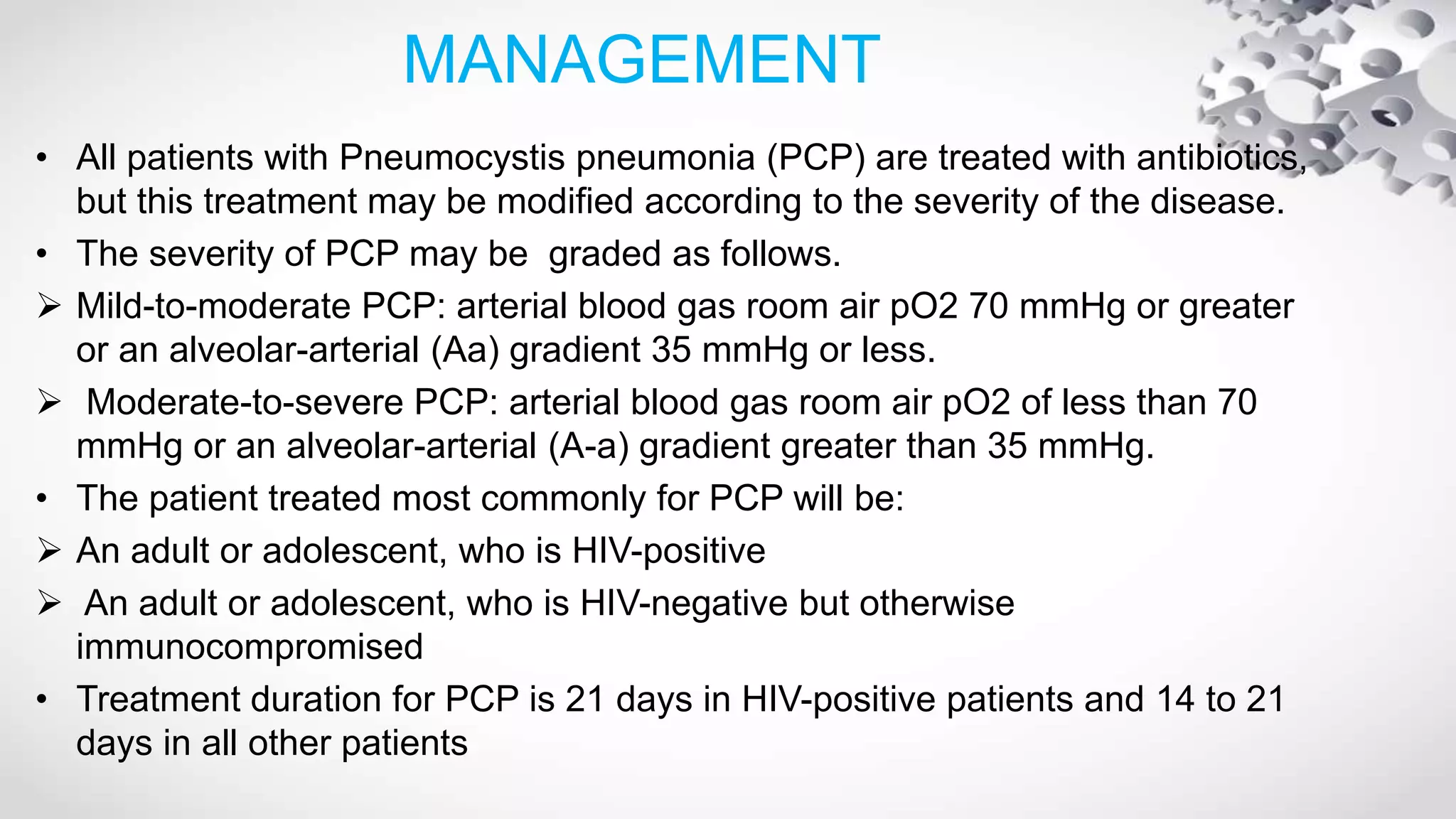
Pneumocystis jirovecii is a fungus that causes pneumonia in immunocompromised patients. It is diagnosed through microscopic visualization of the organism in samples obtained through induced sputum or bronchoscopy with bronchoalveolar lavage. Real-time PCR assays have increased sensitivity over conventional staining but may produce false positives. Risk factors include HIV/AIDS with CD4 count <200 cells/uL, use of immunosuppressive drugs, hematologic malignancies, and organ transplantation. Presentation involves fever, cough, and dyspnea. Treatment involves trimethoprim-sulfamethoxazole.


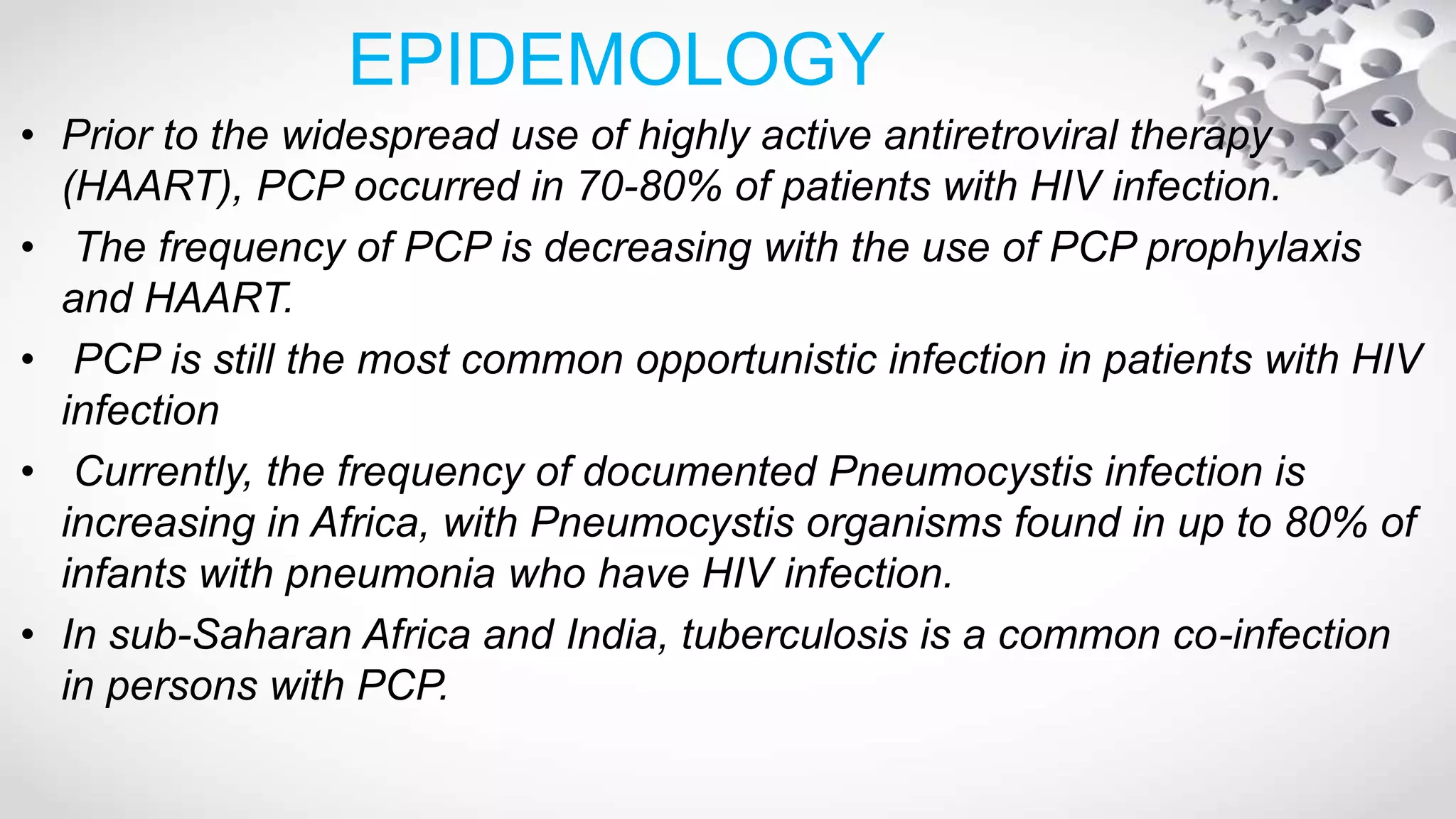


![POLYMERASE CHAIN REACTION• In recent years, real-time PCR-based strategies have largely replaced earlier methods of nested
PCR for clinical diagnosis, which may be less specific for active infection leading to higher false-
positive rates
• The use of real-time PCR has also reduced inter run contamination and reduced turnover time
(< 3hours) increasing the specificity of the assay.
• Multiple protocols that use various Pneumocystis gene targets have been developed. The main
targeted genes include the heat shock protein gene (HSP70) [Huggett et al. 2008], the dihydrofolate
reductase gene (DHFR) [Bandt and Monecke, 2007], the dihydropteroate synthase gene (DHPS)
[Alvarez-Martinez et al.2006], and the cell division cycle 2 gene (CDC2).
• The LightCycler PCR assay was 21% more sensitive than CW staining and was highly specific
demonstrating no cross-reactivity with other microbial pathogens or human DNA.
• Despite the increased sensitivity and specificityof PCR, the potential remains for this
diagnosticapproach to produce false-positive results. This potential has led some authors to
recommend that PCR detection of P jirovecii be regarded in the same manner as detection of
Streptococcuspneumoniae in the upper airway,a transient colonizer with the potential of causing
pneumonia in immunocompromised individuals](https://image.slidesharecdn.com/pcp-180606052936/75/Pcp-33-2048.jpg)


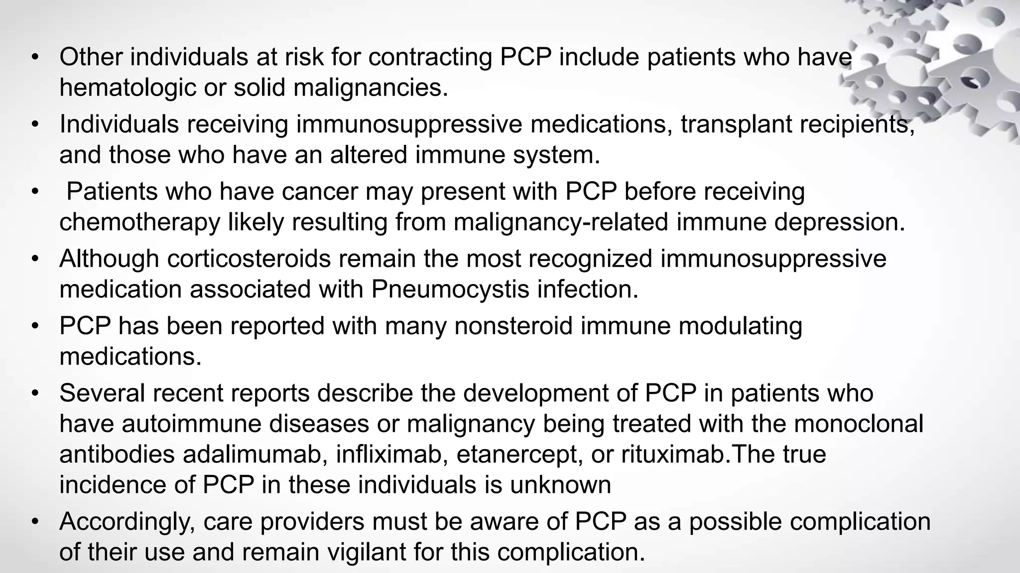
![FIRST LINE TREATMENT
• Historically, the mainstay of treatment for Pneumocystis pneumonia has been
trimethoprim-sulfamethoxazole (TMP-SMX).
• Despite the existence of other drugs to treat Pneumocystis pneumonia,
TMP-SMX is still the recommended first-line therapy for patients with mild,
moderate, and severe disease [Huang et al.2006; Benson et al. 2004]
• The standard dose for both the pediatric (older than 2 months of age) and
adult population is 15-20 mg/kg/day of TMP and 75-100 mg/kg/day of SMX
administered in divided doses.
• For severe cases, the intravenous (IV) form is preferred over the oral
formulation. However, IV can be switched to oral once clinical improvement is
achieved.
• Dose adjustments are necessary for patients with renal and liver failure since
SMX is extensively metabolized in the liver and renally excreted.](https://image.slidesharecdn.com/pcp-180606052936/75/Pcp-37-2048.jpg)
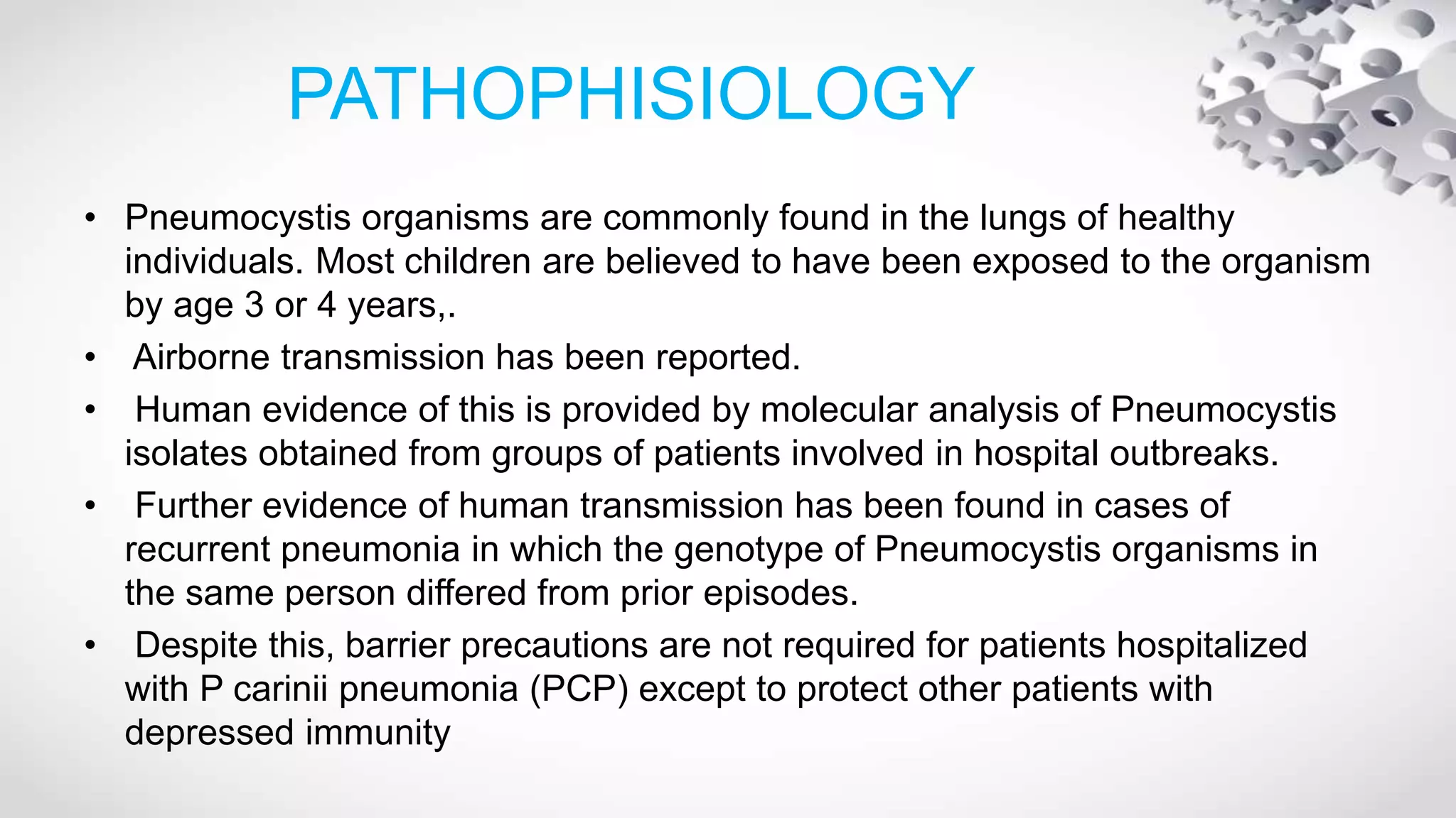
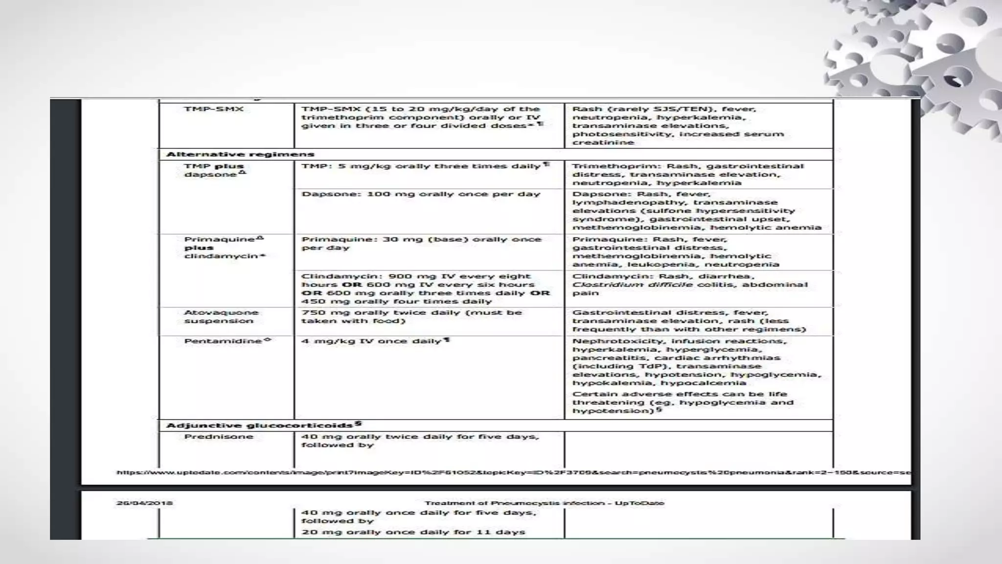
![ALTERNATIVE REGIMEN FOR MILD-MODERATE
The following oral regimens should be administered for 21 days, and are listed
in order of our preference:
●Trimethoprim-dapsone – Oral trimethoprim is administered at a dose of 5
mg/kg (typically rounded to the nearest 100 milligrams) three times per day with
dapsone 100 mg per day. Dapsone is a sulfone that is usually tolerated by
persons who have adverse reactions to TMP-SMX(*G6PD DEFICENCY)
●Clindamycin-primaquine – Oral clindamycin-primaquine is administered as
clindamycin (450 mg every six hours or 600 mg every eight hours) along with
primaquine base 30 mg per day.(* G6PD DEFICENCY)
●Atovaquone – Atovaquone suspension can be used for the treatment of mild
PCP . The dose is 750 mg twice daily and should be taken with food [12]. In
general, this agent is not used for the initial treatment of moderate disease
since atovaquone was less effective than TMP/SMX in a comparative clinical
trial However, we may switch a patient with moderate to severe disease to
atovaquone if they are clinically improved on a more potent agent, but have
developed an adverse reaction.](https://image.slidesharecdn.com/pcp-180606052936/75/Pcp-38-2048.jpg)


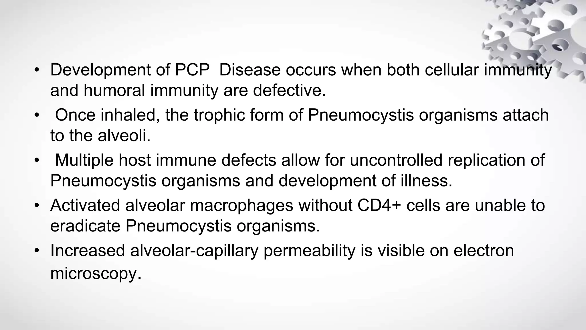
![• Unfortunately, the data for non-HIV patients are far less clear.
• For instance, a review of 31 non-HIV patients with histologically confirmed
Pneumocystis pneumonia and hypoxemia found that those that received higher
dose of steroids(prednisone equivalent >60 mg/day) had a shorter duration for
mechanical ventilation, ICU stay, and oxygen use [Pareja et al. 1998].
• However, another similar study was unable to show improvement in survival
[Delclaux et al.1999]. .
• Taking the sparsely available data into consideration, the recommendation of
adjunctive therapy in non-HIV patient must be individualized.
• Corticosteroids should not be first-line adjunctive therapy in patients with mild to
moderate disease with no respiratory failure.
• However, adjunctive corticosteroids should be seriously considered in non-HIV-
infected patients with severe Pneumocystis pneumonia, particularly if hypoxemia
is present.](https://image.slidesharecdn.com/pcp-180606052936/75/Pcp-42-2048.jpg)


![Pneumocystis_jirovecii_pneumonia[1].pptx](https://cdn.slidesharecdn.com/ss_thumbnails/malamulo-pneumocystisjiroveciipneumonia1-241127111242-957cc6eb-thumbnail.jpg?width=640&height=640&fit=bounds)


