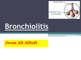
Bronchiolitis
Jwan Ali AlSofi
Objectives:-
① Discuss the epidemiology, etiology and clinical
manifestations of acute bronchiolitis.
② Discuss the differential diagnosis.
③ Discuss the diagnostic evaluation for recurrent
wheezing.
④ Discuss the treatment of acute bronchiolitis.
Definition:-
• Bronchiolitis is a disease of small bronchioles with
increased mucus production and occasional
bronchospasm, sometimes leading to airway obstruction.
• It is an acute viral lower respiratory tract infection.
• Bronchiolitis is most commonly seen in infants and young
children.
• Most severe cases occurring among infants.
• Respiratory syncytial virus (RSV) is a primary cause of
bronchiolitis.
• Bronchiolitis is potentially life-threatening.
① Respiratory syncytial virus (RSV) – is a primary cause of
bronchiolitis
② Human metapneumovirus.
③ Parainfluenza viruses.
④ Pnfluenza viruses.
⑤ Adenoviruses.
⑥ Rhinoviruses.
⑦ Coronaviruses.
⑧ Mycoplasma pneumoniae.
Etiology:-
• Viral bronchiolitis is
extremely contagious
and is spread by
contact with infected
respiratory secretions.
• Hand carriage of
contaminated secretions
is the most frequent
mode of transmission.
Transmission:-
Children who are at increased risk of
severe, potentially fatal disease, include:-
1. premature infants
2. children with chronic lung disease of prematurity,
3. hemodynamically significant congenital heart disease,
4. neuromuscular weakness,
5. immunodeficiency
9
Epidemiology:-
• Bronchiolitis is the most common serious respiratory infection
of infancy.
• Bronchiolitis is a leading cause of hospitalization of infants.
• Bronchiolitis occurs almost exclusively during the first 2
years of life.
• Peak age at 2 to 6 months.
• In the US, annual peaks are usually in the late winter months
from December through March.
• Bronchiolitis classically presents as a progressive respiratory
illness .
• Bronchiolitis caused by RSV has an incubation period of 4 to 6
days.
• its early phase started by cough and rhinorrhea.
• It progresses over 3 to 7 days to :
① Noisy, and raspy breathing.
② Audible wheezing.
③ Low-grade fever.
④ Irritability – may reflect the increased work of breathing
⑤ Decreased oral intake.
• Young infants infected with RSV may have apnea as the first
sign of infection.
Clinical Manifestations:-
• Physical signs include :
① Nasal flaring.
② Suprasternal and Intercostal retractions.
③ Air trapping with hyperexpansion of the lungs.
④ percussion of the chest usually reveals only hyperresonance
⑤ Prolongation of the expiratory phase of breathing
⑥ Diffuse Wheezing and crackles throughout the breathing cycle.
⑦ Poorly audible breath sounds suggest severe disease with nearly
complete bronchiolar obstruction.
⑧ With more severe disease, grunting and cyanosis may be present.
Clinical Manifestations:-
The natural
history of
bronchiolitis:-
Happy wheezer:-
• Bronchiolitis, when noted in an otherwise healthy child beyond
age 3 months, is often called the “disease of the happy
wheezer,” where the child demonstrates considerable cough and
wheezing while generally appearing only mildly ill.
17
Laboratory and Imaging:-
• Routine laboratory tests are not required to confirm the diagnosis.
① Pulse oximetry is adequate for monitoring oxygen saturation.
② Frequent, regular assessments and cardiorespiratory
monitoring of infants are necessary because respiratory failure
may develop.
③ Antigen tests / PCR of nasopharyngeal secretions for RSV,
parainfluenza viruses, influenza viruses, and adenoviruses are
sensitive tests to confirm the infection.
• Chest radiographs frequently show signs of lung
hyperinflation, including :
① Increased lung lucency.
② Flattened or depressed diaphragms.
• Areas of increased density may represent:-
1. viral pneumonia
2. localized atelectasis.
Laboratory and Imaging:-
Differential Diagnosis:-
•Asthma :
①Age of presentation.
②Presence of fever.
③Absence of personal or family history of
asthma.
Bronchiolitis Asthma
< 2 years > 2 years
Fever
No fever, unless a RTI is the
trigger for the asthma
exacerbation.
Viral URI symptoms
preceding presentation
Sudden onset of symptoms
No allergy triad Allergic history
Absence of personal or
family history of asthma
Presence of personal or
family history of asthma
Response to
bronchodilators – negative
Response to
bronchodilators – positive
• Airway foreign body.
▫ Suggested by a focal area on radiography that remains
inflated despite changes in position
• Cystic fibrosis.
▫ is associated with poor growth, chronic diarrhea, and a
positive family history.
• GERD.
▫ Wheezing is likely to be chronic or recurrent, and the
patient may have a history of frequent emesis.
• Cardiogenic asthma.
▫ Is wheezing associated with pulmonary congestion
secondary to left-sided heart failure.
• Congenital airway obstructive lesion.
• Exacerbation of chronic lung disease.
• Viral or bacterial pneumonia.
Differential Diagnosis:-
Treatment:-
•Supportive therapy :
① Oxygen administration, if needed.
② Respiratory monitoring for apnea
③ Control of fever.
④ Hydration.
⑤ Upper airway suctioning.
⑥ Bronchodilators, corticosteroids, chest physiotherapy,
and hypertonic saline are seldom effective and are not
generally recommended.
⑦ Antibiotics should be avoided unless there is strong
suspicion for concomitant bacterial infection.
• Indications for hospitalization :
① Moderate to marked respiratory distress.
② Hypoxemia.
③ Apnea.
④ Inability to tolerate oral feeding.
⑤ Lack of appropriate care available at home.
⑥ High-risk children.
Treatment:-
Complications and Prognosis
• Most hospitalized children show marked improvement
in 2 to 5 days.
• Tachypnea and hypoxia may progress to respiratory
failure requiring assisted ventilation.
• Most cases of bronchiolitis resolve completely.
• Recurrence is common but tends to be mild and
should be assessed and treated similarly to the first
episode.
• The incidence of asthma seems to be higher for children
hospitalized for bronchiolitis as infants.
• There is a 1% to 2% mortality rate, highest among infants
with preexisting cardiopulmonary or immunologic
impairment.
• Rarely, following adenovirus infection, the illness may
result in permanent damage to the airways (bronchiolitis
obliterans).
Complications and Prognosis
Prevention:-
• Monthly injections of palivizumab, an RSV specific
monoclonal antibody.
• Initiated just before the onset of the RSV season.
• Indications :
▫ Infants under 2 years old with :
① chronic lung disease with prematurity.
② Very low birth weight.
③ Hemodynamically significant cyanotic and acyanotic congenital
heart disease.
• Immunization with influenza vaccine is recommended
for all children older than 6 months and may prevent
influenza-associated disease.
Reference
Bronchiolitis.pptx

Bronchiolitis.pptx

  • 1.
  • 2.
    Objectives:- ① Discuss theepidemiology, etiology and clinical manifestations of acute bronchiolitis. ② Discuss the differential diagnosis. ③ Discuss the diagnostic evaluation for recurrent wheezing. ④ Discuss the treatment of acute bronchiolitis.
  • 3.
    Definition:- • Bronchiolitis isa disease of small bronchioles with increased mucus production and occasional bronchospasm, sometimes leading to airway obstruction. • It is an acute viral lower respiratory tract infection. • Bronchiolitis is most commonly seen in infants and young children. • Most severe cases occurring among infants. • Respiratory syncytial virus (RSV) is a primary cause of bronchiolitis. • Bronchiolitis is potentially life-threatening.
  • 7.
    ① Respiratory syncytialvirus (RSV) – is a primary cause of bronchiolitis ② Human metapneumovirus. ③ Parainfluenza viruses. ④ Pnfluenza viruses. ⑤ Adenoviruses. ⑥ Rhinoviruses. ⑦ Coronaviruses. ⑧ Mycoplasma pneumoniae. Etiology:-
  • 8.
    • Viral bronchiolitisis extremely contagious and is spread by contact with infected respiratory secretions. • Hand carriage of contaminated secretions is the most frequent mode of transmission. Transmission:-
  • 9.
    Children who areat increased risk of severe, potentially fatal disease, include:- 1. premature infants 2. children with chronic lung disease of prematurity, 3. hemodynamically significant congenital heart disease, 4. neuromuscular weakness, 5. immunodeficiency 9
  • 10.
    Epidemiology:- • Bronchiolitis isthe most common serious respiratory infection of infancy. • Bronchiolitis is a leading cause of hospitalization of infants. • Bronchiolitis occurs almost exclusively during the first 2 years of life. • Peak age at 2 to 6 months. • In the US, annual peaks are usually in the late winter months from December through March.
  • 11.
    • Bronchiolitis classicallypresents as a progressive respiratory illness . • Bronchiolitis caused by RSV has an incubation period of 4 to 6 days. • its early phase started by cough and rhinorrhea. • It progresses over 3 to 7 days to : ① Noisy, and raspy breathing. ② Audible wheezing. ③ Low-grade fever. ④ Irritability – may reflect the increased work of breathing ⑤ Decreased oral intake. • Young infants infected with RSV may have apnea as the first sign of infection. Clinical Manifestations:-
  • 12.
    • Physical signsinclude : ① Nasal flaring. ② Suprasternal and Intercostal retractions. ③ Air trapping with hyperexpansion of the lungs. ④ percussion of the chest usually reveals only hyperresonance ⑤ Prolongation of the expiratory phase of breathing ⑥ Diffuse Wheezing and crackles throughout the breathing cycle. ⑦ Poorly audible breath sounds suggest severe disease with nearly complete bronchiolar obstruction. ⑧ With more severe disease, grunting and cyanosis may be present. Clinical Manifestations:-
  • 16.
  • 17.
    Happy wheezer:- • Bronchiolitis,when noted in an otherwise healthy child beyond age 3 months, is often called the “disease of the happy wheezer,” where the child demonstrates considerable cough and wheezing while generally appearing only mildly ill. 17
  • 18.
    Laboratory and Imaging:- •Routine laboratory tests are not required to confirm the diagnosis. ① Pulse oximetry is adequate for monitoring oxygen saturation. ② Frequent, regular assessments and cardiorespiratory monitoring of infants are necessary because respiratory failure may develop. ③ Antigen tests / PCR of nasopharyngeal secretions for RSV, parainfluenza viruses, influenza viruses, and adenoviruses are sensitive tests to confirm the infection.
  • 19.
    • Chest radiographsfrequently show signs of lung hyperinflation, including : ① Increased lung lucency. ② Flattened or depressed diaphragms. • Areas of increased density may represent:- 1. viral pneumonia 2. localized atelectasis. Laboratory and Imaging:-
  • 20.
    Differential Diagnosis:- •Asthma : ①Ageof presentation. ②Presence of fever. ③Absence of personal or family history of asthma.
  • 21.
    Bronchiolitis Asthma < 2years > 2 years Fever No fever, unless a RTI is the trigger for the asthma exacerbation. Viral URI symptoms preceding presentation Sudden onset of symptoms No allergy triad Allergic history Absence of personal or family history of asthma Presence of personal or family history of asthma Response to bronchodilators – negative Response to bronchodilators – positive
  • 22.
    • Airway foreignbody. ▫ Suggested by a focal area on radiography that remains inflated despite changes in position • Cystic fibrosis. ▫ is associated with poor growth, chronic diarrhea, and a positive family history. • GERD. ▫ Wheezing is likely to be chronic or recurrent, and the patient may have a history of frequent emesis. • Cardiogenic asthma. ▫ Is wheezing associated with pulmonary congestion secondary to left-sided heart failure. • Congenital airway obstructive lesion. • Exacerbation of chronic lung disease. • Viral or bacterial pneumonia. Differential Diagnosis:-
  • 23.
    Treatment:- •Supportive therapy : ①Oxygen administration, if needed. ② Respiratory monitoring for apnea ③ Control of fever. ④ Hydration. ⑤ Upper airway suctioning. ⑥ Bronchodilators, corticosteroids, chest physiotherapy, and hypertonic saline are seldom effective and are not generally recommended. ⑦ Antibiotics should be avoided unless there is strong suspicion for concomitant bacterial infection.
  • 24.
    • Indications forhospitalization : ① Moderate to marked respiratory distress. ② Hypoxemia. ③ Apnea. ④ Inability to tolerate oral feeding. ⑤ Lack of appropriate care available at home. ⑥ High-risk children. Treatment:-
  • 25.
    Complications and Prognosis •Most hospitalized children show marked improvement in 2 to 5 days. • Tachypnea and hypoxia may progress to respiratory failure requiring assisted ventilation. • Most cases of bronchiolitis resolve completely. • Recurrence is common but tends to be mild and should be assessed and treated similarly to the first episode.
  • 26.
    • The incidenceof asthma seems to be higher for children hospitalized for bronchiolitis as infants. • There is a 1% to 2% mortality rate, highest among infants with preexisting cardiopulmonary or immunologic impairment. • Rarely, following adenovirus infection, the illness may result in permanent damage to the airways (bronchiolitis obliterans). Complications and Prognosis
  • 27.
    Prevention:- • Monthly injectionsof palivizumab, an RSV specific monoclonal antibody. • Initiated just before the onset of the RSV season. • Indications : ▫ Infants under 2 years old with : ① chronic lung disease with prematurity. ② Very low birth weight. ③ Hemodynamically significant cyanotic and acyanotic congenital heart disease. • Immunization with influenza vaccine is recommended for all children older than 6 months and may prevent influenza-associated disease.
  • 28.

Editor's Notes

  • #6 Infection is acquired by inoculation of the nasal or conjunctival mucosa with contaminated secretions or by inhalation of large (>5 μm in diameter), virus-containing respiratory droplets within 2 m of an infectious patient. After an incubation period of 4 to 6 days, viral replication in the nasal epithelium results in congestion, rhinorrhea, irritability, and poor feeding. Fever occurs in approximately 50% of infected infants. Once in the lower respiratory tract, the virus infects the ciliated epithelial cells of the mucosa of the bronchioles and pneumocytes in the alveoli. Two RSV surface glycoproteins, F and G, mediate viral attachment to the glycocalyx of the target cell. Viral attachment initiates a conformational change in F protein to a postfusion structure that facilitates fusion of the viral envelope and the plasma membrane of the host cell, resulting in viral entry into the cell. Viral replication initiates an influx of natural killer cells, helper CD4+ and cytotoxic CD8+ T lymphocytes, and activated granulocytes. Cellular infiltration of the peribronchiolar tissue, edema, increased mucous secretion, sloughing of infected epithelial cells, and impaired ciliary beating cause varying degrees of intraluminal obstruction. During inspiration, negative intrapleural pressure is generated and air flows past the obstruction. The positive pressure of expiration further narrows the airways, producing greater obstruction, which causes wheezing. Innate and adaptive immune responses are involved in viral clearance, and most hospitalized children are discharged after 2 to 3 days. Regeneration of the bronchiolar epithelium begins within 3 to 4 days after the resolution of symptoms. ICU denotes intensive care unit.
  • #17 Infection is acquired by inoculation of the nasal or conjunctival mucosa with contaminated secretions or by inhalation of large (>5 μm in diameter), virus-containing respiratory droplets within 2 m of an infectious patient. After an incubation period of 4 to 6 days, viral replication in the nasal epithelium results in congestion, rhinorrhea, irritability, and poor feeding. Fever occurs in approximately 50% of infected infants. Once in the lower respiratory tract, the virus infects the ciliated epithelial cells of the mucosa of the bronchioles and pneumocytes in the alveoli. Two RSV surface glycoproteins, F and G, mediate viral attachment to the glycocalyx of the target cell. Viral attachment initiates a conformational change in F protein to a postfusion structure that facilitates fusion of the viral envelope and the plasma membrane of the host cell, resulting in viral entry into the cell. Viral replication initiates an influx of natural killer cells, helper CD4+ and cytotoxic CD8+ T lymphocytes, and activated granulocytes. Cellular infiltration of the peribronchiolar tissue, edema, increased mucous secretion, sloughing of infected epithelial cells, and impaired ciliary beating cause varying degrees of intraluminal obstruction. During inspiration, negative intrapleural pressure is generated and air flows past the obstruction. The positive pressure of expiration further narrows the airways, producing greater obstruction, which causes wheezing. Innate and adaptive immune responses are involved in viral clearance, and most hospitalized children are discharged after 2 to 3 days. Regeneration of the bronchiolar epithelium begins within 3 to 4 days after the resolution of symptoms. ICU denotes intensive care unit.