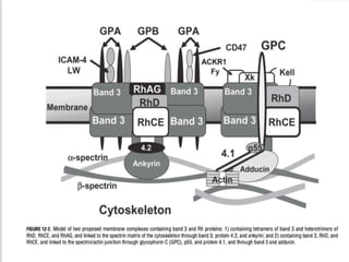
RBC MEMBRANE
STRUCTURE AND ITS
DISORDERS
DR. SAMHATI TRIPATHY
PG 1ST
YEAR RESIDENT
DEPT OF TRANSFUSION MEDICINE
S.C.B. M.C.H.
INTRODUCTION
 Biconcave--40% excess surface area allows it
to stretch upto 2.5 times
 Volume 90 fl
 RBC membrane represents a semipermeable
lipid bilayer supported by a mesh-like
cytoskeleton
2 IMPORTANT CHARACTERISTICS
deformability
•ATP↑
•Ca inside cell ↓
•Phosphorylation of spectrin ↑
permeability
•Freely to water and anions
•Impermeable to cations (Na+ 1:12, K+ 25:1)
•Ca2+
pumped out (calmodulin)
RBC MEMBRANE LIPIDS
 8% carb, 52% proteins, 40% lipids
 Lipid portion contains equal parts of cholesterol and
phospholipids which form a bilayer
 The hydrophilic polar head of PL faces outwards towards
both the aqueous plasma and cytoplasm
 The hydrophobic nonpolar acyl tails of PL arrange to form
a central layer
 The esterified cholesterol being hydrophobic lies parallel
to the acyl tails of phospholipids and confers tensile
strength
 Membrane enzymes maintain the cholesterol conc.(LCAT-
lecithin cholesterol acyltransferase)
 Rise in cholesterol conc increases strength but reduces
deformabilty
 The PL are asymmetrically distributed
 Outer layer – phosphatidylcholine &
sphingomyelin
 Inner layer – phosphatidylserine &
phosphatidylethanolamine
 Membrane PL and cholesterol may also
redistribute laterally to deform in response
to stress
RBC MEMBRANE PROTEINS
Transmembrane/integral
(Serve as transport and adhesion sites and signalling receptors)
Band 3
Glycophorins
Rh proteins
Kell & duffy Ags
ATPase(Na-K, Ca, Mg)
Glycophorins
 Alpha helix made of hydrophobic amino acids
with hydrophilic (negatively charged) sialic acid-
rich intra and extra cellular domain. This –ve
charge prevents clumping of RBCs to each other
Act as transmembrane receptor for entry of
plasmodium into RBC
So, if absent----protected from malaria
Also carry blood group antigens-
A,B- MNSs
C,D- Gerbich
 Band 3
Mediates exchange of Cl- ions(in) with
HCO3- (out) across membrane
Diego antigen
Age related marker on RBC- aggregation
of band 3—autologous IgGs bind to it-
triggers phagocytosis by liver and spleen
macrophages
 CR1(complement receptor) aka CD35-
-regulator of complement system(avoid
autologous complement attack due to deposition of
naturally occuring IgG autoAbs
-phagocytosis of ring stage Pf by complement
activation
 CD55/DAF- pf attachment to RBC surface. CD55 null
RBCs are refractory to invasion
 Duffy- receptor for pv---absent--resistant
 Kidd- functions as urea transporter
 Basigin- recirculating mature RBCs from spleen, Ok
antigen
CYTOSKELETAL PROTEINS
 Supporting framework for the lipid bilayer---
give it shape to make it biconcave
 Protein 4.1
 Protein 4.2
 Actin
 Contraction and relaxation of membrane
Spectrin
 helical flexible heterodimers made of 2 polypeptide
chains- α and β
 Each actin oligomer can bind multiple spectrin
tetramers creating a 2D spectrin-actin skeleton that is
connected to the CM by 2 distinct tethering interactions-
ankyrin and band 4.2 protein 4.1
(binds spectrin to band 3) (binds tail of spectrin to GP)
HEREDITARY SPHEROCYTOSIS
 Inherited intrinsic RBC membrane defect that
render red cells spheroid, less deformable
and vulnerable to splenic sequestration and
destruction
 Northern Europe – 1:5000
 75% AD, 25% AR(protein 4.2 , Japan)
 Rest are compound heterozygosity for 2
different defects---more severe, marked
jaundice at birth, requires exchange
transfusion
DIAGNOSIS
 Family history
 CBC—Hb,MCV MCHC,RDW
 Peripheral blood smear-
 Reticulocyte count↑
 S. indirect bilirubin↑
 S. LDH↑↑
 S. haptoglobin↓↓
 OFT, AGLT(acidified glycerol lysis test)—uses hypotonic soln
 Cryohemolysis test—uses hypertonic solution
 EMA(eosin-5-maleimide binding test-
flowcytometric test to observe CM protein
interaction with EMA dye
 SDS-PAGE(sodium dodecyl sulfate
polyacrylamide gel electrophoresis)—to
quantify each protein
 Ektacytometry- reveals characteristic
response of RBC memb to laser diffraction
biomechanical stressors
 LC-MS/MS(mass spectrometry)—analyse the
protein composition of RBC memb
HISTOPATH. FINDINGS
 Spherocytosis---most specific, but npt
pathognomonic
 Anisocytosis, poikilocytosis, target cells
 Reticulocytosis---compensatory change—5-20%
 Marrow erythroid hyperplasia
 Hemosiderosis—repeated BT
 Howell Jolly bodies (small dark nuclear
remnants) in splenectomised patients
 Osmotic fragility test
 MCHC , RDW↑
 Schistocytes on PBS,
Spherocytes
SCHISTOCYTES - Helmet cells,
bite cells, etc
SDS- PAGE
CLINICAL FEATURES
 chronic hemolytic anemia of mild to mod
severity---pallor, fatigue,dyspnea,
tachycardia
 splenomegaly---mod-—early satiety, LUQ pain
 Jaundice
 Cholelithiasis—pigment stones—40-50%
COMPLICATIONS
 Aplastic crisis—triggered by parvovirus B19
infection---because it infects and kills red
cell progenitors
 Hemolytic crisis—increased splenic
destruction----trigerred by infectious
mononucleosis infection
 Neonatal jaundice, non-immune hydrops
fetalis -----with fetal death if severe
TREATMENT
 Phototherapy, exchange transfusion
 Folic acid supplements
 Blood transfusion
 Splenectomy-anemia is corrected but
spherocytes persist
 Cholecystectomy—for GB stones
 Iron chelation therapy—for hemosiderosis
HEREDITARY ELLIPTOCYTOSIS
 Loss of the normal elastic recoil property in
RBCs within the peripheral circulation,
resulting in their distinct elliptical shape—
extravascular hemolysis
 African & mediterranean
 Defect in spectrin>ankyrin,4.1, band 3, actin
 AD
 A subtype of HE- hereditary
pyropoikilocytosis(HPP) has AR inheritance
PBS
Cigar shaped elliptocytes
Spherocytes
Stomatocytes
Fragmented red cells
TYPES OF HE
BASED ON variatons in RBC morphology and degree of
hemolysis--
 Common hereditary elliptocytosis—m/c, asymptomatic.
Neonates may have transient hemolysis which resolves
within first year
 Hereditary pyropoikilocytosis(HPP)—most severe,
uniquely susceptible to thermal stressors.
Neonatal jaundice and persistent hemolytic anemia
throughout life.
On PBS- microspherocytes , poikilocytes with rare
elliptocytes
 Southeast asian ovalocytosis(SAO)—seen in malaria
endemic regions—confers some protection
mild to no hemolysis
band 3 defect
PBS- stomatocytes with longitudinal slits
 Spherocytic elliptocytosis(SE)– Italian,
mod hemolysis
 Majority are asymptomatic-- don’t need any t/t
 Folate supplementation
 If symp– BT, splenectomy
OTHER RBC MEMBRANE
DISORDERS
HEREDITARY
STOMATOCYTOSIS/XEROCYTOSIS
 Mutations in PIEZO1 gene
 Increased permeability to cations
 Greater efflux of K+
than Na+
----net decrease in
cation content---osmotically resistant xerocytes—
rigid RBCs ---destroyed by spleen—
hemolysis(compensated)
 AD
 PBS – macrocytic stomatocytosis
 Decreased OF
 C/F-fatigue, jaundice, pallor, dark urine esp during
intense physical activity
 T/t not needed
CRYOHYDROCYTOSIS
 Mutation in band 3---convert the anion
exchanger into a nonselective cation channel
 Lyse when stored at 4 ̊C
 AD
 Pseudohyperkalemia due to K+
leakage
THANK YOU

RBC MEMBRANE STRUCTURE AND ITS DISORDERS.pptx

  • 1.
    RBC MEMBRANE STRUCTURE ANDITS DISORDERS DR. SAMHATI TRIPATHY PG 1ST YEAR RESIDENT DEPT OF TRANSFUSION MEDICINE S.C.B. M.C.H.
  • 2.
    INTRODUCTION  Biconcave--40% excesssurface area allows it to stretch upto 2.5 times  Volume 90 fl  RBC membrane represents a semipermeable lipid bilayer supported by a mesh-like cytoskeleton
  • 4.
    2 IMPORTANT CHARACTERISTICS deformability •ATP↑ •Cainside cell ↓ •Phosphorylation of spectrin ↑ permeability •Freely to water and anions •Impermeable to cations (Na+ 1:12, K+ 25:1) •Ca2+ pumped out (calmodulin)
  • 7.
    RBC MEMBRANE LIPIDS 8% carb, 52% proteins, 40% lipids  Lipid portion contains equal parts of cholesterol and phospholipids which form a bilayer  The hydrophilic polar head of PL faces outwards towards both the aqueous plasma and cytoplasm  The hydrophobic nonpolar acyl tails of PL arrange to form a central layer  The esterified cholesterol being hydrophobic lies parallel to the acyl tails of phospholipids and confers tensile strength  Membrane enzymes maintain the cholesterol conc.(LCAT- lecithin cholesterol acyltransferase)  Rise in cholesterol conc increases strength but reduces deformabilty
  • 8.
     The PLare asymmetrically distributed  Outer layer – phosphatidylcholine & sphingomyelin  Inner layer – phosphatidylserine & phosphatidylethanolamine  Membrane PL and cholesterol may also redistribute laterally to deform in response to stress
  • 9.
    RBC MEMBRANE PROTEINS Transmembrane/integral (Serveas transport and adhesion sites and signalling receptors) Band 3 Glycophorins Rh proteins Kell & duffy Ags ATPase(Na-K, Ca, Mg)
  • 10.
    Glycophorins  Alpha helixmade of hydrophobic amino acids with hydrophilic (negatively charged) sialic acid- rich intra and extra cellular domain. This –ve charge prevents clumping of RBCs to each other Act as transmembrane receptor for entry of plasmodium into RBC So, if absent----protected from malaria Also carry blood group antigens- A,B- MNSs C,D- Gerbich
  • 11.
     Band 3 Mediatesexchange of Cl- ions(in) with HCO3- (out) across membrane Diego antigen Age related marker on RBC- aggregation of band 3—autologous IgGs bind to it- triggers phagocytosis by liver and spleen macrophages
  • 12.
     CR1(complement receptor)aka CD35- -regulator of complement system(avoid autologous complement attack due to deposition of naturally occuring IgG autoAbs -phagocytosis of ring stage Pf by complement activation  CD55/DAF- pf attachment to RBC surface. CD55 null RBCs are refractory to invasion  Duffy- receptor for pv---absent--resistant  Kidd- functions as urea transporter  Basigin- recirculating mature RBCs from spleen, Ok antigen
  • 14.
    CYTOSKELETAL PROTEINS  Supportingframework for the lipid bilayer--- give it shape to make it biconcave  Protein 4.1  Protein 4.2  Actin  Contraction and relaxation of membrane
  • 15.
    Spectrin  helical flexibleheterodimers made of 2 polypeptide chains- α and β  Each actin oligomer can bind multiple spectrin tetramers creating a 2D spectrin-actin skeleton that is connected to the CM by 2 distinct tethering interactions- ankyrin and band 4.2 protein 4.1 (binds spectrin to band 3) (binds tail of spectrin to GP)
  • 16.
    HEREDITARY SPHEROCYTOSIS  Inheritedintrinsic RBC membrane defect that render red cells spheroid, less deformable and vulnerable to splenic sequestration and destruction  Northern Europe – 1:5000  75% AD, 25% AR(protein 4.2 , Japan)  Rest are compound heterozygosity for 2 different defects---more severe, marked jaundice at birth, requires exchange transfusion
  • 19.
    DIAGNOSIS  Family history CBC—Hb,MCV MCHC,RDW  Peripheral blood smear-  Reticulocyte count↑  S. indirect bilirubin↑  S. LDH↑↑  S. haptoglobin↓↓  OFT, AGLT(acidified glycerol lysis test)—uses hypotonic soln  Cryohemolysis test—uses hypertonic solution
  • 20.
     EMA(eosin-5-maleimide bindingtest- flowcytometric test to observe CM protein interaction with EMA dye  SDS-PAGE(sodium dodecyl sulfate polyacrylamide gel electrophoresis)—to quantify each protein  Ektacytometry- reveals characteristic response of RBC memb to laser diffraction biomechanical stressors  LC-MS/MS(mass spectrometry)—analyse the protein composition of RBC memb
  • 21.
    HISTOPATH. FINDINGS  Spherocytosis---mostspecific, but npt pathognomonic  Anisocytosis, poikilocytosis, target cells  Reticulocytosis---compensatory change—5-20%  Marrow erythroid hyperplasia  Hemosiderosis—repeated BT  Howell Jolly bodies (small dark nuclear remnants) in splenectomised patients  Osmotic fragility test  MCHC , RDW↑  Schistocytes on PBS,
  • 22.
  • 23.
    SCHISTOCYTES - Helmetcells, bite cells, etc
  • 24.
  • 25.
    CLINICAL FEATURES  chronichemolytic anemia of mild to mod severity---pallor, fatigue,dyspnea, tachycardia  splenomegaly---mod-—early satiety, LUQ pain  Jaundice  Cholelithiasis—pigment stones—40-50%
  • 26.
    COMPLICATIONS  Aplastic crisis—triggeredby parvovirus B19 infection---because it infects and kills red cell progenitors  Hemolytic crisis—increased splenic destruction----trigerred by infectious mononucleosis infection  Neonatal jaundice, non-immune hydrops fetalis -----with fetal death if severe
  • 27.
    TREATMENT  Phototherapy, exchangetransfusion  Folic acid supplements  Blood transfusion  Splenectomy-anemia is corrected but spherocytes persist  Cholecystectomy—for GB stones  Iron chelation therapy—for hemosiderosis
  • 28.
    HEREDITARY ELLIPTOCYTOSIS  Lossof the normal elastic recoil property in RBCs within the peripheral circulation, resulting in their distinct elliptical shape— extravascular hemolysis  African & mediterranean  Defect in spectrin>ankyrin,4.1, band 3, actin  AD  A subtype of HE- hereditary pyropoikilocytosis(HPP) has AR inheritance
  • 29.
  • 30.
    TYPES OF HE BASEDON variatons in RBC morphology and degree of hemolysis--  Common hereditary elliptocytosis—m/c, asymptomatic. Neonates may have transient hemolysis which resolves within first year  Hereditary pyropoikilocytosis(HPP)—most severe, uniquely susceptible to thermal stressors. Neonatal jaundice and persistent hemolytic anemia throughout life. On PBS- microspherocytes , poikilocytes with rare elliptocytes
  • 31.
     Southeast asianovalocytosis(SAO)—seen in malaria endemic regions—confers some protection mild to no hemolysis band 3 defect PBS- stomatocytes with longitudinal slits  Spherocytic elliptocytosis(SE)– Italian, mod hemolysis  Majority are asymptomatic-- don’t need any t/t  Folate supplementation  If symp– BT, splenectomy
  • 32.
  • 33.
    HEREDITARY STOMATOCYTOSIS/XEROCYTOSIS  Mutations inPIEZO1 gene  Increased permeability to cations  Greater efflux of K+ than Na+ ----net decrease in cation content---osmotically resistant xerocytes— rigid RBCs ---destroyed by spleen— hemolysis(compensated)  AD  PBS – macrocytic stomatocytosis  Decreased OF  C/F-fatigue, jaundice, pallor, dark urine esp during intense physical activity  T/t not needed
  • 34.
    CRYOHYDROCYTOSIS  Mutation inband 3---convert the anion exchanger into a nonselective cation channel  Lyse when stored at 4 ̊C  AD  Pseudohyperkalemia due to K+ leakage
  • 35.