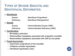
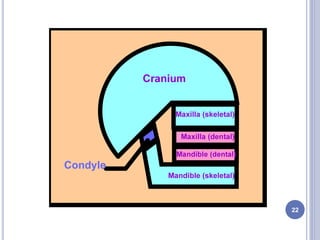
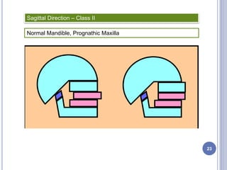
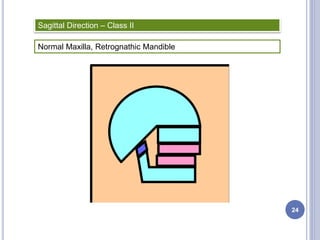
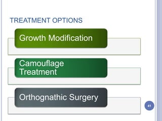
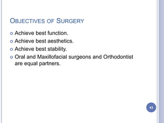
MANDIBULAR
ORTHOGNATHIC
SURGERIES
Dr. KUNAAL AGRAWAL
PG STUDENT
DEPT. OF ORTHODONTICS
GDCRI, BANGALORE
1
INTRODUCTION
 The concept of beauty is central to all
human cultures regardless of race, age and
sex and it is deeply rooted in the nature of
man.
 In various ways, human esthetics has been
woven into the tradition of human
civilization. Physical appearance has always
played a significant role in the development
of self-conceptualization and self esteem, in
the establishment of inter personal
relationship, in employment opportunities
and in quality of life. 2
WHAT IS ORTHOGNATHIC SURGERY?
 A procedure by which dento-facial deformities and
malocclusions are corrected with orthodontics
combined with the surgical modification of the facial
morphology and various soft tissue structures.
 The term orthognathic originates from the Greek
words “Orthos”, meaning straight, and “Gnathos”,
meaning jaw.
3
 As orthodontists, we look at both the occlusion and
the esthetics when evaluating the success of a
surgery. However, the best occlusion will not satisfy
a patient who is unhappy with the esthetic outcome.
 The amount of esthetic change desired by a patient
can dramatically alter the treatment plan. It can
determine the need for bimaxillary versus single-
jaw procedures and whether adjunctive procedures
such as genioplasty, rhinoplasty, or liposuction are
necessary.
4
HISTORICAL DEVELOPMENT
 Historically, the ability to reposition the mandible in a stable
manner long preceded the ability to reposition the maxilla.
As a consequence, many patients underwent only
mandibular surgery to correct a primary maxillary deformity.
 The specialty of orthognathic surgery did not fully develop
until Obwegesser demonstrated the possibility of
repositioning the maxilla in a stable consistent manner in
1965 and reported simultaneous repositioning of the
maxilla and mandible in 1970.
 Before 1960’s, the surgical correction of dentofacial
deformities was done either without patient ever having
orthodontic treatment, after orthodontic appliances had
been removed, or, occasionally before any orthodontics
was begun. 5
 Orthodontics is an essential part of modern orthognathic
surgery was stressed by the surgeon Converse and the
orthodontist Horowitz in 1969.
 At that time, rigid arch bar constructions, familiar from
trauma surgery, were used perioperatively when
needed. Technical development of orthodontic brackets
and steel rectangular wires, edgewise technique, could
give excellent and sufficiently rigid control of occlusion to
be utilized also in surgery.
 The introduction of occlusal wafer splint was an
important step in allowing surgery to occur before
orthodontic detailing of the occlusion was completed.
Consequently, the total treatment time reduced
significantly, when some type of tooth movements could
be more efficiently accomplished postoperatively. (Proffit
& White 1991).
6
MILESTONES…
 First mandibular osteotomy: HULLIHEN (1849) -
done to correct a protrusive malposition of a
mandibular alveolar segment caused by a burn
 Mandibular body osteotomy: VILRAY BLAIR (1897)
- done to correct mandibular prognathism (St. Louis
Operation)
 The beginning of the early orthognathic surgery
was in St. Louis where the orthodontist Edward
Angle and the surgeon Blair worked together.
7
8
9
10
PATIENT SELECTION
 An orthodontist must determine at an early stage
why the patient is seeking treatment and what the
patient hopes to achieve .The surgeon must then
decide whether this demand can be met surgically.
 LAVEL emphasized that satisfaction begins with
selection of appropriate patients. The selection can
be represented by the acronym SAFE.
S - Self assessment of attractiveness
A - Anxiety
F - Fear
E - Expectation 11
 Dentofacial deformity  Developmental problem
 Occasionally the deformity is due to a single
specific cause, much more frequently they result
from a complex interaction among multiple factors
that influence growth and development.
ETIOLOGIC FACTORS
ETIOLOGY
KNOWN SPECIFIC
CAUSE
HEREDITARY
FACTORS
ENVIRONMENTAL
INFLUENCES
12
13
FACIAL SYNDROMES AND
CONGENITAL
DEFECTS, WHOSE ETIOLOGY
IS PRE-NATAL
POST NATAL GROWTH
DISTURBANCES OF
KNOWN ORIGIN, INCLUDING THE
EFFECT OF TRAUMA
SPECIFIC CAUSES
FAS AND RELATED PROBLEMS
ANORMALITIES OF NEURAL REST
CELL ORIGIN AND MIGRATION :
Hemifacial microsomia
Mandibulofacial dysostosis
FACIAL CLEFTING SYNDROME
ACHONDROPLASIA
PREMATURE FUSION OF CRANIAL
AND FACIAL SUTURES:
Plagiocephaly
Crouzon’s syndrome
Apert’s syndrome
TRAUMA:
Maxillary trauma
Mandibular trauma (functional ankylosis)
MUSCLE DISTURBANCES (TORTICOLLIS)
CONDYLAR HYPERPLASIA
 Malocclusion is much more common now than it was in
primitive human populations. It seems logical that one
effect of increased intermarriage among previously
isolated population subgroups would be an increased
number of individuals requiring orthodontic–surgical
treatment.
 The influence of inherited tendencies seems to be
particularly strong for mandibular prognathism.
 Craniofacial anomalies often have a genetic
background. Recent advances in molecular genetics
have revealed a genetic explanation for conditions that
do not even appear to be genetic in origin.
HEREDITARY FACTORS
14
Craniosynostosis MSX-2
Tricho-dento-osseous syndrome DLX-3 and DLX-7
Cleidocranial dysplasia CDFA1
Treacher-Collins syndrome Long arm of chromosome 5
Holoprosencephaly HPE3
Cleft lip and palate MSX-1 and TGFB-3
Crouzon syndrome FGFR-2
15
 Environmental influences on dentofacial development
includes obvious external influences such as trauma,
but more importantly, this category includes the group of
etiologic factors related to function.
 The importance of posture in controlling soft-tissue
pressure
 The form-function interaction includes both the effects of
active movement and the subtle but long-lasting effect of
the soft tissue on the developing skeletal and dental
structures.
ENVIRONMENTAL INFLUENCES
Form Function
16
 Tongue habits, particularly tongue thrust
swallowing, have been blamed for many instances
of protrusion of incisors and anterior open bite.
 Soft tissues of the lips, cheeks and the tongue exert
pressure against the teeth and alveolar process
while the tissues are at rest as well as when they
are moving in function.
 Although these resting pressures are small in the
range of 5 to 15 grams, they are large enough to
cause tooth movement and remodeling of the
alveolar process.
17
 One characteristic of patients with the long-face
condition is that the posterior teeth erupt further than
normal. Conversely, in short face patients, the teeth are
infra-erupted.
 It seems obvious that biting force, which opposes
eruption, should be involved in its control.
 It is possible that difference in biting strength and
therefore in biting force, is involved in the etiology of
long- and short-face problems.
 The relationship between facial morphology and
occlusal forces does not prove a cause and effect
relationship.
Biting Force and Jaw Morphology
18
 3 Possibilities :
 Muscle weakness and thus low occlusal force may
allow the teeth to erupt too much and cause the
mandible to rotate down and back
 Excessive eruption of teeth may cause the
mandible to rotate down and back, putting
muscles at a mechanical disadvantage that
reduces occlusal force
 The long face pattern and the decrease in occlusal
force are both caused by something else and are
not necessarily related
19
 Mouth breathing has been blamed for altered
dentofacial development
 Harvold et al showed that total blocking of the nares led
to various moderate to severe malocclusions (AJODO,
1979-81). Because the lower jaw was positioned
forward, the deformity always included a component of
mandibular prognathism along with various
displacements of teeth.
 Effects :
 Total nasal obstruction
 Downward-backward rotation
 Long-face deformity
Respiratory Influence
20
TYPES OF SEVERE SKELETAL AND
DENTOFACIAL DEFORMITIES
 Mandibular
Excess : Mandibular Prognathism
Deficiency : Mandibular Retrognathism
 Maxilla
Excess : Vertical Maxillary Excess (VME)
Deficiency : Vertical Maxillary Deficiency (VMD)
 Combination
 Bimaxillary protrusion
 Nasomaxillary hypoplasia associated with prognathic mandible
 Nasomaxillary hypoplasia associated with cleft lip and palate
 Facial Asymmetry
 Asymmetric prognathism of the mandible
 Unilateral condylar hyperplasia
 Hemifacial hypertrophy (rare) 21
22
Sagittal Direction – Class II
Normal Mandible, Prognathic Maxilla
23
Normal Maxilla, Retrognathic Mandible
Sagittal Direction – Class II
24
Sagittal Direction – Class II
Prognathic Maxilla, Retrognathic Mandible
25
Sagittal Direction – Class III
Retrognathic Maxilla, Prognathic Mandible or a Combination of the
two
26
 Discrepancies in the vertical dimension occurs in
the form of a long face or a short face syndrome.
 The rotation of the mandible due to vertical growth
discrepancies also has to be distinguished.
 For eg., a class I skeletal case can be rotated into
class II skeletal conditions due to rapid downward
descent of the maxilla.
 Forward and backward rotation of the mandible
occur in both class II and class III skeletal
conditions.
Vertical Direction
27
 Transverse discrepancies can be of two types,
laterognathy and laterocclusion.
 Laterognathy –
 When the dental and skeletal midlines do not coincide
both at rest and in occlusion
 This condition could be due to basal skeletal
abnormalities such as a unilateral condylar hyperplasia
Transverse Direction
28
29
 Laterocclusion –
 When the midlines coincide at rest but the mandible
deviates during closure to the right or the left side
 Laterocclusion develops when the maxillary arch is
severely constricted
 On closure, since the mandibular dentition cannot be
accommodated within the maxillary arch, the mandible
deviates in a convenience bite to one side
 It is very critical to distinguish between these two
because the treatment varies depending on the
problem.
 In the former situation, a predominantly surgical
approach is required.
 In the latter, however, an expansion of the
constricted maxilla will usually take care of the
mandibular deviation. 30
31
CLINICAL FEATURES
32
1. MANDIBULAR EXCESS
 Facial Features –
1. Prominent chin is the dominant feature.
2. A concave profile.
3. Lip incompetence.
4. Obtuse gonial angle.
5. Middle third of the face appears to be deficient.
6. Labiomental fold may be diminished / absent.
7. Nasolabial angle may be acute.
8. Anterior facial height may be increased.
33
 Dental Features –
1. Angle’s class III malocclusion will be seen.
2. Reverse horizontal overjet in the incisor area.
3. Posterior cross bite.
4. Maxillary teeth may be protrusive.
5. Mandibular anterior teeth may be tilted lingually.
6. An anterior open bite may be seen.
34
2. MANDIBULAR DEFICIENCY
 Facial Features –
1. Convex profile.
2. Bird face deformity.
3. Short upper lip.
4. Everted lower lip.
5. Acute gonial angle.
6. Lip strain evident
during closure of
mouth.
35
 Dental Features –
1. Angle’s class II molar malocclusion.
2. Increased overjet.
3. Accentuated curve of Spee of lower anterior.
4. Fanning of lower anterior teeth or crowding.
5. Skeletal deep bite may be present.
36
3. CONDITIONS WITH FACIAL ASYMMETRY
 Asymmetrical mandibular prognathism
 With anterior open bite
 Without anterior open bite
 Unilateral condylar hyperplasia
 Hemimandibular elongation
 Hemimandibular hypoplasia
 Hemifacial hypertrophy (rare)
37
 With anterior open bite –
 Severe facial asymmetry
 Eccentric bilateral mandibular protrusion.
 Deviation of the chin.
 High gonial angle.
 Midline of mandibular arch shifted.
 Without anterior open bite
 Eccentric bilateral mandibular protrusion.
 Deviation of chin.
 Class III dental malocclusion.
 Associated mandibular hypoplasia.
Asymmetrical Mandibular Prognathism
38
 Hemimandibular Elongation –
 Horizontal displacement of mandible & chin to
unaffected side.
 Lateral crossbite on unaffected side.
 Occlusal plane slopes upward to the unaffected
side.
 Sever cases – Lateral open bite on the affected
side.
 IOPA, OPG – Elongation of the condyle.
 Hemimandibular Hyperplasia –
 One side of face enlarged.
 Unilateral bowing of inf. Border of mandible.
 Lip line slopes downward on affected side.
 Associated TMJ pain symptoms on the affected
side.
 RADIOLOGICALLY – Enlarged hemimandible on
the affected side.
39
Unilateral Condylar Hyperplasia
ASSOCIATED PROBLEM LIST
 Esthetic problem.
 Functional problems.
 Psychological problems.
 Impairment of mastication.
 Associated speech problems.
 Susceptibility to caries and periodontal problems.
 Possible TMJ joint pain dysfunction.
 Impact on digestion – general health.
40
TREATMENT OPTIONS
Growth Modification
Camouflage
Treatment
Orthognathic Surgery
41
 Indications –
 Moderate basal bone discrepancy.
 Double jaw involvement in the discrepancy; that is, 50 % of
the skeletal problem is due to maxillary antero-posterior
excess, and the remaining 50% of the anteroposterior
problem is related to mandibular deficiency.
 Adequate alveolar bone and gingiva for incisor
reangulation.
 Contraindications –
 When single jaw imbalances are severe.
 Skeletal Class II cases with maxillary and mandibular
deficiencies (relative to nasal superimposition).
42
Camouflage Treatment
OBJECTIVES OF SURGERY
 Achieve best function.
 Achieve best aesthetics.
 Achieve best stability.
 Oral and Maxillofacial surgeons and Orthodontist
are equal partners.
43
GOALS OF SURGERY
 Produce a concise list of patient’s problems.
 Synthesize the various treatment possibilities into a
rational plan that gives maximum benefit to patient.
44
TIMING OF SURGERY
 Can be done only when the patient is in actively
growing stage.
 Must be warned about the second surgery later on.
 Best timing is when the growth potential of
patient is over.
45
SEQUENTIAL STEPS
UNDERTAKEN IN AN
ORTHODONTIC-SURGICAL
CASE
46
1. Pre-treatment records
2. Multidisciplinary diagnosis and
treatment planning
3. Presurgical decompensation
4. Presurgical records
5. Cephalometric prediction tracing
6. Model surgery and construction of
surgical splint
7. Surgical procedure
8. Postsurgical orthodontics 47
1. Personal data
2. Facial esthetic analysis
3. Lateral cephalometric analysis
4. Occlusal and model analysis
1. Dental arch form
2. Dental alignment
3. Dental occlusion
4. Tooth mass relation
5. Final treatment plan
1. Presurgical orthodontics
2. Surgery plan
3. Postsurgical orthodontics
4. Maintenance
48
49
PRE-SURGICAL DATABASE
 Primary components of the pre surgical database are the
clinical examination, lateral and anteroposterior cephalometric
radiographs, and the articulator-mounted models.
 Secondary components are panoramic and periapical
radiographs and facial and intraoral photographs.
 Functional problems related to the patient -
Temporomandibular joints and malocclusion are recorded.
 In the frontal plane, the clinician establishes, a clinical midline
and integrates diagnostic information in relation to that
reference line. Upper lip support is an important clinical
feature. An assessment of upper lip support will provide the
clinician with information regarding the proper antero-posterior
position of maxilla relative to facial soft tissues. 50
SYSTEMATIC PATIENT EVALUATION
1. General patient evaluation
* Medical history, Dental evaluation
2. Social and Psychologic evaluation
3. Esthetic facial evaluation
* Front face analysis and Profile analysis
4. Cephalometric evaluation
* Soft tissue, Skeletal relations, Dental relations
5. Panoramic and full mouth periapical evaluations
6. Occlusal evaluation
* Functional (Dynamic) and Static
7. Masticatory muscle and TMJ evaluation
* Masticatory muscles and Mandibular movements
* TMJ signs & symptoms
51
52
53
DIAGNOSIS
54
55
CEPHALOMETRIC PREDICTION TRACING
Why do prediction tracings for mandibular surgery?
1) to accurately assess the profile esthetic results which
will result from the proposed surgery
2) to consider the desirability of simultaneous adjunctive
procedures such as genioplasty, suprahyoid myotomy,
etc.
3) to help determine the sequencing of surgery and
orthodontics
4) to help decide what type of orthodontics might best be
employed (i.e., extraction versus non-extraction)
5) to determine the anchorage requirements should
extraction treatment be chosen
56
Epker and Fish, JCO 1980
 Bell, Profitt and White (1980), advocated use of the
cephalometric prediction with templates to double check
the model surgery changes, to predict changes in bony
relationships not seen on the dental casts, and to predict
soft tissue changes.
a. Prediction analysis presents a simple and accurate
method of predicting results of surgical orthodontic
treatment.
b. Quantification of the surgical movements necessary to
correct the deformity is also possible.
c. It can also accurately predict the resultant facial profile.
d. Provides a visual aid with a single overlay.
e. It can be also used for comparing with actual
postsurgical cephalometric tracings for re-evaluating the
surgical results.
57
 When making a prediction tracing, the planning surgeon
and orthodontist will manipulate two-dimensional
drawings, or “cut outs”, of the maxilla or mandible or,
both.
 In the case of isolated mandibular surgery, the distal
mandible is positioned in its estimated post surgical
position with the maxilla.
 In isolated maxillary surgery, the estimated occlusal
relationship between the maxilla and mandible is
established, and the maxilla is auto rotated on
mandibular tracings, around the hinge axis of the
mandible, to its planned vertical position.
 In two-jaw surgery, the surgeon first places the maxilla in
its new position and then brings the mandible to it.
58
Predicting lower lip and chin response to mandibular
advancement and genioplasty
Trevor & Peter H. Buschang, AJODO Dec ’02
This retrospective study were to examine the soft tissue
changes associated with mandibular advancement and
genioplasty and to develop predictive models.
Longitudinal lateral cephalograms of 62 non-growing
patients (27 men and 35 women) were taken in centric
relation with the lips in repose within 4 weeks before
surgery and at least 6 months postoperatively.
The mandibular incisor and pogonion were advanced
surgically approximately 6 mm and 11 mm, respectively.
The lower lip lengthened slightly (2.5 - 3.8 mm), and its
surface contour straightened because of thinning at
labrale inferioris (2.8 - 2.0 mm); there was a slight
thickening at the labiomental fold (1.0 - 2.3 mm) and a
slight thinning at soft tissue pogonion (0.8 - 2.2 mm). 59
Multiple regression models showed that soft tissue
response to advancement surgery depended on
pretreatment tissue thickness, horizontal skeletal
movement, vertical skeletal movement, and the position
of the maxillary incisors.
Lines and Steinhauser, who were among the first to
attempt prediction in this area, concluded that the lower
lip advanced at a 0.66:1 ratio to the mandibular incisor
advancement, and soft tissue pogonion advanced at a
simple 1:1 ratio to hard tissue pogonion advancement.
Other studies have confirmed the 1:1 ratio for soft tissue
pogonion, but predictive ratios for the lower lip have
been highly variable, ranging from 0.26:1 to 0.85:1.
60
Mounting dental models for surgery
The use of anatomic articulator in treatment planning
allows the manipulation of the maxillary and mandibular
models in three planes of space within the articulator.
When the models are correctly mounted, this
manipulation will be analogous to surgical movements of
the jaws within the facial skeleton.
An anatomic articulator is the 3 dimensional analogue of
the 2 dimensional cephalometric radiograph. Currently,
work is being done on correlating model surgery
movements with computerized cephalometric analysis
and prediction tracing programs.
61
62
 In mounting dental models on an anatomic articulator,
the purpose of any face bow transfer procedure is to
reproduce accurately the functional and spatial
relationship of the jaws.
 With a hinge-axis face bow transfer, special techniques
are used to ensure that the intercondylar axis of the
patient coincides with the intercondylar axis of the
articulator.
 Some clinicians would argue that a hinge-axis transfer
should be done in all cases of total maxillary surgery,
whether isolated or in combination with mandibular
surgery.
 The greater vertical change (vertical maxillary change
and increased mandibular autorotation), the more
important a hinge-axis mounting becomes.
63
 Mount the maxillary model as close as possible to true
natural horizontal plane (HP; HP = FH). This practice will
allow a common reference plane for interrelating the
mounted models with the cephalometric prediction
tracing.
 The mandibular cast must always be mounted to
(related to) the maxillary cast, with attention paid to the
position of the mandibular condyles.
 A centric occlusion position - a solely tooth-dictated
position is frequently habitual rather than anatomic and
is insufficient. When mounting the mandibular model, a
wax-bite registration is taken that relates the mandible to
the maxilla independent of the occlusion.
64
Orthognathic Splint Construction
 A true hinge facebow recording is taken with a SAM
Axiograph.
 A set of models mounted on a SAM II Articulator, using
the Axiograph facebow axis recording, using accurate
centric relation interocclusal registration.
 Centric relation is recorded with the "Roth Power Centric
Technique", which relies on the patient's own jaw-
seating musculature to help seat and center the
condyle-disc assemblies in their respective fossae.
 Delar blue wax strips are used for bite registration.
65
PREDICTING SURGICAL OUTCOMES
 There are five general methods of visualizing, planning,
and predicting surgical orthodontic outcomes (AJO 1997
Dec):
 Manual acetate tracing "cut and paste“ techniques as
described by Cohen, McNeill et al., and Henderson.
 Manipulation of patient photographs to illustrate treatment
goals.
 Computerized diagnostic and planning software that produces
a soft tissue profile "line drawing"; as a result of manipulation
of digitized structures of lateral cephalometric radiographs.
 Computerized diagnostic and planning software that
integrates video images with the patient's lateral cephalogram
to aid in planning and predicting surgical orthodontic
procedures (Videocephalometrics).
 Three-dimensional computer technology for planning and
predicting orthognathic surgery.
66
PREDICTION CEPHALOMETRIC
TRACING
 The most important aim of the prediction tracing is
to asses the esthetic profile result after the surgery.
 4 types :
 TRACING OVERLAY METHOD
 TEMPLATE METHOD
 PHOTOGRAPHIC METHOD
 COMPUTER METHOD
67
1. TRACING OVERLAY METHOD
 The tracing overlay approach is the simplest way
to simulate the effects of the mandibular surgery.
 The final prediction tracing is produced without
any intermediate tracings.
 This method is limited to surgery that does not
affect the vertical position of the maxilla (i.e., the
mandible does not rotate around the condylar axis).
68
69
Original tracing Tracing of the structures that will not
be changed by mandibular surgery
70
Slide overlay tracing so that the mandibular teeth can be
seen through it in the desired post-surgical position and
trace the lower teeth and the jaw
71
Measurements are made to find how far the lower incisor
has moved forward by superimposing the overlay back to
the cranial base
(lower lip will move forward by 2/3rd and is marked)
72
Superimpose again on the mandible. Draw the soft tissue
chin and complete the soft tissue profile
73
Superimpose again on the cranial base and complete the
soft tissue profile with the help of table
2. TEMPLATE METHOD
 Templates can be used for any type of prediction
surgery.
 Disadvantage – Time consuming.
 The use of templates for intermediate tracings
between the original and the final tracing is
mandatory when the maxilla will be repositioned
vertically, repositioning of the chin and in cases
where major teeth movements have to be carried
out.
74
 Special considerations –
1. Color coding of templates.
2. Use of different colors for the structures to be
repositioned.
3. When mandibular template is prepared, the approximate
center of the condyle on the original tracing should be
marked, and this mark is transferred to the template.
The mandibular template can be rotated around this
point.
75
76
Template for maxillary teeth
77
Two mandibular templates are prepared – one without
extraction and second one with extraction (crowding
resolved)
78Ready templates
79
Place the upper anterior template in the desired position
approximately 2 mm below the lip line
80
Check for the better fit of the mandibular teeth by placing
either of mandibular templates. It is clear that the
prominence of the upper anterior teeth will be a function of
how much the mandibular incisors are retracted and how far
up the maxilla is moved
81
Position the upper posterior template
82
Complete the prediction tracing on a fresh tracing paper
3. PHOTOGRAPHIC METHOD
 Is an attempt to improve communication with
patients. This was proposed, as a method of
illustrating to the patient, the soft-tissue results of
the suggested plan.
 Method - The photographs are physically sectioned;
the cut-outs represent the parts that will be moved
in the planned osteotomies and are arranged to
simulate surgical movements.
 Advantages - It gives the patient a better
visualization of the profile changes than a acetate
tracing does.
83
 Disadvantages –
1.Does not permit change to soft
tissue contours that occurs
with treatment.
2.Unavoidable gaps in photo
have an unnatural
appearance.
3.An experienced clinician with
artistic skills is essential with
this methodology.
84
4. COMPUTER METHOD
 The first step in using a computer program for
cephalometric prediction is to enter the digital
model of the patient’s tracing in to computer
memory.
 Rocky mountain data systems has developed the
computerized “visual norm” based on the size, age,
sex and race. (JCO 1977)
 Using this data and Broadbent’s template method,
surgical VTO can be constructed.
85
86
87
The final red surgical VTO is prepared showing the skeletal,
dental, and soft tissue changes
VIDEOCEPHALOMETRY
 Video imaging technology is a method in which orthodontist
gathers facial frontal, profile, and dental images and modify
them to project potential esthetic treatment goals (David M.
Sarver).
 Video cephalometric prediction methodology is virtually
identical to the cephalometric tracing method.
 Hence the difficulties encountered is similar to the tracing
method (except the improved visualization and recognition of
facial profile changes).
 Video cephalometry technique helps in quantifying treatment
plans. In other words, co-ordination of calibrated profile
images with facial profile images permits precise
measurement of bony and dental movements, and through the
application of algorithmic prediction ratios, images are
produced that express the expected surgical and/or
orthodontic outcome.
88
89
90
91
Surgical predictions for a patient with skeletal Class II problem, with mild maxillary retrusion, severe
mandibular retrotrusion and inadequate projection of the chin. A, The initial tracing linked to the profile
photograph. B, Simulation of 5 mm mandibular advancement, an amount of advancement
corresponding to the initial overjet. C, 5 mm mandibular advancement plus genioplasty to improve chin
projection relative to the lower incisor. D, 6 mm maxilla advancement, 11 mm mandibular advancement,
and rhinoplasty. The maxilla was advanced to increase support of upper lip and allow for greater
mandibular advancement. E, Presurgical retraction of the lower incisors after lower premolar
extraction, creating more overjet and allowing a 9 mm mandibular advancement. F, Retraction of lower
incisors, 9 mm advancement, and rhinoplasty. The rhinoplasty changes were subtle, and it was not
recommended. After the patient and her parents viewed the simulations and discussed it with the
orthodontist, E was selected as the plan.
MODEL SURGERY
 Model surgery simulates actual surgery, in the
dental arch models of the patient. It gives the three
dimensional understanding of the post operative
relationship of the jaws.
 Aims –
1.To get the definite idea about the extent of bone / arch
advancement or reduction required in the surgery.
2.To get a post-operative relationship of the jaws,
dentition and occlusion.
3.To decide about the post-surgical orthodontic treatment.
4.As a vehicle for fabrication of splints for stabilization
after surgery.
92
DIAGNOSTIC SETUP
 A diagnostic set up is employed to be sure that it
will be possible to get the teeth to fit together if a
given orthodontic treatment plan is employed.
 Individually remove the teeth from the dental casts
and reset the teeth in soft wax so that their
alignment and interdigitation can be observed.
93
94Diagnostic pre-orthodontic set-up showing the proposed
extractions and tooth movements
95
96
97
B
A
98Marked models with the recorded distances
Vm
VbVc
Vm
Vb
Vc
99
Transverse changes are recorded by the inter-canine and
inter-molar distances measured across the palate and
recorded by taking reference points on the canine tips and
the mesiobuccal cusp of the first molars
100
Interrupted line is the proposed osteotomy site
101
Maxilla is reassembled with the wax after the osteotomy
cuts. Mandible closes in to the intermediate occlusal
relationship
Intermediate wafer is made at this stage
102
Lower segmental set-down of 3mm is carried out with the
forward slide of 5mm to correct the inter-arch occlusal
relationship
103
Anterior view: models showing the upper midline split to
widen the intercanine width and the lower anterior set-down
MANDIBULAR SURGERIES
Mandibular Ramus
Osteotomies
Mandibular Body
Osteotomies
104
105
SOFT TISSUE INCISIONS
 Mandibular body surgeries - Degloving vestibular
incision intraorally.
 Extraoral ramus osteotomies – submandibular
Ridson’s incision and postramal Hind’s incision.
 Intraoral ramus osteotomies – incision similar to 3rd
molar extraction.
106
1. ANTERIOR BODY OSTEOTOMY
 Indications –
 Mandibular prognathism with functional posterior
occlusion
 Class III malocculsion with or without anterior open bite
where the posterior teeth cross bite is dental in nature
(can be corrected by orthodontics).
107
2. POSTERIOR BODY OSTEOTOMY
 Indications –
 Missing Posterior teeth
 Class III deformity
 For correction of Cross Bite
108
3. MIDSYMPHYSEAL OSTEOTOMY
 The complete vestibular incision can be planned if it
is combined with posterior or anterior body
osteotomy.
109
110
The surgical movements that are possible in the transverse dimension are
shown on this posteroanterior illustration of the skull. The solid arrows
indicate that the maxilla can be expanded laterally or constricted with
reasonable stability. The smaller size of the arrows pointing to the midline
represents the fact that the amount of constriction possible is somewhat
less than the range of expansion. The only transverse movement easily
achieved in the mandible is constriction, although limited expansion now is
possible with distraction osteogenesis.
111
4. SEGMENTAL SUBAPICAL MANDIBULAR
SURGERIES
 Used to reposition anterior, posterior or the entire
mandibular dentoalveolar segment
 Ant. Subapical mandibular osteotomy
 Post. Subapical mandibular osteotomy
 Total Subapical mandibular osteotomy
112
4.1. ANTERIOR SUBAPICAL MANDIBULAR
OSTEOTOMY
 Indications –
 Correcting mandibular dentoalveolar proclination
 Closing mild anterior open bite
 Leveling an accentuated curve of Spee
 Correcting mandibular dental arch asymmetry
 Used as an adjunctive with other surgical
procedures –
 With anterior maxillary osteotomy to correct bimaxillary protrusion
 With mandibular advancement to level the curve of Spee
 With genioplasty procedure
113
114
115
116
4.2. POSTERIOR SUBAPICAL MANDIBULAR
OSTEOTOMY
 Indications –
 Uprighting the posterior segment which is in extreme
linguoversion or buccoversion
 Closing a premolar or molar space
 Levelling supraerupted posterior teeth
117
118
119
120
121
4.3. TOTAL SUBAPICAL MANDIBULAR
OSTEOTOMY
 Indications –
 It can be used to reposition entire mandibular
dentoalveolar segment anteriorly, posteriorly or
superiorly.
 For lengthening of lower third of the face or for
advancing the mandibular dentoalveolar segment, etc.
 However, it is not performed routinely due to high
chances of damage to neurovascular bundles.
122
123
124
125
5. GENIOPLASTIES
 Genioplasty can be used as a
single procedure or it can be used
as an adjunctive procedure along
with other major osteotomies of
the jaw bone.
 Deformities of the chin should be
considered in all 3 planes,
 AP
 Vertical
 Transverse
 It can be used to augment,
reduce, straighten or lengthen the
chin. 126
The chin can be sectioned anterior to the mental foramen and
repositioned in all three planes of space. The lingual surface remains
attached to muscles in the floor of the mouth, which provide the
blood supply. Moving the chin anteriorly, upward, or laterally usually
produces highly favorable esthetic results. Moving it back or down
may produce a “boxy” appearance. 127
5.1. AUGMENTATION GENIOPLASTY
 Used to increase the chin projection.
 Sliding horizontal osteotomy of the symphysis region.
 Autogenous bone graft.
 Alloplastic material – silastic, hydroxyapatite.
128
129
5.2. REDUCTION GENIOPLASTY
 Reduction of the symphysis region can be achieved
both in the anteroposterior and vertical planes or in
both planes depending on the need of the patient.
130
131
132
133
134
5.3. STRAIGHTENING GENIOPLASTY
 Indications –
 In facial asymmetry, where the complete correction of
the asymmetry cannot be achieved by appropriate jaw
osteotomies. Eg, TM joint ankylosis.
 The horizontal osteotomy is done and segment is
shifted laterally and then contoured to get desired
result.
135
5.4. LENGTHENING GENIOPLASTY
 Indications –
 Indicated in patients with short vertical facial height with
Class I and Class II deep bites.
 After horizontal osteotomy, the osteotomized
segment is pushed inferiorly and bone graft is
sandwiched in between to increase the height.
136
6. SUBCONDYLAR VERTICAL OSTEOTOMY
 It was proposed by Caldwell – Letterman in 1954.
 The indications for extraoral subsigmoid vertical
ramus osteotomy are,
 Major setback of mandible more than 10 mm.
 Asymmetric setback of the mandible.
 Reoperation of previously operated case.
137
138
139
140
141
142
7. INTRAORAL MODIFIED SAGITTAL SPLIT
OSTEOTOMY
 Also called as - Bilateral Sagittal Split Osteotomy
 It is performed on the mandibular ramus and body.
 First described by Obwegesser and Trauner and later
modified by Dal Pont, Hunsuck and Epker.
 Transoral incision, similar to that used for IVRO.
 The osteotomy splits the ramus & the posterior body of the
mandible sagittally, which allows either setback or
advancement.
 This is a highly cosmetic procedure, as it is done intraorally,
plus, there is broader bony contact of the osteotomised
segments, ensuring good healing.
Drawback:
* High level of operative skill is required.
* Experience to minimize the surgical complication.
143
144
145
146
147
148
149
150
151
152
153
154
155
156
157
158
159
160
161
162
163
CONCLUSIONS
164
165
The maxilla and mandible can be moved anteriorly and posteriorly as indicated by the
red arrows in these line drawings. Anterior movements of the mandible greater than
approximately 10 mm create considerable tension in the investing soft tissues and
tend to be unstable. Anterior movement of the maxilla is similarly limited to 6 to 8 mm
in most circumstances—the possibility of relapse or speech alteration from
nasopharyngeal incompetence increases with larger movements. Posterior movement
of the entire maxilla, though possible, is difficult and usually unnecessary. Instead,
posterior movement of protruding incisors up to the width of a premolar is
accomplished by removal of a premolar tooth on each side, followed by segmentation
of the maxilla. The major limitation of posterior movement of the mandible is its effect
on the appearance of the throat. When the mandible is moved back, the tongue moves
down as the airway is maintained, and a “turkey gobbler” prominence appears below
the chin
BIBLIOGRAPHY
166
Mandibular orthognathic surgeries

Mandibular orthognathic surgeries

  • 1.
    MANDIBULAR ORTHOGNATHIC SURGERIES Dr. KUNAAL AGRAWAL PGSTUDENT DEPT. OF ORTHODONTICS GDCRI, BANGALORE 1
  • 2.
    INTRODUCTION  The conceptof beauty is central to all human cultures regardless of race, age and sex and it is deeply rooted in the nature of man.  In various ways, human esthetics has been woven into the tradition of human civilization. Physical appearance has always played a significant role in the development of self-conceptualization and self esteem, in the establishment of inter personal relationship, in employment opportunities and in quality of life. 2
  • 3.
    WHAT IS ORTHOGNATHICSURGERY?  A procedure by which dento-facial deformities and malocclusions are corrected with orthodontics combined with the surgical modification of the facial morphology and various soft tissue structures.  The term orthognathic originates from the Greek words “Orthos”, meaning straight, and “Gnathos”, meaning jaw. 3
  • 4.
     As orthodontists,we look at both the occlusion and the esthetics when evaluating the success of a surgery. However, the best occlusion will not satisfy a patient who is unhappy with the esthetic outcome.  The amount of esthetic change desired by a patient can dramatically alter the treatment plan. It can determine the need for bimaxillary versus single- jaw procedures and whether adjunctive procedures such as genioplasty, rhinoplasty, or liposuction are necessary. 4
  • 5.
    HISTORICAL DEVELOPMENT  Historically,the ability to reposition the mandible in a stable manner long preceded the ability to reposition the maxilla. As a consequence, many patients underwent only mandibular surgery to correct a primary maxillary deformity.  The specialty of orthognathic surgery did not fully develop until Obwegesser demonstrated the possibility of repositioning the maxilla in a stable consistent manner in 1965 and reported simultaneous repositioning of the maxilla and mandible in 1970.  Before 1960’s, the surgical correction of dentofacial deformities was done either without patient ever having orthodontic treatment, after orthodontic appliances had been removed, or, occasionally before any orthodontics was begun. 5
  • 6.
     Orthodontics isan essential part of modern orthognathic surgery was stressed by the surgeon Converse and the orthodontist Horowitz in 1969.  At that time, rigid arch bar constructions, familiar from trauma surgery, were used perioperatively when needed. Technical development of orthodontic brackets and steel rectangular wires, edgewise technique, could give excellent and sufficiently rigid control of occlusion to be utilized also in surgery.  The introduction of occlusal wafer splint was an important step in allowing surgery to occur before orthodontic detailing of the occlusion was completed. Consequently, the total treatment time reduced significantly, when some type of tooth movements could be more efficiently accomplished postoperatively. (Proffit & White 1991). 6
  • 7.
    MILESTONES…  First mandibularosteotomy: HULLIHEN (1849) - done to correct a protrusive malposition of a mandibular alveolar segment caused by a burn  Mandibular body osteotomy: VILRAY BLAIR (1897) - done to correct mandibular prognathism (St. Louis Operation)  The beginning of the early orthognathic surgery was in St. Louis where the orthodontist Edward Angle and the surgeon Blair worked together. 7
  • 8.
  • 9.
  • 10.
  • 11.
    PATIENT SELECTION  Anorthodontist must determine at an early stage why the patient is seeking treatment and what the patient hopes to achieve .The surgeon must then decide whether this demand can be met surgically.  LAVEL emphasized that satisfaction begins with selection of appropriate patients. The selection can be represented by the acronym SAFE. S - Self assessment of attractiveness A - Anxiety F - Fear E - Expectation 11
  • 12.
     Dentofacial deformity Developmental problem  Occasionally the deformity is due to a single specific cause, much more frequently they result from a complex interaction among multiple factors that influence growth and development. ETIOLOGIC FACTORS ETIOLOGY KNOWN SPECIFIC CAUSE HEREDITARY FACTORS ENVIRONMENTAL INFLUENCES 12
  • 13.
    13 FACIAL SYNDROMES AND CONGENITAL DEFECTS,WHOSE ETIOLOGY IS PRE-NATAL POST NATAL GROWTH DISTURBANCES OF KNOWN ORIGIN, INCLUDING THE EFFECT OF TRAUMA SPECIFIC CAUSES FAS AND RELATED PROBLEMS ANORMALITIES OF NEURAL REST CELL ORIGIN AND MIGRATION : Hemifacial microsomia Mandibulofacial dysostosis FACIAL CLEFTING SYNDROME ACHONDROPLASIA PREMATURE FUSION OF CRANIAL AND FACIAL SUTURES: Plagiocephaly Crouzon’s syndrome Apert’s syndrome TRAUMA: Maxillary trauma Mandibular trauma (functional ankylosis) MUSCLE DISTURBANCES (TORTICOLLIS) CONDYLAR HYPERPLASIA
  • 14.
     Malocclusion ismuch more common now than it was in primitive human populations. It seems logical that one effect of increased intermarriage among previously isolated population subgroups would be an increased number of individuals requiring orthodontic–surgical treatment.  The influence of inherited tendencies seems to be particularly strong for mandibular prognathism.  Craniofacial anomalies often have a genetic background. Recent advances in molecular genetics have revealed a genetic explanation for conditions that do not even appear to be genetic in origin. HEREDITARY FACTORS 14
  • 15.
    Craniosynostosis MSX-2 Tricho-dento-osseous syndromeDLX-3 and DLX-7 Cleidocranial dysplasia CDFA1 Treacher-Collins syndrome Long arm of chromosome 5 Holoprosencephaly HPE3 Cleft lip and palate MSX-1 and TGFB-3 Crouzon syndrome FGFR-2 15
  • 16.
     Environmental influenceson dentofacial development includes obvious external influences such as trauma, but more importantly, this category includes the group of etiologic factors related to function.  The importance of posture in controlling soft-tissue pressure  The form-function interaction includes both the effects of active movement and the subtle but long-lasting effect of the soft tissue on the developing skeletal and dental structures. ENVIRONMENTAL INFLUENCES Form Function 16
  • 17.
     Tongue habits,particularly tongue thrust swallowing, have been blamed for many instances of protrusion of incisors and anterior open bite.  Soft tissues of the lips, cheeks and the tongue exert pressure against the teeth and alveolar process while the tissues are at rest as well as when they are moving in function.  Although these resting pressures are small in the range of 5 to 15 grams, they are large enough to cause tooth movement and remodeling of the alveolar process. 17
  • 18.
     One characteristicof patients with the long-face condition is that the posterior teeth erupt further than normal. Conversely, in short face patients, the teeth are infra-erupted.  It seems obvious that biting force, which opposes eruption, should be involved in its control.  It is possible that difference in biting strength and therefore in biting force, is involved in the etiology of long- and short-face problems.  The relationship between facial morphology and occlusal forces does not prove a cause and effect relationship. Biting Force and Jaw Morphology 18
  • 19.
     3 Possibilities:  Muscle weakness and thus low occlusal force may allow the teeth to erupt too much and cause the mandible to rotate down and back  Excessive eruption of teeth may cause the mandible to rotate down and back, putting muscles at a mechanical disadvantage that reduces occlusal force  The long face pattern and the decrease in occlusal force are both caused by something else and are not necessarily related 19
  • 20.
     Mouth breathinghas been blamed for altered dentofacial development  Harvold et al showed that total blocking of the nares led to various moderate to severe malocclusions (AJODO, 1979-81). Because the lower jaw was positioned forward, the deformity always included a component of mandibular prognathism along with various displacements of teeth.  Effects :  Total nasal obstruction  Downward-backward rotation  Long-face deformity Respiratory Influence 20
  • 21.
    TYPES OF SEVERESKELETAL AND DENTOFACIAL DEFORMITIES  Mandibular Excess : Mandibular Prognathism Deficiency : Mandibular Retrognathism  Maxilla Excess : Vertical Maxillary Excess (VME) Deficiency : Vertical Maxillary Deficiency (VMD)  Combination  Bimaxillary protrusion  Nasomaxillary hypoplasia associated with prognathic mandible  Nasomaxillary hypoplasia associated with cleft lip and palate  Facial Asymmetry  Asymmetric prognathism of the mandible  Unilateral condylar hyperplasia  Hemifacial hypertrophy (rare) 21
  • 22.
  • 23.
    Sagittal Direction –Class II Normal Mandible, Prognathic Maxilla 23
  • 24.
    Normal Maxilla, RetrognathicMandible Sagittal Direction – Class II 24
  • 25.
    Sagittal Direction –Class II Prognathic Maxilla, Retrognathic Mandible 25
  • 26.
    Sagittal Direction –Class III Retrognathic Maxilla, Prognathic Mandible or a Combination of the two 26
  • 27.
     Discrepancies inthe vertical dimension occurs in the form of a long face or a short face syndrome.  The rotation of the mandible due to vertical growth discrepancies also has to be distinguished.  For eg., a class I skeletal case can be rotated into class II skeletal conditions due to rapid downward descent of the maxilla.  Forward and backward rotation of the mandible occur in both class II and class III skeletal conditions. Vertical Direction 27
  • 28.
     Transverse discrepanciescan be of two types, laterognathy and laterocclusion.  Laterognathy –  When the dental and skeletal midlines do not coincide both at rest and in occlusion  This condition could be due to basal skeletal abnormalities such as a unilateral condylar hyperplasia Transverse Direction 28
  • 29.
  • 30.
     Laterocclusion – When the midlines coincide at rest but the mandible deviates during closure to the right or the left side  Laterocclusion develops when the maxillary arch is severely constricted  On closure, since the mandibular dentition cannot be accommodated within the maxillary arch, the mandible deviates in a convenience bite to one side  It is very critical to distinguish between these two because the treatment varies depending on the problem.  In the former situation, a predominantly surgical approach is required.  In the latter, however, an expansion of the constricted maxilla will usually take care of the mandibular deviation. 30
  • 31.
  • 32.
  • 33.
    1. MANDIBULAR EXCESS Facial Features – 1. Prominent chin is the dominant feature. 2. A concave profile. 3. Lip incompetence. 4. Obtuse gonial angle. 5. Middle third of the face appears to be deficient. 6. Labiomental fold may be diminished / absent. 7. Nasolabial angle may be acute. 8. Anterior facial height may be increased. 33
  • 34.
     Dental Features– 1. Angle’s class III malocclusion will be seen. 2. Reverse horizontal overjet in the incisor area. 3. Posterior cross bite. 4. Maxillary teeth may be protrusive. 5. Mandibular anterior teeth may be tilted lingually. 6. An anterior open bite may be seen. 34
  • 35.
    2. MANDIBULAR DEFICIENCY Facial Features – 1. Convex profile. 2. Bird face deformity. 3. Short upper lip. 4. Everted lower lip. 5. Acute gonial angle. 6. Lip strain evident during closure of mouth. 35
  • 36.
     Dental Features– 1. Angle’s class II molar malocclusion. 2. Increased overjet. 3. Accentuated curve of Spee of lower anterior. 4. Fanning of lower anterior teeth or crowding. 5. Skeletal deep bite may be present. 36
  • 37.
    3. CONDITIONS WITHFACIAL ASYMMETRY  Asymmetrical mandibular prognathism  With anterior open bite  Without anterior open bite  Unilateral condylar hyperplasia  Hemimandibular elongation  Hemimandibular hypoplasia  Hemifacial hypertrophy (rare) 37
  • 38.
     With anterioropen bite –  Severe facial asymmetry  Eccentric bilateral mandibular protrusion.  Deviation of the chin.  High gonial angle.  Midline of mandibular arch shifted.  Without anterior open bite  Eccentric bilateral mandibular protrusion.  Deviation of chin.  Class III dental malocclusion.  Associated mandibular hypoplasia. Asymmetrical Mandibular Prognathism 38
  • 39.
     Hemimandibular Elongation–  Horizontal displacement of mandible & chin to unaffected side.  Lateral crossbite on unaffected side.  Occlusal plane slopes upward to the unaffected side.  Sever cases – Lateral open bite on the affected side.  IOPA, OPG – Elongation of the condyle.  Hemimandibular Hyperplasia –  One side of face enlarged.  Unilateral bowing of inf. Border of mandible.  Lip line slopes downward on affected side.  Associated TMJ pain symptoms on the affected side.  RADIOLOGICALLY – Enlarged hemimandible on the affected side. 39 Unilateral Condylar Hyperplasia
  • 40.
    ASSOCIATED PROBLEM LIST Esthetic problem.  Functional problems.  Psychological problems.  Impairment of mastication.  Associated speech problems.  Susceptibility to caries and periodontal problems.  Possible TMJ joint pain dysfunction.  Impact on digestion – general health. 40
  • 41.
  • 42.
     Indications – Moderate basal bone discrepancy.  Double jaw involvement in the discrepancy; that is, 50 % of the skeletal problem is due to maxillary antero-posterior excess, and the remaining 50% of the anteroposterior problem is related to mandibular deficiency.  Adequate alveolar bone and gingiva for incisor reangulation.  Contraindications –  When single jaw imbalances are severe.  Skeletal Class II cases with maxillary and mandibular deficiencies (relative to nasal superimposition). 42 Camouflage Treatment
  • 43.
    OBJECTIVES OF SURGERY Achieve best function.  Achieve best aesthetics.  Achieve best stability.  Oral and Maxillofacial surgeons and Orthodontist are equal partners. 43
  • 44.
    GOALS OF SURGERY Produce a concise list of patient’s problems.  Synthesize the various treatment possibilities into a rational plan that gives maximum benefit to patient. 44
  • 45.
    TIMING OF SURGERY Can be done only when the patient is in actively growing stage.  Must be warned about the second surgery later on.  Best timing is when the growth potential of patient is over. 45
  • 46.
    SEQUENTIAL STEPS UNDERTAKEN INAN ORTHODONTIC-SURGICAL CASE 46
  • 47.
    1. Pre-treatment records 2.Multidisciplinary diagnosis and treatment planning 3. Presurgical decompensation 4. Presurgical records 5. Cephalometric prediction tracing 6. Model surgery and construction of surgical splint 7. Surgical procedure 8. Postsurgical orthodontics 47
  • 48.
    1. Personal data 2.Facial esthetic analysis 3. Lateral cephalometric analysis 4. Occlusal and model analysis 1. Dental arch form 2. Dental alignment 3. Dental occlusion 4. Tooth mass relation 5. Final treatment plan 1. Presurgical orthodontics 2. Surgery plan 3. Postsurgical orthodontics 4. Maintenance 48
  • 49.
  • 50.
    PRE-SURGICAL DATABASE  Primarycomponents of the pre surgical database are the clinical examination, lateral and anteroposterior cephalometric radiographs, and the articulator-mounted models.  Secondary components are panoramic and periapical radiographs and facial and intraoral photographs.  Functional problems related to the patient - Temporomandibular joints and malocclusion are recorded.  In the frontal plane, the clinician establishes, a clinical midline and integrates diagnostic information in relation to that reference line. Upper lip support is an important clinical feature. An assessment of upper lip support will provide the clinician with information regarding the proper antero-posterior position of maxilla relative to facial soft tissues. 50
  • 51.
    SYSTEMATIC PATIENT EVALUATION 1.General patient evaluation * Medical history, Dental evaluation 2. Social and Psychologic evaluation 3. Esthetic facial evaluation * Front face analysis and Profile analysis 4. Cephalometric evaluation * Soft tissue, Skeletal relations, Dental relations 5. Panoramic and full mouth periapical evaluations 6. Occlusal evaluation * Functional (Dynamic) and Static 7. Masticatory muscle and TMJ evaluation * Masticatory muscles and Mandibular movements * TMJ signs & symptoms 51
  • 52.
  • 53.
  • 54.
  • 55.
  • 56.
    CEPHALOMETRIC PREDICTION TRACING Whydo prediction tracings for mandibular surgery? 1) to accurately assess the profile esthetic results which will result from the proposed surgery 2) to consider the desirability of simultaneous adjunctive procedures such as genioplasty, suprahyoid myotomy, etc. 3) to help determine the sequencing of surgery and orthodontics 4) to help decide what type of orthodontics might best be employed (i.e., extraction versus non-extraction) 5) to determine the anchorage requirements should extraction treatment be chosen 56 Epker and Fish, JCO 1980
  • 57.
     Bell, Profittand White (1980), advocated use of the cephalometric prediction with templates to double check the model surgery changes, to predict changes in bony relationships not seen on the dental casts, and to predict soft tissue changes. a. Prediction analysis presents a simple and accurate method of predicting results of surgical orthodontic treatment. b. Quantification of the surgical movements necessary to correct the deformity is also possible. c. It can also accurately predict the resultant facial profile. d. Provides a visual aid with a single overlay. e. It can be also used for comparing with actual postsurgical cephalometric tracings for re-evaluating the surgical results. 57
  • 58.
     When makinga prediction tracing, the planning surgeon and orthodontist will manipulate two-dimensional drawings, or “cut outs”, of the maxilla or mandible or, both.  In the case of isolated mandibular surgery, the distal mandible is positioned in its estimated post surgical position with the maxilla.  In isolated maxillary surgery, the estimated occlusal relationship between the maxilla and mandible is established, and the maxilla is auto rotated on mandibular tracings, around the hinge axis of the mandible, to its planned vertical position.  In two-jaw surgery, the surgeon first places the maxilla in its new position and then brings the mandible to it. 58
  • 59.
    Predicting lower lipand chin response to mandibular advancement and genioplasty Trevor & Peter H. Buschang, AJODO Dec ’02 This retrospective study were to examine the soft tissue changes associated with mandibular advancement and genioplasty and to develop predictive models. Longitudinal lateral cephalograms of 62 non-growing patients (27 men and 35 women) were taken in centric relation with the lips in repose within 4 weeks before surgery and at least 6 months postoperatively. The mandibular incisor and pogonion were advanced surgically approximately 6 mm and 11 mm, respectively. The lower lip lengthened slightly (2.5 - 3.8 mm), and its surface contour straightened because of thinning at labrale inferioris (2.8 - 2.0 mm); there was a slight thickening at the labiomental fold (1.0 - 2.3 mm) and a slight thinning at soft tissue pogonion (0.8 - 2.2 mm). 59
  • 60.
    Multiple regression modelsshowed that soft tissue response to advancement surgery depended on pretreatment tissue thickness, horizontal skeletal movement, vertical skeletal movement, and the position of the maxillary incisors. Lines and Steinhauser, who were among the first to attempt prediction in this area, concluded that the lower lip advanced at a 0.66:1 ratio to the mandibular incisor advancement, and soft tissue pogonion advanced at a simple 1:1 ratio to hard tissue pogonion advancement. Other studies have confirmed the 1:1 ratio for soft tissue pogonion, but predictive ratios for the lower lip have been highly variable, ranging from 0.26:1 to 0.85:1. 60
  • 61.
    Mounting dental modelsfor surgery The use of anatomic articulator in treatment planning allows the manipulation of the maxillary and mandibular models in three planes of space within the articulator. When the models are correctly mounted, this manipulation will be analogous to surgical movements of the jaws within the facial skeleton. An anatomic articulator is the 3 dimensional analogue of the 2 dimensional cephalometric radiograph. Currently, work is being done on correlating model surgery movements with computerized cephalometric analysis and prediction tracing programs. 61
  • 62.
  • 63.
     In mountingdental models on an anatomic articulator, the purpose of any face bow transfer procedure is to reproduce accurately the functional and spatial relationship of the jaws.  With a hinge-axis face bow transfer, special techniques are used to ensure that the intercondylar axis of the patient coincides with the intercondylar axis of the articulator.  Some clinicians would argue that a hinge-axis transfer should be done in all cases of total maxillary surgery, whether isolated or in combination with mandibular surgery.  The greater vertical change (vertical maxillary change and increased mandibular autorotation), the more important a hinge-axis mounting becomes. 63
  • 64.
     Mount themaxillary model as close as possible to true natural horizontal plane (HP; HP = FH). This practice will allow a common reference plane for interrelating the mounted models with the cephalometric prediction tracing.  The mandibular cast must always be mounted to (related to) the maxillary cast, with attention paid to the position of the mandibular condyles.  A centric occlusion position - a solely tooth-dictated position is frequently habitual rather than anatomic and is insufficient. When mounting the mandibular model, a wax-bite registration is taken that relates the mandible to the maxilla independent of the occlusion. 64
  • 65.
    Orthognathic Splint Construction A true hinge facebow recording is taken with a SAM Axiograph.  A set of models mounted on a SAM II Articulator, using the Axiograph facebow axis recording, using accurate centric relation interocclusal registration.  Centric relation is recorded with the "Roth Power Centric Technique", which relies on the patient's own jaw- seating musculature to help seat and center the condyle-disc assemblies in their respective fossae.  Delar blue wax strips are used for bite registration. 65
  • 66.
    PREDICTING SURGICAL OUTCOMES There are five general methods of visualizing, planning, and predicting surgical orthodontic outcomes (AJO 1997 Dec):  Manual acetate tracing "cut and paste“ techniques as described by Cohen, McNeill et al., and Henderson.  Manipulation of patient photographs to illustrate treatment goals.  Computerized diagnostic and planning software that produces a soft tissue profile "line drawing"; as a result of manipulation of digitized structures of lateral cephalometric radiographs.  Computerized diagnostic and planning software that integrates video images with the patient's lateral cephalogram to aid in planning and predicting surgical orthodontic procedures (Videocephalometrics).  Three-dimensional computer technology for planning and predicting orthognathic surgery. 66
  • 67.
    PREDICTION CEPHALOMETRIC TRACING  Themost important aim of the prediction tracing is to asses the esthetic profile result after the surgery.  4 types :  TRACING OVERLAY METHOD  TEMPLATE METHOD  PHOTOGRAPHIC METHOD  COMPUTER METHOD 67
  • 68.
    1. TRACING OVERLAYMETHOD  The tracing overlay approach is the simplest way to simulate the effects of the mandibular surgery.  The final prediction tracing is produced without any intermediate tracings.  This method is limited to surgery that does not affect the vertical position of the maxilla (i.e., the mandible does not rotate around the condylar axis). 68
  • 69.
    69 Original tracing Tracingof the structures that will not be changed by mandibular surgery
  • 70.
    70 Slide overlay tracingso that the mandibular teeth can be seen through it in the desired post-surgical position and trace the lower teeth and the jaw
  • 71.
    71 Measurements are madeto find how far the lower incisor has moved forward by superimposing the overlay back to the cranial base (lower lip will move forward by 2/3rd and is marked)
  • 72.
    72 Superimpose again onthe mandible. Draw the soft tissue chin and complete the soft tissue profile
  • 73.
    73 Superimpose again onthe cranial base and complete the soft tissue profile with the help of table
  • 74.
    2. TEMPLATE METHOD Templates can be used for any type of prediction surgery.  Disadvantage – Time consuming.  The use of templates for intermediate tracings between the original and the final tracing is mandatory when the maxilla will be repositioned vertically, repositioning of the chin and in cases where major teeth movements have to be carried out. 74
  • 75.
     Special considerations– 1. Color coding of templates. 2. Use of different colors for the structures to be repositioned. 3. When mandibular template is prepared, the approximate center of the condyle on the original tracing should be marked, and this mark is transferred to the template. The mandibular template can be rotated around this point. 75
  • 76.
  • 77.
    77 Two mandibular templatesare prepared – one without extraction and second one with extraction (crowding resolved)
  • 78.
  • 79.
    79 Place the upperanterior template in the desired position approximately 2 mm below the lip line
  • 80.
    80 Check for thebetter fit of the mandibular teeth by placing either of mandibular templates. It is clear that the prominence of the upper anterior teeth will be a function of how much the mandibular incisors are retracted and how far up the maxilla is moved
  • 81.
    81 Position the upperposterior template
  • 82.
    82 Complete the predictiontracing on a fresh tracing paper
  • 83.
    3. PHOTOGRAPHIC METHOD Is an attempt to improve communication with patients. This was proposed, as a method of illustrating to the patient, the soft-tissue results of the suggested plan.  Method - The photographs are physically sectioned; the cut-outs represent the parts that will be moved in the planned osteotomies and are arranged to simulate surgical movements.  Advantages - It gives the patient a better visualization of the profile changes than a acetate tracing does. 83
  • 84.
     Disadvantages – 1.Doesnot permit change to soft tissue contours that occurs with treatment. 2.Unavoidable gaps in photo have an unnatural appearance. 3.An experienced clinician with artistic skills is essential with this methodology. 84
  • 85.
    4. COMPUTER METHOD The first step in using a computer program for cephalometric prediction is to enter the digital model of the patient’s tracing in to computer memory.  Rocky mountain data systems has developed the computerized “visual norm” based on the size, age, sex and race. (JCO 1977)  Using this data and Broadbent’s template method, surgical VTO can be constructed. 85
  • 86.
  • 87.
    87 The final redsurgical VTO is prepared showing the skeletal, dental, and soft tissue changes
  • 88.
    VIDEOCEPHALOMETRY  Video imagingtechnology is a method in which orthodontist gathers facial frontal, profile, and dental images and modify them to project potential esthetic treatment goals (David M. Sarver).  Video cephalometric prediction methodology is virtually identical to the cephalometric tracing method.  Hence the difficulties encountered is similar to the tracing method (except the improved visualization and recognition of facial profile changes).  Video cephalometry technique helps in quantifying treatment plans. In other words, co-ordination of calibrated profile images with facial profile images permits precise measurement of bony and dental movements, and through the application of algorithmic prediction ratios, images are produced that express the expected surgical and/or orthodontic outcome. 88
  • 89.
  • 90.
  • 91.
    91 Surgical predictions fora patient with skeletal Class II problem, with mild maxillary retrusion, severe mandibular retrotrusion and inadequate projection of the chin. A, The initial tracing linked to the profile photograph. B, Simulation of 5 mm mandibular advancement, an amount of advancement corresponding to the initial overjet. C, 5 mm mandibular advancement plus genioplasty to improve chin projection relative to the lower incisor. D, 6 mm maxilla advancement, 11 mm mandibular advancement, and rhinoplasty. The maxilla was advanced to increase support of upper lip and allow for greater mandibular advancement. E, Presurgical retraction of the lower incisors after lower premolar extraction, creating more overjet and allowing a 9 mm mandibular advancement. F, Retraction of lower incisors, 9 mm advancement, and rhinoplasty. The rhinoplasty changes were subtle, and it was not recommended. After the patient and her parents viewed the simulations and discussed it with the orthodontist, E was selected as the plan.
  • 92.
    MODEL SURGERY  Modelsurgery simulates actual surgery, in the dental arch models of the patient. It gives the three dimensional understanding of the post operative relationship of the jaws.  Aims – 1.To get the definite idea about the extent of bone / arch advancement or reduction required in the surgery. 2.To get a post-operative relationship of the jaws, dentition and occlusion. 3.To decide about the post-surgical orthodontic treatment. 4.As a vehicle for fabrication of splints for stabilization after surgery. 92
  • 93.
    DIAGNOSTIC SETUP  Adiagnostic set up is employed to be sure that it will be possible to get the teeth to fit together if a given orthodontic treatment plan is employed.  Individually remove the teeth from the dental casts and reset the teeth in soft wax so that their alignment and interdigitation can be observed. 93
  • 94.
    94Diagnostic pre-orthodontic set-upshowing the proposed extractions and tooth movements
  • 95.
  • 96.
  • 97.
  • 98.
    98Marked models withthe recorded distances Vm VbVc Vm Vb Vc
  • 99.
    99 Transverse changes arerecorded by the inter-canine and inter-molar distances measured across the palate and recorded by taking reference points on the canine tips and the mesiobuccal cusp of the first molars
  • 100.
    100 Interrupted line isthe proposed osteotomy site
  • 101.
    101 Maxilla is reassembledwith the wax after the osteotomy cuts. Mandible closes in to the intermediate occlusal relationship Intermediate wafer is made at this stage
  • 102.
    102 Lower segmental set-downof 3mm is carried out with the forward slide of 5mm to correct the inter-arch occlusal relationship
  • 103.
    103 Anterior view: modelsshowing the upper midline split to widen the intercanine width and the lower anterior set-down
  • 104.
  • 105.
  • 106.
    SOFT TISSUE INCISIONS Mandibular body surgeries - Degloving vestibular incision intraorally.  Extraoral ramus osteotomies – submandibular Ridson’s incision and postramal Hind’s incision.  Intraoral ramus osteotomies – incision similar to 3rd molar extraction. 106
  • 107.
    1. ANTERIOR BODYOSTEOTOMY  Indications –  Mandibular prognathism with functional posterior occlusion  Class III malocculsion with or without anterior open bite where the posterior teeth cross bite is dental in nature (can be corrected by orthodontics). 107
  • 108.
    2. POSTERIOR BODYOSTEOTOMY  Indications –  Missing Posterior teeth  Class III deformity  For correction of Cross Bite 108
  • 109.
    3. MIDSYMPHYSEAL OSTEOTOMY The complete vestibular incision can be planned if it is combined with posterior or anterior body osteotomy. 109
  • 110.
    110 The surgical movementsthat are possible in the transverse dimension are shown on this posteroanterior illustration of the skull. The solid arrows indicate that the maxilla can be expanded laterally or constricted with reasonable stability. The smaller size of the arrows pointing to the midline represents the fact that the amount of constriction possible is somewhat less than the range of expansion. The only transverse movement easily achieved in the mandible is constriction, although limited expansion now is possible with distraction osteogenesis.
  • 111.
  • 112.
    4. SEGMENTAL SUBAPICALMANDIBULAR SURGERIES  Used to reposition anterior, posterior or the entire mandibular dentoalveolar segment  Ant. Subapical mandibular osteotomy  Post. Subapical mandibular osteotomy  Total Subapical mandibular osteotomy 112
  • 113.
    4.1. ANTERIOR SUBAPICALMANDIBULAR OSTEOTOMY  Indications –  Correcting mandibular dentoalveolar proclination  Closing mild anterior open bite  Leveling an accentuated curve of Spee  Correcting mandibular dental arch asymmetry  Used as an adjunctive with other surgical procedures –  With anterior maxillary osteotomy to correct bimaxillary protrusion  With mandibular advancement to level the curve of Spee  With genioplasty procedure 113
  • 114.
  • 115.
  • 116.
  • 117.
    4.2. POSTERIOR SUBAPICALMANDIBULAR OSTEOTOMY  Indications –  Uprighting the posterior segment which is in extreme linguoversion or buccoversion  Closing a premolar or molar space  Levelling supraerupted posterior teeth 117
  • 118.
  • 119.
  • 120.
  • 121.
  • 122.
    4.3. TOTAL SUBAPICALMANDIBULAR OSTEOTOMY  Indications –  It can be used to reposition entire mandibular dentoalveolar segment anteriorly, posteriorly or superiorly.  For lengthening of lower third of the face or for advancing the mandibular dentoalveolar segment, etc.  However, it is not performed routinely due to high chances of damage to neurovascular bundles. 122
  • 123.
  • 124.
  • 125.
  • 126.
    5. GENIOPLASTIES  Genioplastycan be used as a single procedure or it can be used as an adjunctive procedure along with other major osteotomies of the jaw bone.  Deformities of the chin should be considered in all 3 planes,  AP  Vertical  Transverse  It can be used to augment, reduce, straighten or lengthen the chin. 126
  • 127.
    The chin canbe sectioned anterior to the mental foramen and repositioned in all three planes of space. The lingual surface remains attached to muscles in the floor of the mouth, which provide the blood supply. Moving the chin anteriorly, upward, or laterally usually produces highly favorable esthetic results. Moving it back or down may produce a “boxy” appearance. 127
  • 128.
    5.1. AUGMENTATION GENIOPLASTY Used to increase the chin projection.  Sliding horizontal osteotomy of the symphysis region.  Autogenous bone graft.  Alloplastic material – silastic, hydroxyapatite. 128
  • 129.
  • 130.
    5.2. REDUCTION GENIOPLASTY Reduction of the symphysis region can be achieved both in the anteroposterior and vertical planes or in both planes depending on the need of the patient. 130
  • 131.
  • 132.
  • 133.
  • 134.
  • 135.
    5.3. STRAIGHTENING GENIOPLASTY Indications –  In facial asymmetry, where the complete correction of the asymmetry cannot be achieved by appropriate jaw osteotomies. Eg, TM joint ankylosis.  The horizontal osteotomy is done and segment is shifted laterally and then contoured to get desired result. 135
  • 136.
    5.4. LENGTHENING GENIOPLASTY Indications –  Indicated in patients with short vertical facial height with Class I and Class II deep bites.  After horizontal osteotomy, the osteotomized segment is pushed inferiorly and bone graft is sandwiched in between to increase the height. 136
  • 137.
    6. SUBCONDYLAR VERTICALOSTEOTOMY  It was proposed by Caldwell – Letterman in 1954.  The indications for extraoral subsigmoid vertical ramus osteotomy are,  Major setback of mandible more than 10 mm.  Asymmetric setback of the mandible.  Reoperation of previously operated case. 137
  • 138.
  • 139.
  • 140.
  • 141.
  • 142.
  • 143.
    7. INTRAORAL MODIFIEDSAGITTAL SPLIT OSTEOTOMY  Also called as - Bilateral Sagittal Split Osteotomy  It is performed on the mandibular ramus and body.  First described by Obwegesser and Trauner and later modified by Dal Pont, Hunsuck and Epker.  Transoral incision, similar to that used for IVRO.  The osteotomy splits the ramus & the posterior body of the mandible sagittally, which allows either setback or advancement.  This is a highly cosmetic procedure, as it is done intraorally, plus, there is broader bony contact of the osteotomised segments, ensuring good healing. Drawback: * High level of operative skill is required. * Experience to minimize the surgical complication. 143
  • 144.
  • 145.
  • 146.
  • 147.
  • 148.
  • 149.
  • 150.
  • 151.
  • 152.
  • 153.
  • 154.
  • 155.
  • 156.
  • 157.
  • 158.
  • 159.
  • 160.
  • 161.
  • 162.
  • 163.
  • 164.
  • 165.
    165 The maxilla andmandible can be moved anteriorly and posteriorly as indicated by the red arrows in these line drawings. Anterior movements of the mandible greater than approximately 10 mm create considerable tension in the investing soft tissues and tend to be unstable. Anterior movement of the maxilla is similarly limited to 6 to 8 mm in most circumstances—the possibility of relapse or speech alteration from nasopharyngeal incompetence increases with larger movements. Posterior movement of the entire maxilla, though possible, is difficult and usually unnecessary. Instead, posterior movement of protruding incisors up to the width of a premolar is accomplished by removal of a premolar tooth on each side, followed by segmentation of the maxilla. The major limitation of posterior movement of the mandible is its effect on the appearance of the throat. When the mandible is moved back, the tongue moves down as the airway is maintained, and a “turkey gobbler” prominence appears below the chin
  • 166.