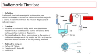
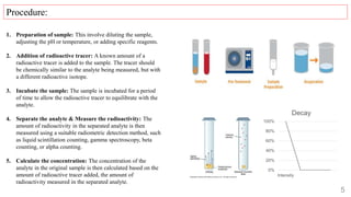
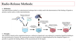
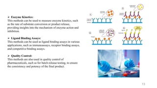
RADIOMETRIC TITRATIONS AND
RADIO-RELEASE METHODS
CONTENTS
2
 Introduction of Radiometric Titration.
 Introduction of Radio Release Methods.
 Applications.
 Reference.
3
 Definition:
Radiometric titration is an analytical technique that uses
radioactive isotopes to measure the concentration of an analyte in
a sample. It is a form of titration that relies on the principle of
radioactive decay.
 Principle:
o It is based on radioactive decay, which is the spontaneous
breakdown of an unstable atomic nucleus into a more stable
nucleus, emitting radiation in the process.
o The rate of radioactive decay is proportional to the number of
radioactive atoms present in the sample, and this can be used to
determine the concentration of the analyte being measured.
 Radioactive Isotopes :
o Carbon-14, Tritium.
o Phosphorus-32, Sulfur-35.
o Iodine-125
Radiometric Titration:
4
Types Of Radiometric Titration:
 Liquid Scintillation Counting: It involves adding a small
amount of a scintillation fluid to the sample, which absorbs
radiation emitted by the radioactive isotopes in the sample and
produces light.
 Gamma Spectroscopy: It measure the energy and intensity of
gamma rays emitted by radioactive isotopes in the sample. It is
directly proportional to the concentration of the analyte being
measured.
 Beta Counting: It involves using a beta counter to measure
the number of beta particles emitted by radioactive isotopes in
the sample.
 Alpha Counting: It involves using an alpha counter to
measure the number of alpha particles emitted by radioactive
isotopes in the sample.
5
1. Preparation of sample: This involve diluting the sample,
adjusting the pH or temperature, or adding specific reagents.
2. Addition of radioactive tracer: A known amount of a
radioactive tracer is added to the sample. The tracer should
be chemically similar to the analyte being measured, but with
a different radioactive isotope.
3. Incubate the sample: The sample is incubated for a period
of time to allow the radioactive tracer to equilibrate with the
analyte.
4. Separate the analyte & Measure the radioactivity: The
amount of radioactivity in the separated analyte is then
measured using a suitable radiometric detection method, such
as liquid scintillation counting, gamma spectroscopy, beta
counting, or alpha counting.
5. Calculate the concentration: The concentration of the
analyte in the original sample is then calculated based on the
amount of radioactive tracer added, the amount of
radioactivity measured in the separated analyte.
Procedure:
0%
20%
40%
60%
80%
100%
Intensity
Decay
6
 High sensitivity and accuracy:
It is useful for measuring trace amounts of analytes in complex
matrices.
 Can measure trace amounts of analytes in complex matrices:
Measuring organic compounds to studying biological molecules.
 They are a powerful analytical tool for a variety of fields, including
chemistry, biochemistry, and environmental science.
 Require specialized equipment and trained personnel
 Handling of radioactive materials
 Potential safety risks
Advantages:
Disadvantages:
7
 Defination:
The Radio Release method is a radiochemical technique that is widely used in the determination of the binding of ligands to
proteins, nucleic acids, or other macromolecules.
 Principle :
This method is based on the competition between a radioactive ligand and a non-radioactive competitor ligand for binding to
the macromolecule of interest. By measuring the amount of radioactive ligand displaced by the competitor ligand, it is
possible to quantify the binding affinity of the macromolecule for the ligand.
Radio-Release Methods:
8
 Filter binding assay:
It involves the filtration of a reaction mixture through a filter, which retains the
protein-bound radioactive ligand. The amount of radioactivity retained on the
filter is then measured and compared.
 Centrifugation assay:
It involves a reaction mixture containing the protein-bound radioactive ligand is
centrifuged through a medium, such as a sucrose gradient or a gel. The protein-
bound radioactive ligand is retained in the pellet, while the non-bound ligand is
found in the supernatant. The amount of radioactivity in each fraction is then
measured and compared.
 Microfiltration assay:
It involves the use of a microfilter to retain the protein-bound radioactive ligand.
The radioactivity of the retained ligand is then measured and compared.
 Solid-phase assay:
It involves the protein-bound radioactive ligand is immobilized on a solid
support, such as a membrane or a bead. The non-bound ligand is then removed
by washing, and the amount of retained radioactivity is measured and compared.
Types radio-release methods:
9
1. Preparation of Macromolecule Sample :
Macromolecule is get isolate and purified on the basis of
its concentration and purity. The macromolecule is then
diluted to the desired concentration in the assay buffer.
2. Preparation of Radiolabeled Ligand : It involves
labeling the ligand with a radioactive isotope, such as
tritium or carbon-14, using standard labeling techniques.
The radiolabeled ligand is then purified to remove any
unincorporated radioactive isotope and is diluted to the
desired concentration in the assay buffer.
3. Incubation of Macromolecule and Radiolabeled
Ligand :The macromolecule sample and the radiolabeled
ligand are mixed together in the assay buffer and
incubated for a specific period of time.
Procedure:
10
4. Addition of Non-Radiolabeled Competitor Ligand :
After the incubation period, a non-radiolabeled competitor
ligand is added to the mixture. It is typically similar
molecule that competes with the radiolabeled ligand for
binding to the macromolecule.
5. Separation of Bound and Free Ligand :
This can be done as centrifugation, filtration, or
chromatography.
6. Measurement of Radioactivity:
This can be done using a scintillation counter, which
detects the emitted radiation from the radioactive isotope.
By comparing the amount of radioactivity in the bound and free
ligand fractions, it is possible to calculate the percentage of
radiolabeled ligand bound to the macromolecule.
This data is then used to determine the binding affinity and other
parameters of the macromolecule-ligand interaction.
11
 High Sensitivity
 Rapid:
 Versatile:
 Quantitative:
 No Labeling of Macromolecule Required:
 Radioactivity Potential Interference:
 Non-physiological Conditions:
 Limited Information:
 Potential Artifacts:
Advantages:
Disadvantages:
12
 Drug Discovery and Development:
This methods widely used in drug discovery and development
to evaluate the binding affinity and kinetics of potential drug
candidates with target macromolecules.
 Biochemical Studies:
This methods also useful in biochemical studies to investigate
the binding interactions between macromolecules, such as
enzymes, receptors, and antibodies, and their ligands,
substrates, or inhibitors.
 Protein-Protein Interactions:
This methods can be used to study protein-protein
interactions, such as interactions between signaling proteins,
transcription factors, or protein complexes.
Applications:
 Enzyme Kinetics:
This methods can be used to measure enzyme kinetics, such
as the rate of substrate conversion or product release,
providing insights into the mechanism of enzyme action and
inhibition.
 Ligand Binding Assays:
This methods can be used as ligand binding assays in various
applications, such as immunoassays, receptor binding assays,
and competitive binding assays.
 Quality Control:
This methods are also used in quality control of
pharmaceuticals, such as for batch release testing, to ensure
the consistency and potency of the final product.
13
14
 Bio-Rad Laboratories. (2021). Radiometric Titrations: Principles and Applications.
https://www.bio-rad.com/webroot/web/pdf/lsr/literature/LIT665A.pdf
 Newman, D. J., & Cragg, G. M. (2016). Natural products as sources of new drugs over the 30 years from 1981 to 2010. Journal of
natural products, 79(3), 629-661.
 Shanmugam, G., & Latha, P. (2019). Radiometric titration: An overview. Journal of Advanced Chemical Sciences, 5(3), 363-369.
 Harris, J. L., Backes, B. J., Leonetti, F., Mahrus, S., & Ellman, J. A. (2000). Rapid and general profiling of protease specificity by
using combinatorial fluorogenic substrate libraries. Proceedings of the National Academy of Sciences, 97(14), 7754-7759.
 Kameyama, K., & Kikuchi, K. (2019). Recent advances in the development of activity-based probes for proteases. Journal of
Biochemistry, 165(3), 211-219.
 Kim, H. R., Kim, S., & Ko, Y. G. (2020). Radioactive isotopes in biomedical research. Applied Sciences, 10(9), 3096.
 Neumann, T., & Junker, H. D. (2018). Radioimmunoassay and related techniques in medical research. Methods in molecular
biology, 1797, 59-84.
Reference.
15
Thank You.

Radiometric titrations and radio-release methods

  • 1.
  • 2.
    CONTENTS 2  Introduction ofRadiometric Titration.  Introduction of Radio Release Methods.  Applications.  Reference.
  • 3.
    3  Definition: Radiometric titrationis an analytical technique that uses radioactive isotopes to measure the concentration of an analyte in a sample. It is a form of titration that relies on the principle of radioactive decay.  Principle: o It is based on radioactive decay, which is the spontaneous breakdown of an unstable atomic nucleus into a more stable nucleus, emitting radiation in the process. o The rate of radioactive decay is proportional to the number of radioactive atoms present in the sample, and this can be used to determine the concentration of the analyte being measured.  Radioactive Isotopes : o Carbon-14, Tritium. o Phosphorus-32, Sulfur-35. o Iodine-125 Radiometric Titration:
  • 4.
    4 Types Of RadiometricTitration:  Liquid Scintillation Counting: It involves adding a small amount of a scintillation fluid to the sample, which absorbs radiation emitted by the radioactive isotopes in the sample and produces light.  Gamma Spectroscopy: It measure the energy and intensity of gamma rays emitted by radioactive isotopes in the sample. It is directly proportional to the concentration of the analyte being measured.  Beta Counting: It involves using a beta counter to measure the number of beta particles emitted by radioactive isotopes in the sample.  Alpha Counting: It involves using an alpha counter to measure the number of alpha particles emitted by radioactive isotopes in the sample.
  • 5.
    5 1. Preparation ofsample: This involve diluting the sample, adjusting the pH or temperature, or adding specific reagents. 2. Addition of radioactive tracer: A known amount of a radioactive tracer is added to the sample. The tracer should be chemically similar to the analyte being measured, but with a different radioactive isotope. 3. Incubate the sample: The sample is incubated for a period of time to allow the radioactive tracer to equilibrate with the analyte. 4. Separate the analyte & Measure the radioactivity: The amount of radioactivity in the separated analyte is then measured using a suitable radiometric detection method, such as liquid scintillation counting, gamma spectroscopy, beta counting, or alpha counting. 5. Calculate the concentration: The concentration of the analyte in the original sample is then calculated based on the amount of radioactive tracer added, the amount of radioactivity measured in the separated analyte. Procedure: 0% 20% 40% 60% 80% 100% Intensity Decay
  • 6.
    6  High sensitivityand accuracy: It is useful for measuring trace amounts of analytes in complex matrices.  Can measure trace amounts of analytes in complex matrices: Measuring organic compounds to studying biological molecules.  They are a powerful analytical tool for a variety of fields, including chemistry, biochemistry, and environmental science.  Require specialized equipment and trained personnel  Handling of radioactive materials  Potential safety risks Advantages: Disadvantages:
  • 7.
    7  Defination: The RadioRelease method is a radiochemical technique that is widely used in the determination of the binding of ligands to proteins, nucleic acids, or other macromolecules.  Principle : This method is based on the competition between a radioactive ligand and a non-radioactive competitor ligand for binding to the macromolecule of interest. By measuring the amount of radioactive ligand displaced by the competitor ligand, it is possible to quantify the binding affinity of the macromolecule for the ligand. Radio-Release Methods:
  • 8.
    8  Filter bindingassay: It involves the filtration of a reaction mixture through a filter, which retains the protein-bound radioactive ligand. The amount of radioactivity retained on the filter is then measured and compared.  Centrifugation assay: It involves a reaction mixture containing the protein-bound radioactive ligand is centrifuged through a medium, such as a sucrose gradient or a gel. The protein- bound radioactive ligand is retained in the pellet, while the non-bound ligand is found in the supernatant. The amount of radioactivity in each fraction is then measured and compared.  Microfiltration assay: It involves the use of a microfilter to retain the protein-bound radioactive ligand. The radioactivity of the retained ligand is then measured and compared.  Solid-phase assay: It involves the protein-bound radioactive ligand is immobilized on a solid support, such as a membrane or a bead. The non-bound ligand is then removed by washing, and the amount of retained radioactivity is measured and compared. Types radio-release methods:
  • 9.
    9 1. Preparation ofMacromolecule Sample : Macromolecule is get isolate and purified on the basis of its concentration and purity. The macromolecule is then diluted to the desired concentration in the assay buffer. 2. Preparation of Radiolabeled Ligand : It involves labeling the ligand with a radioactive isotope, such as tritium or carbon-14, using standard labeling techniques. The radiolabeled ligand is then purified to remove any unincorporated radioactive isotope and is diluted to the desired concentration in the assay buffer. 3. Incubation of Macromolecule and Radiolabeled Ligand :The macromolecule sample and the radiolabeled ligand are mixed together in the assay buffer and incubated for a specific period of time. Procedure:
  • 10.
    10 4. Addition ofNon-Radiolabeled Competitor Ligand : After the incubation period, a non-radiolabeled competitor ligand is added to the mixture. It is typically similar molecule that competes with the radiolabeled ligand for binding to the macromolecule. 5. Separation of Bound and Free Ligand : This can be done as centrifugation, filtration, or chromatography. 6. Measurement of Radioactivity: This can be done using a scintillation counter, which detects the emitted radiation from the radioactive isotope. By comparing the amount of radioactivity in the bound and free ligand fractions, it is possible to calculate the percentage of radiolabeled ligand bound to the macromolecule. This data is then used to determine the binding affinity and other parameters of the macromolecule-ligand interaction.
  • 11.
    11  High Sensitivity Rapid:  Versatile:  Quantitative:  No Labeling of Macromolecule Required:  Radioactivity Potential Interference:  Non-physiological Conditions:  Limited Information:  Potential Artifacts: Advantages: Disadvantages:
  • 12.
    12  Drug Discoveryand Development: This methods widely used in drug discovery and development to evaluate the binding affinity and kinetics of potential drug candidates with target macromolecules.  Biochemical Studies: This methods also useful in biochemical studies to investigate the binding interactions between macromolecules, such as enzymes, receptors, and antibodies, and their ligands, substrates, or inhibitors.  Protein-Protein Interactions: This methods can be used to study protein-protein interactions, such as interactions between signaling proteins, transcription factors, or protein complexes. Applications:
  • 13.
     Enzyme Kinetics: Thismethods can be used to measure enzyme kinetics, such as the rate of substrate conversion or product release, providing insights into the mechanism of enzyme action and inhibition.  Ligand Binding Assays: This methods can be used as ligand binding assays in various applications, such as immunoassays, receptor binding assays, and competitive binding assays.  Quality Control: This methods are also used in quality control of pharmaceuticals, such as for batch release testing, to ensure the consistency and potency of the final product. 13
  • 14.
    14  Bio-Rad Laboratories.(2021). Radiometric Titrations: Principles and Applications. https://www.bio-rad.com/webroot/web/pdf/lsr/literature/LIT665A.pdf  Newman, D. J., & Cragg, G. M. (2016). Natural products as sources of new drugs over the 30 years from 1981 to 2010. Journal of natural products, 79(3), 629-661.  Shanmugam, G., & Latha, P. (2019). Radiometric titration: An overview. Journal of Advanced Chemical Sciences, 5(3), 363-369.  Harris, J. L., Backes, B. J., Leonetti, F., Mahrus, S., & Ellman, J. A. (2000). Rapid and general profiling of protease specificity by using combinatorial fluorogenic substrate libraries. Proceedings of the National Academy of Sciences, 97(14), 7754-7759.  Kameyama, K., & Kikuchi, K. (2019). Recent advances in the development of activity-based probes for proteases. Journal of Biochemistry, 165(3), 211-219.  Kim, H. R., Kim, S., & Ko, Y. G. (2020). Radioactive isotopes in biomedical research. Applied Sciences, 10(9), 3096.  Neumann, T., & Junker, H. D. (2018). Radioimmunoassay and related techniques in medical research. Methods in molecular biology, 1797, 59-84. Reference.
  • 15.