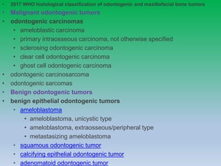
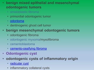
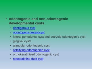
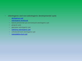
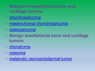
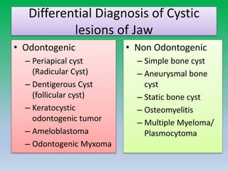
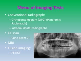
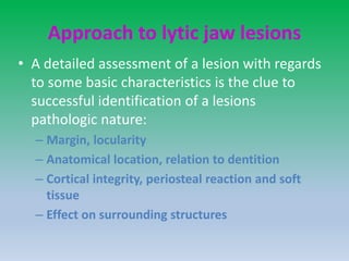
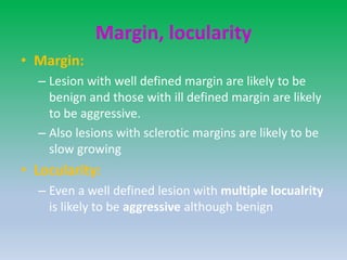
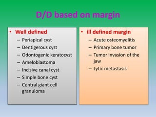
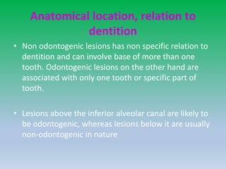
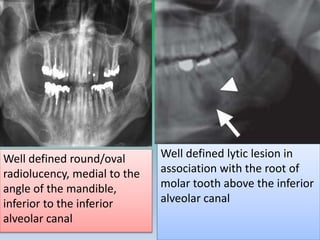
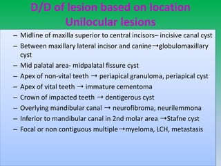
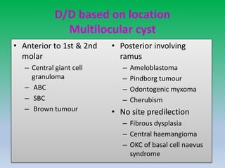
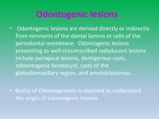
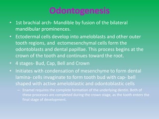
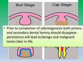
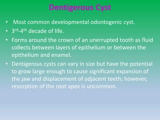
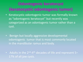
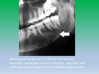
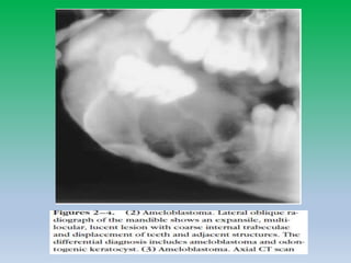
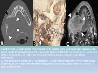
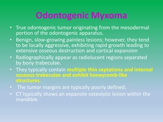
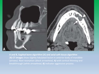
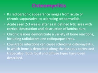
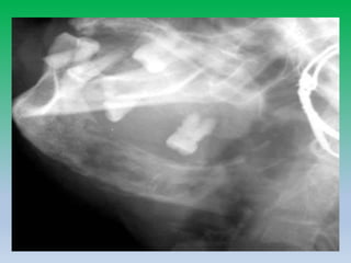
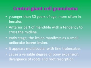
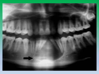
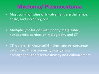
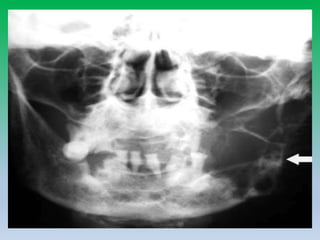
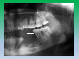
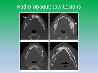
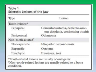
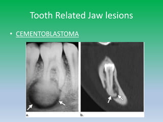
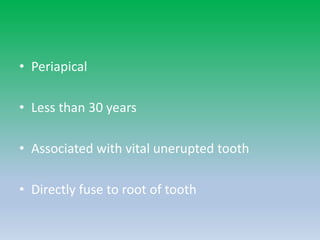
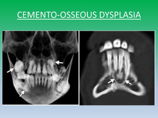
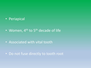
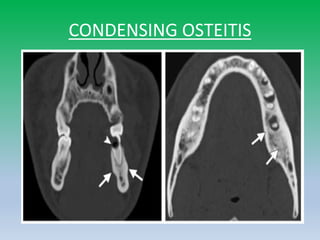
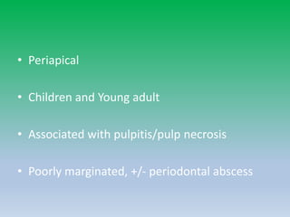
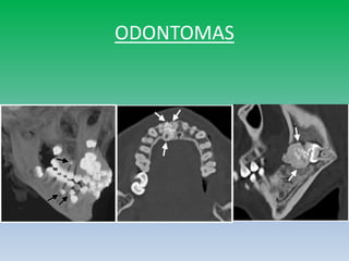
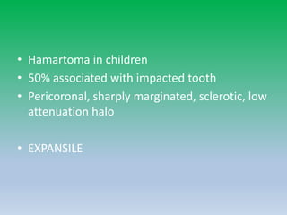
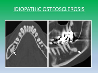
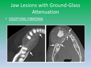
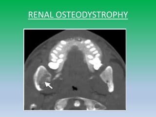
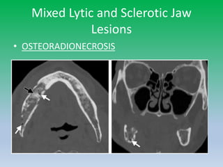
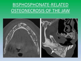
• 2017 WHO histological classification of odontogenic and maxillofacial bone tumors
• Malignant odontogenic tumors
• odontogenic carcinomas
• ameloblastic carcinoma
• primary intraosseous carcinoma, not otherwise specified
• sclerosing odontogenic carcinoma
• clear cell odontogenic carcinoma
• ghost cell odontogenic carcinoma
• odontogenic carcinosarcoma
• odontogenic sarcomas
• Benign odontogenic tumors
• benign epithelial odontogenic tumors
• ameloblastoma
• ameloblastoma, unicystic type
• ameloblastoma, extraosseous/peripheral type
• metastasizing ameloblastoma
• squamous odontogenic tumor
• calcifying epithelial odontogenic tumor
• adenomatoid odontogenic tumor
• benign mixed epithelial and mesenchymal
odontogenic tumors
• ameloblastic fibroma
• primordial odontogenic tumor
• odontoma
• dentinogenic ghost cell tumor
• benign mesenchymal odontogenic tumors
• odontogenic fibroma
• odontogenic myxoma/myxofibroma
• cementoblastoma
• cemento-ossifying fibroma
• Odontogenic cyst
• odontogenic cysts of inflammatory origin
• radicular cyst
• inflammatory collateral cysts
• odontogenic and non-odontogenic
developmental cysts
• dentigerous cyst
• odontogenic keratocyst
• lateral periodontal cyst and botryoid odontogenic cyst
• gingival cysts
• glandular odontogenic cyst
• calcifying odontogenic cyst
• orthokeratinized odontogenic cyst
• nasopalatine duct cyst
• odontogenic and non-odontogenic developmental cysts
• dentigerous cyst
• odontogenic keratocyst
• lateral periodontal cyst and botryoid odontogenic cyst
• gingival cysts
• glandular odontogenic cyst
• calcifying odontogenic cyst
• orthokeratinized odontogenic cyst
• nasopalatine duct cyst
• Malignant maxillofacial bone and
cartilage tumors
• chondrosarcoma
• mesenchymal chondrosarcoma
• osteosarcoma
• Benign maxillofacial bone and cartilage
tumors
• chondroma
• osteoma
• melanotic neuroectodermal tumor
Differential Diagnosis of Cystic
lesions of Jaw
• Odontogenic
– Periapical cyst
(Radicular Cyst)
– Dentigerous Cyst
(follicular cyst)
– Keratocystic
odontogenic tumor
– Ameloblastoma
– Odontogenic Myxoma
• Non Odontogenic
– Simple bone cyst
– Aneurysmal bone
cyst
– Static bone cyst
– Osteomyelitis
– Multiple Myeloma/
Plasmocytoma
Menu of Imaging Tests
• Conventional radiograph:
– Orthopantomogram (OPG) (Panoramic
Radiograph)
– Intraoral dental radiographs
• CT scan
– Cone beam CT
• MRI
• Fusion imaging:
– PET/CT
Approach to lytic jaw lesions
• A detailed assessment of a lesion with regards
to some basic characteristics is the clue to
successful identification of a lesions
pathologic nature:
– Margin, locularity
– Anatomical location, relation to dentition
– Cortical integrity, periosteal reaction and soft
tissue
– Effect on surrounding structures
Margin, locularity
• Margin:
– Lesion with well defined margin are likely to be
benign and those with ill defined margin are likely
to be aggressive.
– Also lesions with sclerotic margins are likely to be
slow growing
• Locularity:
– Even a well defined lesion with multiple locualrity
is likely to be aggressive although benign
Well circumscribed cytic space
around the crown of an
unerrupted third molar
extending to the cemento-
enamel junction. Dentigerous
Cyst
Expansile, multilocular, lucent
lesion with coarse internal
trabeculae and displacement of
teeth and adjacent structures-
Ameloblastoma
D/D based on margin
• Well defined
– Periapical cyst
– Dentigerous cyst
– Odontogenic keratocyst
– Ameloblastoma
– Incisive canal cyst
– Simple bone cyst
– Central giant cell
granuloma
• ill defined margin
– Acute osteomyelitis
– Primary bone tumor
– Tumor invasion of the
jaw
– Lytic metastasis
Anatomical location, relation to
dentition
• Non odontogenic lesions has non specific relation to
dentition and can involve base of more than one
tooth. Odontogenic lesions on the other hand are
associated with only one tooth or specific part of
tooth.
• Lesions above the inferior alveolar canal are likely to
be odontogenic, whereas lesions below it are usually
non-odontogenic in nature
Well defined round/oval
radiolucency, medial to the
angle of the mandible,
inferior to the inferior
alveolar canal
Well defined lytic lesion in
association with the root of
molar tooth above the inferior
alveolar canal
D/D of lesion based on location
Unilocular lesions
– Midline of maxilla superior to central incisors– incisive canal cyst
– Between maxillary lateral incisor and canine➝globulomaxillary
cyst
– Mid palatal area- midpalatal fissure cyst
– Apex of non-vital teeth ➝ periapical granuloma, periapical cyst
– Apex of vital teeth ➝ immature cementoma
– Crown of impacted teeth ➝ dentigerous cyst
– Overlying mandibular canal ➝ neurofibroma, neurilemmona
– Inferior to mandibular canal in 2nd molar area ➝Stafne cyst
– Focal or non contiguous multiple➝myeloma, LCH, metastasis
D/D based on location
Multilocular cyst
• Anterior to 1st & 2nd
molar
– Central giant cell
granuloma
– ABC
– SBC
– Brown tumour
• Posterior involving
ramus
– Ameloblastoma
– Pindborg tumour
– Odontogenic myxoma
– Cherubism
• No site predilection
– Fibrous dysplasia
– Central haemangioma
– OKC of basal cell naevus
syndrome
Cortical integrity, periosteal
reaction and soft tissue
• Cortical expansion is caused by slow growing
benign lesion whereas cortical breech and
destruction is a feature of aggressive
inflammatory or tumoral lesion.
• Periosteal reaction suggest infective or
neoplastic etiology. E.g sunburst type of
periosteal reaction in osteosarcoma.
Mixed lytic and sclerotic ill defined
lesion in the body of mandible with
cortical destruction and spicualted
periosteal reaction
Bone window and soft tissue window CT of
mandible showing lucent lesion in body of
mandible with cortical destruction and
periosteal reaction- osteomyelitis
Q-Effect on surrounding structure
• Displacement of tooth
– slow-growing, space-occupying lesions
– epicentre above the crown of the tooth (i.e., dentigerous
cyst and occasionally odontomas) displace the tooth
apically.
– Lesion that start in the ramus, such as cherubism, may push
teeth in the anterior direction
• Resorption of tooth
– more chronic and slow-growing processes; however,
malignant lesions also occasionally resorb teeth.
• irregular widening and destruction of lamina dura- malignant
lesion
• Widening of mandibular canal – neurogenic or vascular lesion
• Odontogenic
cyst
displacing
the molar
tooth crown
apically
Odontogenic lesions
• Odontogenic lesions are derived directly or indirectly
from remnants of the dental lamina or cells of the
periodontal membrane. Odontogenic lesions
presenting as well circumscribed radiolucent lesions
include periapical lesions, dentigerous cysts,
odoontogenic keratocyst, cysts of the
globullomaxillary region, and ameloblastomas.
• Basics of Odontogenesis is required to understand
the origin of odontogenic lesions
Odontogenesis
• 1st brachial arch- Mandible by fusion of the bilateral
mandibular prominences.
• Ectodermal cells develop into ameloblasts and other outer
tooth regions, and ectomesenchymal cells form the
odontoblasts and dental papillae. This process begins at the
crown of the tooth and continues toward the root.
• 4 stages- Bud, Cap, Bell and Crown
• Initiates with condensation of mesenchyme to form dental
lamina- cells invaginate to form tooth bud with cap- bell
shaped with active ameloblastic and odontoblastic cells
– Enamel requires the complete formation of the underlying dentin. Both of
these processes are completed during the crown stage, as the tooth enters the
final stage of development.
• Prior to completion of odontogenesis both pimary
and secondary dental lamina should disappear-
persistence will lead to benign and malignant
lesion later in life.
Dentigerous Cyst
• Most common developmental odontogenic cyst.
• 3rd-4th decade of life.
• Forms around the crown of an unerrupted tooth as fluid
collects between layers of epithelium or between the
epithelium and enamel.
• Dentigerous cysts can vary in size but have the potential
to grow large enough to cause significant expansion of
the jaw and displacement of adjacent teeth; however,
resorption of the root apex is uncommon.
Dentigerous cyst. Lateral oblique radiograph shows a well-
defined expansile lucent lesion in the left mandible. Note
unerrupted third molar with the crown lying inside the cyst
(arrow).
Dentigerous cyst in a 40-year-old man. Coronal reformatted CT image
reveals a cystic lesion with an unerupted tooth in the right molar
region (arrow). The crown of the tooth is contained within the lesion.
Note the presence of bone remodeling rather than expansion.
Radicular cyst
• Most common type (about 70 %) of odontogenic cyst.
These lesions have a slight male predominance.
• 30 -50 years.
• Periapical cysts generally originate after trauma or
dental caries. Dental caries cause inflammation of the
pulp cavity leading to pulp necrosis. The infection then
spreads to the apex causing periapical periodontitis,
which leads to either an acute abscess or a chronic
granuloma. Persistent chronic infection can lead to
formation of a periapical cyst.
• 42-year-old woman
with periapicalcyst.
• Cropped panoramic
radiograph shows
radiolucent
lesion(arrowhead) in
posterior body of
ramus of mandible
with displacement of
mandibular canal
(arrow).
25-year-old man with periapical cyst.
A–C, Axial (A), sagittal (B), and volume-rendering 3D (C) CT images show well-
circumscribed radiolucent lesions surrounding apex of first (white arrows, A and B) and
second (black arrows, B and C) molars (teeth 18 and 19). Note large cavities in affected
teeth with mild root resorption (arrowheads, B and C).
A, Coronal T2-weighted MR image shows high signal in anterior body of mandible
(arrow).
B, Axial gadolinium-enhanced fat-suppressed T1- weighted image shows
enhancement of cyst wall (arrow); this finding is consistent with periapical cyst.
Odontogenic keratocyst
(keratocystic odontogenic tumor)
• Keratocystic odontogenic tumor was formally known
as “odontogenic keratocyst” but recently was
categorized as an odontogenic tumor rather than a
cyst.
• Benign but locally aggressive developmental
odontogenic tumor that is most commonly located
in the mandibular ramus and body.
• Adults in the 2nd-4th decades of life and represent 5–
17% of all jaw cysts.
Odontogenic keratocyst in a 36-year-old woman.
Panoramic radiograph shows an ellipsoid, expansile, well-
corticated, lucent lesion in left mandibular body (arrow).
• CT shows a unilocular or multilocular cyst with corticated
margins in the body and ramus of the mandible in association
with an impacted tooth.
• MRI can help distinguish keratocystic odontogenic tumor from
ameloblastoma:
– Ameloblastoma mixed pattern of solid and cystic components,
irregular thick walls, and avid enhancement of solid components
24-year-old woman with keratocystic odontogenic tumor.
A, Oblique sagittal reconstructed CT image obtained using bone algorithm shows large
radiolucent lesion (arrows) in posterior body of mandibular ramus, with scalloping of
cortex.
B, Axial contrast-enhanced CT image obtained using soft-tissue algorithm shows cystic
lesion with mild peripheral enhancement (arrow) breaking through cortex and extending
into left masseter muscle.
Ameloblastoma
• Benign but locally aggressive epithelial odontogenic
tumor.
• 10% of odontogenic tumors, and 80% are located
within the mandible.
• Most commonly found in adults in the 3rd-4th decade
without sex predominance.
• Malignant ameloblastoma is a term given to tumors
with metastasis, even those with a histologically
benign appearance.
• Ameloblastic carcinoma is a term given to tumors
that display histologically malignant features with or
without metastasis .
• On the basis of its clinical behavior, anatomic location, radiographic
appearance, and histologic characteristics, ameloblastoma can be
grouped into four main forms
Types Radiological findings
Multicystic
multiloculated with internal septations manifested by a honeycomb or
soap-bubble appearance. This variant can be confused with large
keratocystic odontogenic tumors .
Unicystic
Unilocular, well-circumscribed, and well-corticated lucent lesion often
associated with the crown of an unerupted or impacted tooth. This
variant can resemble a dentigerous Cyst.
Extraosseous
The peripheral ameloblastoma variant represents a soft tissue tumor
that is histologically identical to an intraosseous ameloblastoma but
occurs over the tooth-bearing parts of the jaw. These lesions appear
solid on imaging, have a benign clinical course, and can be treated with
local excision.
Desmoplastic
The desmoplastic variant can be distinguished from other variants by
the presence of multiple coarse internal calcifications with significant
surrounding cortical destruction.
A and B, Axial (A) and volume-rendering 3D (B) CT images show large destructive radiolucent
lesion (white arrowheads) centered in posterior body extending to manibular ramus, significant
cortical destruction, and root erosion that significantly displaces second molar (black
arrowhead).
C, Axial gadolinium-enhanced fat-suppressed T1-weighted MR image shows heterogeneously
enhancing lesion (white arrowheads) with regions of decreased signal that corresponded to
cystic degeneration.
Odontogenic Myxoma
• True odontogenic tumor originating from the mesodermal
portion of the odontogenic apparatus.
• Benign, slow-growing painless lesions; however, they tend
to be locally aggressive, exhibiting rapid growth leading to
extensive osseous destruction and cortical expansion
• Radiographically appear as radiolucent regions separated
by bony trabeculae.
• They typically contain multiple thin septations and internal
osseous trabeculae and exhibit honeycomb-like
structures.
• The tumor margins are typically poorly defined.
• CT typically shows an expansile osteolytic lesion within the
mandible.
A and B, Sagittal bone algorithm (A) and axial soft tissue algorithm
(B) CT images show slightly lobulated lesion in anterior body of mandible
(arrows). Root resorption (black arrowhead, A) with cortical thinning and
breakthrough (white arrowhead, B) indicates aggressive process.
Odontogenic myxoma: Bubbly lytic lesion in the rt side of maxilla
with multiple thin internal
septations
Non Odontogenic lesions
• They may be lesions
– Arising from non dental elements e.g fissural cysts
– Lytic lesions arising in the bone
Stafne bone cyst
• Static bone cavity is also known as stafne cyst. However, this
condition is not a cyst, but a focal cortical bone defect.
• The location the lingual aspect of the angle or posterior body
of the mandible, below the mandibular canal, is unique
enough to make the diagnosis.
• Clasically it has been described that the submandibular gland
tissue is seen in the defect, however often only fatty tissue is
identified in the cavity.
Cropped panoramic radiograph
showing
well defined corticated radiolucency
in the posterior
body of mandible
Axial CT demonstrates a well defined
lingual cortical defect in the posterio
mandible
Primordial Cyst
• A less common cystic lesion is the primordial
cyst, which develops instead of a tooth.
• It is believed that the dental follicle forms but
subsequently undergoes cystic degeneration
before completion of odontogenesis.
• The non expansile lesion is well defined and
radiolucent, without an associated tooth.
Primordial cyst. Panoramic radiograph shows a welldefined
radiolucency in the location of the right third molar which has
failed to develop (arrow).
Nasopalatine duct cyst
• A non odontogenic developmental cyst or fissural
cyst arising from epithelial remnants of the
nasopalatine duct.
• Located in anterior hard palate at midline.
• Commonest nonodontogenic cyst.
• On imaging, they appear as round, ovoid, well
defined lucent lesion in the anterior maxilla. The
margin is well defined , smooth and dense rim of
cortical bone is often seen along the periphery.
large midline radiolucent lesion in the anterior portion of
the hard palate causing destruction of the vomer and
the anterior part of the nasal bony septum (arrows).
Solitary Bone Cyst (Traumatic, Simple,
Hemorrhagic Bone Cyst)
• A solitary bone cyst results from trauma, which leads
to intramedullary hemorrhage and subsequent
resorption.
• These pseudocysts are most commonly located in the
posterior marrow space of the mandible and appear
slightly irregular with poorly defined borders.
• They have a characteristic scalloped superior margin
extending between the roots of adjacent teeth.
• In addition, the mandibular cortex may be thinned
secondary to osseous expansion.
A and B, Axial (A) and coronal (B) CT images show wellcircumscribed radiolucent lesion
(arrows) with cortical scalloping in anterior body of mandible without root resorption.
Prominent expansile change is not typical of simple bone cyst.
Aneurysmal bone cyst
• Rare lesions that are most common within the
mandible of children with a slight female
predominance
• Clinically these lesions present with rapid
painless facial swelling that can be disfiguring.
• The origin of ABCs is controversial: Some
investigators believe ABCs are primary congenital
lesions or vascular malformations, whereas
others believe they are acquired and arise after
local hemodynamic disturbances from trauma.
The radiograph revealed a well-defined unilocular expansile radiolucent lesion in the
posterior mandible. The lesion has caused displacement and impaction of teeth. ABC was
confirmed histopathologically.
Axial CT of the mandible demonstrates the full extent of the lesion
which extends buccally and lingually with thinning and perforation of
the cortex. Note the delicate septa.
Osteomyelitis
• Its radiographic appearance ranges from acute or
chronic suppurative to sclerosing osteomyelitis.
• Acute seen 2-3 weeks after as ill defined lytic area with
cortical destruction and destruction of lamina dura
• Chronic lesions demonstrate a variety of bone reactions,
including radiolucent and radiopaque areas.
• Low-grade infections can cause sclerosing osteomyelitis,
in which bone is deposited along the osseous cortex and
trabeculae. Both focal and diffuse types have been
described.
Central giant cell granuloma
• younger than 30 years of age, more often in
females
• Anterior part of mandible with a tendency to
cross the midline
• early stage, the lesion manifests as a small
unilocular lucent lesion.
• it appears multilocular with fine trabeculae.
• cause a variable degree of bony expansion,
divergence of roots and root resorption
Myeloma/ Plasmocytoma
• Most common sites of involvement are the ramus,
angle, and molar regions
• Multiple lytic lesions with poorly marginated,
nonsclerotic borders on radiography and CT.
• CT is useful to show solid lesions and extraosseous
extension. These lesions typically show
homogeneous soft-tissue density and enhancement
Eosinophilic Granuloma
• Two types:
– Alveolar- multiple lesion in alveolar process. Arise
in midroot region and with progression scooped
out appearance. Bone of the teeth including
lamina dura destroyed- floating teeth
– Intraosseus- solitary in mandibular ramus.
Irregular, oval or round may simulate periosteal
reaction.
Radio-opaque Jaw Lesions
Tooth Related Jaw lesions
• CEMENTOBLASTOMA
• Periapical
• Less than 30 years
• Associated with vital unerupted tooth
• Directly fuse to root of tooth
CEMENTO-OSSEOUS DYSPLASIA
• Periapical
• Women, 4th to 5th decade of life
• Associated with vital tooth
• Do not fuse directly to tooth root
CONDENSING OSTEITIS
• Periapical
• Children and Young adult
• Associated with pulpitis/pulp necrosis
• Poorly marginated, +/- periodontal abscess
ODONTOMAS
• Hamartoma in children
• 50% associated with impacted tooth
• Pericoronal, sharply marginated, sclerotic, low
attenuation halo
• EXPANSILE
IDIOPATHIC OSTEOSCLEROSIS
• 1st and 2nd decade of life, unknown etiology
• 90% in mandible
• Periapical, well defined, spiculations, sclerotic
• NON-EXPANSILE
Sclerotic Non– Tooth-related
Lesions
• OSTEOMAS
• Benign lesion, non-exophytic lesion
• Expansile, non-tooth related
• No perilesional halo
• Multiple osteomas: Gardner syndrome
TORI AND EXOSTOSES
Jaw Lesions with Ground-Glass
Attenuation
• OSSIFYING FIBROMA
• 3rd and 4th decade
• Growth is perpendicular to long axis of bone
• Well defined narrow zone of transition
• Expansile, displace tooth
FIBROUS DYSPLASIA
• Growth along longitudinal axis of bone
• Shape of bone and lamina dura are altered
• Ill defined transition zone
• Encroaches on orbit, sinuses or vascular canals
RENAL OSTEODYSTROPHY
• Loss of cortex and Lamina dura
Mixed Lytic and Sclerotic Jaw
Lesions
• OSTEORADIONECROSIS
• Most common: Mandible, in body and along
buccal cortex.
• Sclerosis, cortical interruptions, bone
fragments, areas of gas attenuation.
• Sometimes: fluid collection, areas of soft
tissue enhancement
BISPHOSPHONATE-RELATED
OSTEONECROSIS OF THE JAW
• Following oral or IV bisphosphonate therapy
• Poorly marginated diffuse area of low
attenuation with bilateral symmetrical
sclerosis.
MANDIBULAR OSTEOMYELITIS
• Chronic osteomyelitis > 1 month.
• Sinuses, fistula
• Sequestra, pathological fracture, areas of gas
attenuation
• Periosteal new bone formation
Thank you
Jaw lesion  radiology ppt

Jaw lesion radiology ppt

  • 2.
    • 2017 WHOhistological classification of odontogenic and maxillofacial bone tumors • Malignant odontogenic tumors • odontogenic carcinomas • ameloblastic carcinoma • primary intraosseous carcinoma, not otherwise specified • sclerosing odontogenic carcinoma • clear cell odontogenic carcinoma • ghost cell odontogenic carcinoma • odontogenic carcinosarcoma • odontogenic sarcomas • Benign odontogenic tumors • benign epithelial odontogenic tumors • ameloblastoma • ameloblastoma, unicystic type • ameloblastoma, extraosseous/peripheral type • metastasizing ameloblastoma • squamous odontogenic tumor • calcifying epithelial odontogenic tumor • adenomatoid odontogenic tumor
  • 3.
    • benign mixedepithelial and mesenchymal odontogenic tumors • ameloblastic fibroma • primordial odontogenic tumor • odontoma • dentinogenic ghost cell tumor • benign mesenchymal odontogenic tumors • odontogenic fibroma • odontogenic myxoma/myxofibroma • cementoblastoma • cemento-ossifying fibroma • Odontogenic cyst • odontogenic cysts of inflammatory origin • radicular cyst • inflammatory collateral cysts
  • 4.
    • odontogenic andnon-odontogenic developmental cysts • dentigerous cyst • odontogenic keratocyst • lateral periodontal cyst and botryoid odontogenic cyst • gingival cysts • glandular odontogenic cyst • calcifying odontogenic cyst • orthokeratinized odontogenic cyst • nasopalatine duct cyst
  • 5.
    • odontogenic andnon-odontogenic developmental cysts • dentigerous cyst • odontogenic keratocyst • lateral periodontal cyst and botryoid odontogenic cyst • gingival cysts • glandular odontogenic cyst • calcifying odontogenic cyst • orthokeratinized odontogenic cyst • nasopalatine duct cyst
  • 6.
    • Malignant maxillofacialbone and cartilage tumors • chondrosarcoma • mesenchymal chondrosarcoma • osteosarcoma • Benign maxillofacial bone and cartilage tumors • chondroma • osteoma • melanotic neuroectodermal tumor
  • 7.
    Differential Diagnosis ofCystic lesions of Jaw • Odontogenic – Periapical cyst (Radicular Cyst) – Dentigerous Cyst (follicular cyst) – Keratocystic odontogenic tumor – Ameloblastoma – Odontogenic Myxoma • Non Odontogenic – Simple bone cyst – Aneurysmal bone cyst – Static bone cyst – Osteomyelitis – Multiple Myeloma/ Plasmocytoma
  • 8.
    Menu of ImagingTests • Conventional radiograph: – Orthopantomogram (OPG) (Panoramic Radiograph) – Intraoral dental radiographs • CT scan – Cone beam CT • MRI • Fusion imaging: – PET/CT
  • 9.
    Approach to lyticjaw lesions • A detailed assessment of a lesion with regards to some basic characteristics is the clue to successful identification of a lesions pathologic nature: – Margin, locularity – Anatomical location, relation to dentition – Cortical integrity, periosteal reaction and soft tissue – Effect on surrounding structures
  • 10.
    Margin, locularity • Margin: –Lesion with well defined margin are likely to be benign and those with ill defined margin are likely to be aggressive. – Also lesions with sclerotic margins are likely to be slow growing • Locularity: – Even a well defined lesion with multiple locualrity is likely to be aggressive although benign
  • 11.
    Well circumscribed cyticspace around the crown of an unerrupted third molar extending to the cemento- enamel junction. Dentigerous Cyst Expansile, multilocular, lucent lesion with coarse internal trabeculae and displacement of teeth and adjacent structures- Ameloblastoma
  • 12.
    D/D based onmargin • Well defined – Periapical cyst – Dentigerous cyst – Odontogenic keratocyst – Ameloblastoma – Incisive canal cyst – Simple bone cyst – Central giant cell granuloma • ill defined margin – Acute osteomyelitis – Primary bone tumor – Tumor invasion of the jaw – Lytic metastasis
  • 13.
    Anatomical location, relationto dentition • Non odontogenic lesions has non specific relation to dentition and can involve base of more than one tooth. Odontogenic lesions on the other hand are associated with only one tooth or specific part of tooth. • Lesions above the inferior alveolar canal are likely to be odontogenic, whereas lesions below it are usually non-odontogenic in nature
  • 14.
    Well defined round/oval radiolucency,medial to the angle of the mandible, inferior to the inferior alveolar canal Well defined lytic lesion in association with the root of molar tooth above the inferior alveolar canal
  • 15.
    D/D of lesionbased on location Unilocular lesions – Midline of maxilla superior to central incisors– incisive canal cyst – Between maxillary lateral incisor and canine➝globulomaxillary cyst – Mid palatal area- midpalatal fissure cyst – Apex of non-vital teeth ➝ periapical granuloma, periapical cyst – Apex of vital teeth ➝ immature cementoma – Crown of impacted teeth ➝ dentigerous cyst – Overlying mandibular canal ➝ neurofibroma, neurilemmona – Inferior to mandibular canal in 2nd molar area ➝Stafne cyst – Focal or non contiguous multiple➝myeloma, LCH, metastasis
  • 16.
    D/D based onlocation Multilocular cyst • Anterior to 1st & 2nd molar – Central giant cell granuloma – ABC – SBC – Brown tumour • Posterior involving ramus – Ameloblastoma – Pindborg tumour – Odontogenic myxoma – Cherubism • No site predilection – Fibrous dysplasia – Central haemangioma – OKC of basal cell naevus syndrome
  • 17.
    Cortical integrity, periosteal reactionand soft tissue • Cortical expansion is caused by slow growing benign lesion whereas cortical breech and destruction is a feature of aggressive inflammatory or tumoral lesion. • Periosteal reaction suggest infective or neoplastic etiology. E.g sunburst type of periosteal reaction in osteosarcoma.
  • 18.
    Mixed lytic andsclerotic ill defined lesion in the body of mandible with cortical destruction and spicualted periosteal reaction Bone window and soft tissue window CT of mandible showing lucent lesion in body of mandible with cortical destruction and periosteal reaction- osteomyelitis
  • 19.
    Q-Effect on surroundingstructure • Displacement of tooth – slow-growing, space-occupying lesions – epicentre above the crown of the tooth (i.e., dentigerous cyst and occasionally odontomas) displace the tooth apically. – Lesion that start in the ramus, such as cherubism, may push teeth in the anterior direction • Resorption of tooth – more chronic and slow-growing processes; however, malignant lesions also occasionally resorb teeth. • irregular widening and destruction of lamina dura- malignant lesion • Widening of mandibular canal – neurogenic or vascular lesion
  • 20.
  • 21.
    Odontogenic lesions • Odontogeniclesions are derived directly or indirectly from remnants of the dental lamina or cells of the periodontal membrane. Odontogenic lesions presenting as well circumscribed radiolucent lesions include periapical lesions, dentigerous cysts, odoontogenic keratocyst, cysts of the globullomaxillary region, and ameloblastomas. • Basics of Odontogenesis is required to understand the origin of odontogenic lesions
  • 22.
    Odontogenesis • 1st brachialarch- Mandible by fusion of the bilateral mandibular prominences. • Ectodermal cells develop into ameloblasts and other outer tooth regions, and ectomesenchymal cells form the odontoblasts and dental papillae. This process begins at the crown of the tooth and continues toward the root. • 4 stages- Bud, Cap, Bell and Crown • Initiates with condensation of mesenchyme to form dental lamina- cells invaginate to form tooth bud with cap- bell shaped with active ameloblastic and odontoblastic cells – Enamel requires the complete formation of the underlying dentin. Both of these processes are completed during the crown stage, as the tooth enters the final stage of development.
  • 23.
    • Prior tocompletion of odontogenesis both pimary and secondary dental lamina should disappear- persistence will lead to benign and malignant lesion later in life.
  • 24.
    Dentigerous Cyst • Mostcommon developmental odontogenic cyst. • 3rd-4th decade of life. • Forms around the crown of an unerrupted tooth as fluid collects between layers of epithelium or between the epithelium and enamel. • Dentigerous cysts can vary in size but have the potential to grow large enough to cause significant expansion of the jaw and displacement of adjacent teeth; however, resorption of the root apex is uncommon.
  • 25.
    Dentigerous cyst. Lateraloblique radiograph shows a well- defined expansile lucent lesion in the left mandible. Note unerrupted third molar with the crown lying inside the cyst (arrow).
  • 26.
    Dentigerous cyst ina 40-year-old man. Coronal reformatted CT image reveals a cystic lesion with an unerupted tooth in the right molar region (arrow). The crown of the tooth is contained within the lesion. Note the presence of bone remodeling rather than expansion.
  • 27.
    Radicular cyst • Mostcommon type (about 70 %) of odontogenic cyst. These lesions have a slight male predominance. • 30 -50 years. • Periapical cysts generally originate after trauma or dental caries. Dental caries cause inflammation of the pulp cavity leading to pulp necrosis. The infection then spreads to the apex causing periapical periodontitis, which leads to either an acute abscess or a chronic granuloma. Persistent chronic infection can lead to formation of a periapical cyst.
  • 28.
    • 42-year-old woman withperiapicalcyst. • Cropped panoramic radiograph shows radiolucent lesion(arrowhead) in posterior body of ramus of mandible with displacement of mandibular canal (arrow).
  • 29.
    25-year-old man withperiapical cyst. A–C, Axial (A), sagittal (B), and volume-rendering 3D (C) CT images show well- circumscribed radiolucent lesions surrounding apex of first (white arrows, A and B) and second (black arrows, B and C) molars (teeth 18 and 19). Note large cavities in affected teeth with mild root resorption (arrowheads, B and C).
  • 30.
    A, Coronal T2-weightedMR image shows high signal in anterior body of mandible (arrow). B, Axial gadolinium-enhanced fat-suppressed T1- weighted image shows enhancement of cyst wall (arrow); this finding is consistent with periapical cyst.
  • 31.
    Odontogenic keratocyst (keratocystic odontogenictumor) • Keratocystic odontogenic tumor was formally known as “odontogenic keratocyst” but recently was categorized as an odontogenic tumor rather than a cyst. • Benign but locally aggressive developmental odontogenic tumor that is most commonly located in the mandibular ramus and body. • Adults in the 2nd-4th decades of life and represent 5– 17% of all jaw cysts.
  • 32.
    Odontogenic keratocyst ina 36-year-old woman. Panoramic radiograph shows an ellipsoid, expansile, well- corticated, lucent lesion in left mandibular body (arrow).
  • 33.
    • CT showsa unilocular or multilocular cyst with corticated margins in the body and ramus of the mandible in association with an impacted tooth. • MRI can help distinguish keratocystic odontogenic tumor from ameloblastoma: – Ameloblastoma mixed pattern of solid and cystic components, irregular thick walls, and avid enhancement of solid components
  • 34.
    24-year-old woman withkeratocystic odontogenic tumor. A, Oblique sagittal reconstructed CT image obtained using bone algorithm shows large radiolucent lesion (arrows) in posterior body of mandibular ramus, with scalloping of cortex. B, Axial contrast-enhanced CT image obtained using soft-tissue algorithm shows cystic lesion with mild peripheral enhancement (arrow) breaking through cortex and extending into left masseter muscle.
  • 35.
    Ameloblastoma • Benign butlocally aggressive epithelial odontogenic tumor. • 10% of odontogenic tumors, and 80% are located within the mandible. • Most commonly found in adults in the 3rd-4th decade without sex predominance. • Malignant ameloblastoma is a term given to tumors with metastasis, even those with a histologically benign appearance. • Ameloblastic carcinoma is a term given to tumors that display histologically malignant features with or without metastasis .
  • 36.
    • On thebasis of its clinical behavior, anatomic location, radiographic appearance, and histologic characteristics, ameloblastoma can be grouped into four main forms Types Radiological findings Multicystic multiloculated with internal septations manifested by a honeycomb or soap-bubble appearance. This variant can be confused with large keratocystic odontogenic tumors . Unicystic Unilocular, well-circumscribed, and well-corticated lucent lesion often associated with the crown of an unerupted or impacted tooth. This variant can resemble a dentigerous Cyst. Extraosseous The peripheral ameloblastoma variant represents a soft tissue tumor that is histologically identical to an intraosseous ameloblastoma but occurs over the tooth-bearing parts of the jaw. These lesions appear solid on imaging, have a benign clinical course, and can be treated with local excision. Desmoplastic The desmoplastic variant can be distinguished from other variants by the presence of multiple coarse internal calcifications with significant surrounding cortical destruction.
  • 38.
    A and B,Axial (A) and volume-rendering 3D (B) CT images show large destructive radiolucent lesion (white arrowheads) centered in posterior body extending to manibular ramus, significant cortical destruction, and root erosion that significantly displaces second molar (black arrowhead). C, Axial gadolinium-enhanced fat-suppressed T1-weighted MR image shows heterogeneously enhancing lesion (white arrowheads) with regions of decreased signal that corresponded to cystic degeneration.
  • 39.
    Odontogenic Myxoma • Trueodontogenic tumor originating from the mesodermal portion of the odontogenic apparatus. • Benign, slow-growing painless lesions; however, they tend to be locally aggressive, exhibiting rapid growth leading to extensive osseous destruction and cortical expansion • Radiographically appear as radiolucent regions separated by bony trabeculae. • They typically contain multiple thin septations and internal osseous trabeculae and exhibit honeycomb-like structures. • The tumor margins are typically poorly defined. • CT typically shows an expansile osteolytic lesion within the mandible.
  • 40.
    A and B,Sagittal bone algorithm (A) and axial soft tissue algorithm (B) CT images show slightly lobulated lesion in anterior body of mandible (arrows). Root resorption (black arrowhead, A) with cortical thinning and breakthrough (white arrowhead, B) indicates aggressive process.
  • 41.
    Odontogenic myxoma: Bubblylytic lesion in the rt side of maxilla with multiple thin internal septations
  • 42.
    Non Odontogenic lesions •They may be lesions – Arising from non dental elements e.g fissural cysts – Lytic lesions arising in the bone
  • 43.
    Stafne bone cyst •Static bone cavity is also known as stafne cyst. However, this condition is not a cyst, but a focal cortical bone defect. • The location the lingual aspect of the angle or posterior body of the mandible, below the mandibular canal, is unique enough to make the diagnosis. • Clasically it has been described that the submandibular gland tissue is seen in the defect, however often only fatty tissue is identified in the cavity.
  • 44.
    Cropped panoramic radiograph showing welldefined corticated radiolucency in the posterior body of mandible Axial CT demonstrates a well defined lingual cortical defect in the posterio mandible
  • 45.
    Primordial Cyst • Aless common cystic lesion is the primordial cyst, which develops instead of a tooth. • It is believed that the dental follicle forms but subsequently undergoes cystic degeneration before completion of odontogenesis. • The non expansile lesion is well defined and radiolucent, without an associated tooth.
  • 46.
    Primordial cyst. Panoramicradiograph shows a welldefined radiolucency in the location of the right third molar which has failed to develop (arrow).
  • 47.
    Nasopalatine duct cyst •A non odontogenic developmental cyst or fissural cyst arising from epithelial remnants of the nasopalatine duct. • Located in anterior hard palate at midline. • Commonest nonodontogenic cyst. • On imaging, they appear as round, ovoid, well defined lucent lesion in the anterior maxilla. The margin is well defined , smooth and dense rim of cortical bone is often seen along the periphery.
  • 48.
    large midline radiolucentlesion in the anterior portion of the hard palate causing destruction of the vomer and the anterior part of the nasal bony septum (arrows).
  • 49.
    Solitary Bone Cyst(Traumatic, Simple, Hemorrhagic Bone Cyst) • A solitary bone cyst results from trauma, which leads to intramedullary hemorrhage and subsequent resorption. • These pseudocysts are most commonly located in the posterior marrow space of the mandible and appear slightly irregular with poorly defined borders. • They have a characteristic scalloped superior margin extending between the roots of adjacent teeth. • In addition, the mandibular cortex may be thinned secondary to osseous expansion.
  • 50.
    A and B,Axial (A) and coronal (B) CT images show wellcircumscribed radiolucent lesion (arrows) with cortical scalloping in anterior body of mandible without root resorption. Prominent expansile change is not typical of simple bone cyst.
  • 51.
    Aneurysmal bone cyst •Rare lesions that are most common within the mandible of children with a slight female predominance • Clinically these lesions present with rapid painless facial swelling that can be disfiguring. • The origin of ABCs is controversial: Some investigators believe ABCs are primary congenital lesions or vascular malformations, whereas others believe they are acquired and arise after local hemodynamic disturbances from trauma.
  • 52.
    The radiograph revealeda well-defined unilocular expansile radiolucent lesion in the posterior mandible. The lesion has caused displacement and impaction of teeth. ABC was confirmed histopathologically.
  • 53.
    Axial CT ofthe mandible demonstrates the full extent of the lesion which extends buccally and lingually with thinning and perforation of the cortex. Note the delicate septa.
  • 54.
    Osteomyelitis • Its radiographicappearance ranges from acute or chronic suppurative to sclerosing osteomyelitis. • Acute seen 2-3 weeks after as ill defined lytic area with cortical destruction and destruction of lamina dura • Chronic lesions demonstrate a variety of bone reactions, including radiolucent and radiopaque areas. • Low-grade infections can cause sclerosing osteomyelitis, in which bone is deposited along the osseous cortex and trabeculae. Both focal and diffuse types have been described.
  • 56.
    Central giant cellgranuloma • younger than 30 years of age, more often in females • Anterior part of mandible with a tendency to cross the midline • early stage, the lesion manifests as a small unilocular lucent lesion. • it appears multilocular with fine trabeculae. • cause a variable degree of bony expansion, divergence of roots and root resorption
  • 58.
    Myeloma/ Plasmocytoma • Mostcommon sites of involvement are the ramus, angle, and molar regions • Multiple lytic lesions with poorly marginated, nonsclerotic borders on radiography and CT. • CT is useful to show solid lesions and extraosseous extension. These lesions typically show homogeneous soft-tissue density and enhancement
  • 61.
    Eosinophilic Granuloma • Twotypes: – Alveolar- multiple lesion in alveolar process. Arise in midroot region and with progression scooped out appearance. Bone of the teeth including lamina dura destroyed- floating teeth – Intraosseus- solitary in mandibular ramus. Irregular, oval or round may simulate periosteal reaction.
  • 63.
  • 65.
    Tooth Related Jawlesions • CEMENTOBLASTOMA
  • 66.
    • Periapical • Lessthan 30 years • Associated with vital unerupted tooth • Directly fuse to root of tooth
  • 67.
  • 68.
    • Periapical • Women,4th to 5th decade of life • Associated with vital tooth • Do not fuse directly to tooth root
  • 69.
  • 70.
    • Periapical • Childrenand Young adult • Associated with pulpitis/pulp necrosis • Poorly marginated, +/- periodontal abscess
  • 71.
  • 72.
    • Hamartoma inchildren • 50% associated with impacted tooth • Pericoronal, sharply marginated, sclerotic, low attenuation halo • EXPANSILE
  • 73.
  • 74.
    • 1st and2nd decade of life, unknown etiology • 90% in mandible • Periapical, well defined, spiculations, sclerotic • NON-EXPANSILE
  • 75.
  • 76.
    • Benign lesion,non-exophytic lesion • Expansile, non-tooth related • No perilesional halo • Multiple osteomas: Gardner syndrome
  • 77.
  • 78.
    Jaw Lesions withGround-Glass Attenuation • OSSIFYING FIBROMA
  • 79.
    • 3rd and4th decade • Growth is perpendicular to long axis of bone • Well defined narrow zone of transition • Expansile, displace tooth
  • 80.
  • 81.
    • Growth alonglongitudinal axis of bone • Shape of bone and lamina dura are altered • Ill defined transition zone • Encroaches on orbit, sinuses or vascular canals
  • 82.
  • 83.
    • Loss ofcortex and Lamina dura
  • 84.
    Mixed Lytic andSclerotic Jaw Lesions • OSTEORADIONECROSIS
  • 85.
    • Most common:Mandible, in body and along buccal cortex. • Sclerosis, cortical interruptions, bone fragments, areas of gas attenuation. • Sometimes: fluid collection, areas of soft tissue enhancement
  • 86.
  • 87.
    • Following oralor IV bisphosphonate therapy • Poorly marginated diffuse area of low attenuation with bilateral symmetrical sclerosis.
  • 88.
  • 89.
    • Chronic osteomyelitis> 1 month. • Sinuses, fistula • Sequestra, pathological fracture, areas of gas attenuation • Periosteal new bone formation
  • 90.

Editor's Notes

  • #24 Drawing illustrate the major stages of tooth development: bud stage, cap stage, bell stage and crown stage. Pink= oral epithelium, brown= dental mesenchyme, dark blue=ameloblasts, light blue=odontoblasts, yellow=dentin, white= enamel, red=pulp.
  • #25 Large lesions can secondarily be infected or be complicated by pathological fracture. The presence of bilateral lesions is extremely rare in isolation and association with a syndrome, such as mucopolysaccharidosis (type 4) or cleidocranial dysplasia, should be suspected. A well circumscribed unilocular radiolucent lesion adjacent to the crown of an unerupted tooth, most commonly the third molar tooth. CT: Large lesions and can show the origin, size, and internal contents of the cyst and evaluate the integrity of the cortical plate and its relationship to the adjacent anatomic structures. Significant cortical expansion or thinning of the buccal and lingual cortical plates may be seen with larger lesions. The roots of the affected tooth are typically outside the lesion. Large lesions can develop undulating borders due to uneven expansion rates and may mimic ameloblastomas and keratocystic odontogenic tumors.
  • #28 Well circumscribed corticated radiolucency at the apex of the non vital tooth. Cortical expansion may be seen with large lesions. Periapical cysts can cause root resorption of the affected tooth and can displace adjacent structures including adjacent teeth and the mandibular canal. MRI of periapical cysts shows high T2 (due to high fluid content) and variable T1 signal intensity. Contrast-enhanced MR images show an enhancing cystic wall consistent with inflammation.
  • #33 Unilocular or multiloculated Extend into the marrow cavity with either a smooth border but without significant cortical expansion. More aggressive pattern with multilocularity, cortical expansion and destruction, root resorption and extrusion of erupted tooth can be seen. Slight to no expansion is seen in the body of mandible but once it reaches the ramus it causes significant expansion. Associated with impacted tooth. Shows more aggressiveness as compared to odontogenic cysts with multilocularity and undulating border D/D ameloblastoma.
  • #38 Hallmark - extensive tooth root absorption. CT- access the extent of the lesion, cortical perforation, and adjacent soft-tissue involvement. Solid enhancing components, papillary projections, extraosseous extension and invasion is suggestive of malignancy ( ameloblastic carcinoma) MRI is useful for evaluation of intra- and extraosseous extension and involvement of adjacent structures. Malignant ameloblastoma and ameloblastic carcinoma show strong FDG avidity on PET/CT, which can be used for initial staging, therapy assessment, and postsurgical surveillance
  • #49 A nasopalatine duct cyst, also known as an incisive canal cyst, is the most common nonodontogenic (fissural) cyst of the maxilla. This midline cyst develops from the proliferation of trapped epithelial cells of the nasopalatine duct (or incisive canal or canalis incisivus) that secrete mucoid material within the incisive canal
  • #53 Uni- or multicystic radiolucencies causing expansion and destruction of the osseous cortex on radiographs. They have a typical meshwork appearance on CT, which reflects the histopathologic appearance of a partially cystic, blood-filled meshwork divided by coarse septa. Other CT features include the presence of an expansile hypoattenuating lesion with numerous fluid-fluid levels.
  • #56 Chronic osteomyelitis Ill defined areas of lysis and sclerosis in the body and ramus of mandible
  • #58 Lytic lesion in the anterior part of body of madible crossing the midline with divergence of root
  • #60 Lytic lesion in the posterior body and ramus of mandible with irregular outline and internal trabeculations
  • #63 Lytic lesion in body of mandible with destruction of alveolar process and loss of bone and lamina dura around the remaining teeth giving floating teeth appearance.
  • #64 Figure 1. Various jaw lesions. (a–c) Axial computed tomographic (CT) images show jaw lesions (arrow) that demonstrate lysis (a), sclerosis (b), and ground-glass attenuation (c). (d) Axial CT image shows a mixed lytic (arrowhead) and sclerotic (arrow) lesion.
  • #66 Cementoblastoma. Bite-wing radiograph obtained in an adult patient (a) and coronal CT image obtained in a 34-year-old woman (b) show a periapical sclerotic lesion with sharp margins and a lucent or low-attenuation halo (arrows) that is fused to the root of the tooth. Cementoblastoma arises in the molar or premolar region in 90% of cases.
  • #68 (4) Cemento-osseous dysplasia. (a) Three-dimensional volume-rendered CT image shows multifocal periapical sclerotic lesions with sharp margins (arrows). Note the coalescence of adjacent lesions. (b) Coronal reformatted CT image shows a cemento-osseous dysplasia lesion (arrows), which does not fuse to the tooth root, unlike cementoblastoma. (5) Early cementoosseous dysplasia. Coronal CT image shows a low-attenuation periapical lesion with central calcification (arrows), a finding indicative of early cemento-osseous dysplasia and that may be confused with a periapical inflammatory lesion. However, periapical inflammatory lesions are unlikely in vital teeth (ie, with no caries or restoration).
  • #70 Condensing osteitis in two patients. (a) Coronal CT image shows a poorly marginated nonexpansile sclerotic lesion (arrows), a finding indicative of condensing osteitis. The lesion is associated with a carious lesion (arrowhead). (b) Axial CT image obtained in a different patient shows two poorly marginated nonexpansile sclerotic lesions (arrows) associated with periapical inflammatory lesions, a finding indicative of condensing osteitis.
  • #72 Odontoma in three patients. (a) Three-dimensional volume-rendered CT image obtained in a child shows several simple odontomas (arrows) that resemble supernumerary teeth. (b) Axial CT image shows multiple small toothlike structures (arrows), a finding indicative of a compound odontoma. (c) Sagittal reformatted CT image shows a conglomerate mass of enamel and dentin surrounding the crown of the tooth (arrows), a finding indicative of a complex odontoma. A low-attenuation halo is also seen, a finding that may help differentiate compound odontoma from osteoma.
  • #74 Idiopathic osteosclerosis in two patients. Axial (a) and sagittal reformatted (b) CT images show a sclerotic eccentric tooth-related lesion (arrow) with sharp, spiculated margins. No low-attneuation rim is seen
  • #76 Osteoma in two patients. (a) Axial CT image shows a well-circumscribed sclerotic mass with smooth margins in the mandibular ramus (arrows). The mass is associated with mild bone expansion. No low-attenuation halo is seen. In the absence of bone expansion, it may not be possible to differentiate osteoma and idiopathic osteosclerosis. (b) Sagittal volume-rendered CT image obtained in a different patient shows an osteoma (arrowheads) in the posterior mandibular body and ramus with associated expansile simple bone cysts (arrows).
  • #78 Exostoses and tori. CT images obtained in three different patients show torus mandibularis (arrows in a), torus palatinus (arrows in b), and torus maxillarus (arrows in c)
  • #79 Ossifying fibroma. Axial CT images obtained in two different patients show well-defined, focally expansile, sharply marginated lesions with predominantly ground-glass attenuation. The presence of a narrow zone of transition (arrows) helps differentiate ossifying fibroma from fibrous dysplasia.
  • #81 (13) Monostotic fibrous dysplasia. (a) Axial CT image shows an expansile lesion with heterogeneous ground-glass attenuation. The transition zone that separates clearly normal bone from abnormal bone is wide and ill defined, and marked cortical thickening is seen along the lingual mandibular cortex (arrows), a finding more commonly seen in fibrous dysplasia than ossifying fibroma. (b, c) Sagittal reformatted (b) and volume-rendered (c) CT images show that expansion of the lesion (arrows) is greater in the longitudinal direction than in the transverse expansion.
  • #83 Renal osteodystrophy in a patient with chronic renal failure. Axial CT image shows an area of diffuse ground-glass attenuation and bone expansion involving the maxilla and mandible and a low-attenuation area in the right mandible (arrow). Although diffuse ground-glass attenuation similar to that in fibrous dysplasia is seen, the maxillary cortex is nearly imperceptible in many places (rather than thickened), a finding indicative of renal osteodystrophy
  • #85 Mandibular osteoradionecrosis in a patient with a history of radiation therapy. Axial (a) and coronal reformatted (b) CT images show sclerosis, loss of trabeculation, cortical interruptions, and areas of gas attenuation (white arrows) in the right mandible, findings consistent with osteoradionecrosis. More sclerotic necrotic bone (black arrow in a) is also seen.
  • #87 BRONJ in two patients. Axial (a) and sagittal reformatted (b) CT images show symmetric, poorly marginated, mixed sclerotic and lytic lesions throughout the mandible with multiple cortical interruptions. The lesions are bilateral. An area of evolving sclerotic necrotic bone (arrow in b) is also seen
  • #89 Mandibular osteomyelitis in two patients. (a) Axial CT image shows sclerotic sequestrum (arrow) with a surrounding low-attenuation zone and a sinus tract through the buccal lingual cortex (arrowhead). (b, c) Axial (b) and coronal reformatted (c) CT images show cloaking periosteal new bone formation (arrows), a finding referred to as “onion skinning,” and sequestrum (arrowhead)