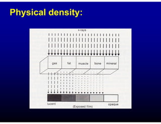
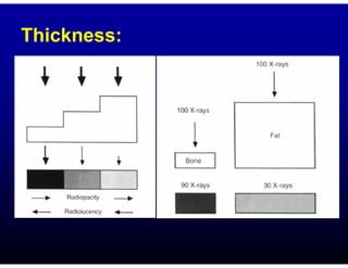
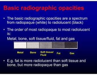
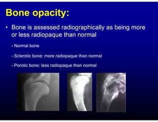
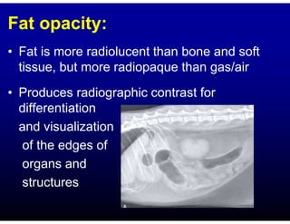
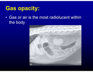
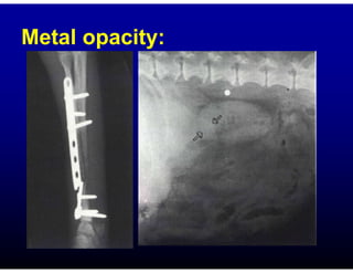
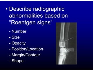
DIAGNOSTIC IMAGING
VM 352
RADIOGRAPHIC
INTERPRETATION
Dr. M. Makungu
Department of Veterinary
Surgery and Theriogenology
2022
X-rays and radiograph
• A radiograph is an image of the number and
distribution of X-rays that pass through the patient
and strike the film
• Areas struck by a large number of X-rays appear
black (radiolucent)
• Areas struck by few X-rays appear white
(radiopaque)
• Between these two extremes is a range of gray film
tones
• X-rays are not absorbed homogenous
by the body- differential absorption
• Factors affecting absorption
- Physical density
- Atomic number
- Thickness
Physical density:
Thickness:
Basic radiographic opacities
• The basic radiographic opacities are a spectrum
from radiopaque (white) to radiolucent (black)
• The order of most radiopaque to most radiolucent
is:
Metal, bone, soft tissue/fluid, fat and gas
• E.g. fat is more radiolucent than soft tissue and
bone, but more radiopaque than gas
Metal Bone Soft tissue/
Fluid
Fat Gas
Bone opacity:
• Bone is assessed radiographically as being more
or less radiopaque than normal
- Normal bone
- Sclerotic bone: more radiopaque than normal
- Porotic bone: less radiopaque than normal
Porotic
Sclerotic
Normal
Soft tissue opacity:
• Includes the radiographic opacities of normal
soft tissue such as muscle, solid and fluid
filled organs
• Variation in volume
and thickness create
a spectrum of varying
opacities on the
radiograph
VD
Fat opacity:
• Fat is more radiolucent than bone and soft
tissue, but more radiopaque than gas/air
• Produces radiographic contrast for
differentiation
and visualization
of the edges of
organs and
structures
Gas opacity:
• Gas or air is the most radiolucent within
the body
Metal opacity:
Interpretation
• Correlate the patient information and radiograph
being evaluated
• At least 2 projections/views perpendicular to
each other
• Evaluate the positioning and technical factors
• Systematically evaluation:
- From outside to inside
- Inside (body systems e.g. respiratory, digestive)
• Describe radiographic
abnormalities based on
“Roentgen signs”
- Number
- Size
- Opacity
- Position/Location
- Margin/Contour
- Shape
• Integrate radiographic findings and clinical
data
• Diagnosis

Radiographic interpretation.pdf

  • 1.
    DIAGNOSTIC IMAGING VM 352 RADIOGRAPHIC INTERPRETATION Dr.M. Makungu Department of Veterinary Surgery and Theriogenology 2022
  • 2.
    X-rays and radiograph •A radiograph is an image of the number and distribution of X-rays that pass through the patient and strike the film • Areas struck by a large number of X-rays appear black (radiolucent) • Areas struck by few X-rays appear white (radiopaque) • Between these two extremes is a range of gray film tones
  • 4.
    • X-rays arenot absorbed homogenous by the body- differential absorption • Factors affecting absorption - Physical density - Atomic number - Thickness
  • 5.
  • 6.
  • 7.
    Basic radiographic opacities •The basic radiographic opacities are a spectrum from radiopaque (white) to radiolucent (black) • The order of most radiopaque to most radiolucent is: Metal, bone, soft tissue/fluid, fat and gas • E.g. fat is more radiolucent than soft tissue and bone, but more radiopaque than gas Metal Bone Soft tissue/ Fluid Fat Gas
  • 8.
    Bone opacity: • Boneis assessed radiographically as being more or less radiopaque than normal - Normal bone - Sclerotic bone: more radiopaque than normal - Porotic bone: less radiopaque than normal Porotic Sclerotic Normal
  • 9.
    Soft tissue opacity: •Includes the radiographic opacities of normal soft tissue such as muscle, solid and fluid filled organs • Variation in volume and thickness create a spectrum of varying opacities on the radiograph VD
  • 10.
    Fat opacity: • Fatis more radiolucent than bone and soft tissue, but more radiopaque than gas/air • Produces radiographic contrast for differentiation and visualization of the edges of organs and structures
  • 11.
    Gas opacity: • Gasor air is the most radiolucent within the body
  • 12.
  • 13.
    Interpretation • Correlate thepatient information and radiograph being evaluated • At least 2 projections/views perpendicular to each other • Evaluate the positioning and technical factors • Systematically evaluation: - From outside to inside - Inside (body systems e.g. respiratory, digestive)
  • 15.
    • Describe radiographic abnormalitiesbased on “Roentgen signs” - Number - Size - Opacity - Position/Location - Margin/Contour - Shape
  • 17.
    • Integrate radiographicfindings and clinical data • Diagnosis