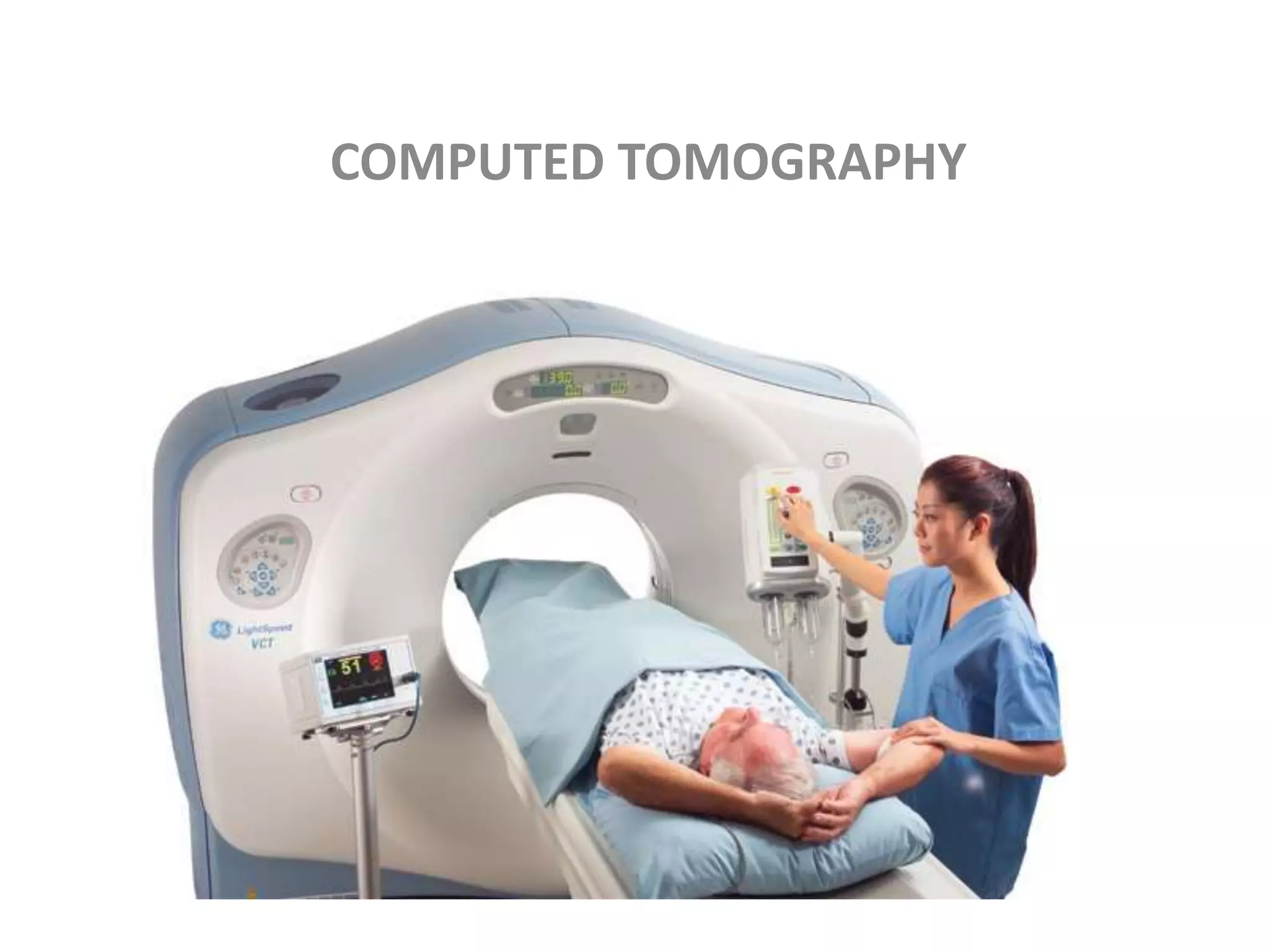
Diagnostic imaging modalities
Prepared by
Muhammad Umar
Radiology
• Radiology: The medical specialty concerned with
radiation for the diagnosis and treatment of disease,
including both ionizing radiation such as X-rays and
nonionizing radiation such as ultrasound. Also known
as roentgenology.
• Wilhelm Conrad Roentgen. Wilhelm Roentgen, a
German professor of physics, was the first person to
discover electromagnetic radiation in a wavelength
range commonly known as X-rays today. Although,
many people had observed the effects of X-ray beams
before, but Roentgen was the first one to study them
systematically.
o Conventional radiography
o Fluoroscopy
o Computed tomography
o Magnetic Resonance imaging
o Ultrasound
o Nuclear medicine
o mammography
Conventional Radiography
o Uses an electro magnetic ionizing radiation
o X-rays : are highly penetrating electromagnetic
ionizing radiation with shorter wavelength and
high frequency that travel through the space
at the speed of light or not at all.
o It always travel in a straight line and cause the
shadow of a radio opaque object place in their
path .
o It has a wavelength range from 10-9 to 10-16 m
Frequency range from 1017 to 1024 Hz.
Energy range from 103 to 1010 ev.
o Conventional radiography provides us 2D
images
Chest PA film
• The plain postero-anterior (PA) chest film i s the
most frequently requested radiological
examination. Visualization of the lungs is
excellent because of the inherent contrast of the
tissues of the thorax. Lateral films should not be
undertaken routinely. Comparison of the current
film with old films is valuable and should always
be undertaken if the old films are available. A
current film is mandatory before proceeding to
more complex investigations.
Chest X-ray
Major limitation of conventional
radiography
Conventional radiographs shows a three-
dimensional object as a two-dimensional
image. This results in overlying tissues being
superimposed on the image, a major
limitation of conventional radiography.
Five basic densities
• 
Black: Air
Dark gray: Fat
Light gray: Soft tissues or water
White: Calcification, bone
Whiter: Metal, contrast
X-RAY
TUBE
IMAGE
INTENSIFIER
TV CAMERA
(for digital screening)
CONTROLS
Fluoroscopy
Allows dynamic imaging of
Blood vessels ( angiography )
and ‘interventional’ procedures
T HE primary function of the fluoroscope is to provide real-time
dynamic viewing of anatomic structures. Dynamic studies are
examinations that show the motion of circulation or
the motion of internal structures. During fluoroscopy, the radiologist
generally uses contrast media to highlight the anatomy. The
radiologist then views a continuous image of the internal
structure while the x-ray tube is energized. If the radiologist
observes something during the fluoroscopic examination and
would like to preserve that image for further study, a radiograph
called a spot film can be taken without interruption of the
dynamic examination. The recent introduction of computer
technology into fluoroscopy and radiography has enhanced the
training and performance demands placed on radiologic
technologists.
ESOPHAGRAM
.
B: A large ulcerated GIST has replaced most of the gastric fundus (arrows).This
appearance would be atypical for an adenocarcinoma.
Upper limb venogram
COMPUTED TOMOGRAPHY
Tomography
The word tomography is derived from Greek
word Tomo mean to cut and Graphy mean to
draw or to image some thing.
The tomographic examination is design to image
only that structures that lies in the plane of
interest while blurring the structures that lies
above and below it . It is also called axial
tomography.
Principal of operation of Computed
Tomography
The simple Computed Tomography system consist of finely collimated
x-ray beam and a single detector.
The x-rays source and detectors moves simultaneously .
When the source –detector assembly makes one sweep or translation
across the patient , the internal structure of the body attenuate the
xrays beam according to their mass density and effective atomic
number.
At the end of this translation the source detector assembly return to
its starting position, and the entire assembly rotates and begin a
second translation .
If this process is repeated many time a large number of projection will
be obtained . These projections are then reconstructed and display
on tv monitor
mammography
• Mammography is the process of using low-
energy X-rays (usually around 30 kVp) to
examine the human breast for diagnosis and
screening. The goal of mammography is the
early detection of breast cancer, typically
through detection of characteristic masses or
microcalcification
• mammograms use doses of ionizing radiation
to create images. These images are then
analyzed for abnormal findings. It is usual to
employ lower-energy X-rays, typically Mo (K-
shell x-ray energies of 17.5 and 19.6 keV) and
Rh (20.2 and 22.7 keV) than those used for
radiography of bones

Medical imaging modalities(2)

  • 1.
    Diagnostic imaging modalities Preparedby Muhammad Umar Radiology
  • 2.
    • Radiology: Themedical specialty concerned with radiation for the diagnosis and treatment of disease, including both ionizing radiation such as X-rays and nonionizing radiation such as ultrasound. Also known as roentgenology. • Wilhelm Conrad Roentgen. Wilhelm Roentgen, a German professor of physics, was the first person to discover electromagnetic radiation in a wavelength range commonly known as X-rays today. Although, many people had observed the effects of X-ray beams before, but Roentgen was the first one to study them systematically.
  • 3.
    o Conventional radiography oFluoroscopy o Computed tomography o Magnetic Resonance imaging o Ultrasound o Nuclear medicine o mammography
  • 4.
    Conventional Radiography o Usesan electro magnetic ionizing radiation o X-rays : are highly penetrating electromagnetic ionizing radiation with shorter wavelength and high frequency that travel through the space at the speed of light or not at all. o It always travel in a straight line and cause the shadow of a radio opaque object place in their path .
  • 5.
    o It hasa wavelength range from 10-9 to 10-16 m Frequency range from 1017 to 1024 Hz. Energy range from 103 to 1010 ev. o Conventional radiography provides us 2D images
  • 6.
    Chest PA film •The plain postero-anterior (PA) chest film i s the most frequently requested radiological examination. Visualization of the lungs is excellent because of the inherent contrast of the tissues of the thorax. Lateral films should not be undertaken routinely. Comparison of the current film with old films is valuable and should always be undertaken if the old films are available. A current film is mandatory before proceeding to more complex investigations.
  • 8.
  • 10.
    Major limitation ofconventional radiography Conventional radiographs shows a three- dimensional object as a two-dimensional image. This results in overlying tissues being superimposed on the image, a major limitation of conventional radiography.
  • 11.
    Five basic densities • Black: Air Dark gray: Fat Light gray: Soft tissues or water White: Calcification, bone Whiter: Metal, contrast
  • 12.
    X-RAY TUBE IMAGE INTENSIFIER TV CAMERA (for digitalscreening) CONTROLS Fluoroscopy Allows dynamic imaging of Blood vessels ( angiography ) and ‘interventional’ procedures
  • 13.
    T HE primaryfunction of the fluoroscope is to provide real-time dynamic viewing of anatomic structures. Dynamic studies are examinations that show the motion of circulation or the motion of internal structures. During fluoroscopy, the radiologist generally uses contrast media to highlight the anatomy. The radiologist then views a continuous image of the internal structure while the x-ray tube is energized. If the radiologist observes something during the fluoroscopic examination and would like to preserve that image for further study, a radiograph called a spot film can be taken without interruption of the dynamic examination. The recent introduction of computer technology into fluoroscopy and radiography has enhanced the training and performance demands placed on radiologic technologists.
  • 14.
  • 17.
    . B: A largeulcerated GIST has replaced most of the gastric fundus (arrows).This appearance would be atypical for an adenocarcinoma.
  • 18.
  • 19.
  • 20.
    Tomography The word tomographyis derived from Greek word Tomo mean to cut and Graphy mean to draw or to image some thing. The tomographic examination is design to image only that structures that lies in the plane of interest while blurring the structures that lies above and below it . It is also called axial tomography.
  • 21.
    Principal of operationof Computed Tomography The simple Computed Tomography system consist of finely collimated x-ray beam and a single detector. The x-rays source and detectors moves simultaneously . When the source –detector assembly makes one sweep or translation across the patient , the internal structure of the body attenuate the xrays beam according to their mass density and effective atomic number. At the end of this translation the source detector assembly return to its starting position, and the entire assembly rotates and begin a second translation . If this process is repeated many time a large number of projection will be obtained . These projections are then reconstructed and display on tv monitor
  • 24.
    mammography • Mammography isthe process of using low- energy X-rays (usually around 30 kVp) to examine the human breast for diagnosis and screening. The goal of mammography is the early detection of breast cancer, typically through detection of characteristic masses or microcalcification
  • 25.
    • mammograms usedoses of ionizing radiation to create images. These images are then analyzed for abnormal findings. It is usual to employ lower-energy X-rays, typically Mo (K- shell x-ray energies of 17.5 and 19.6 keV) and Rh (20.2 and 22.7 keV) than those used for radiography of bones