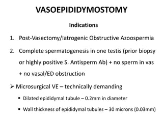
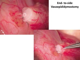
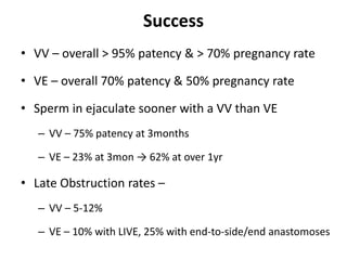
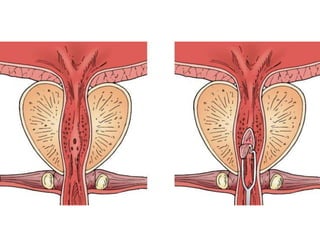
OBSTRUCTIVE AzoospermiA
Surgical Management
Operative Chat
- Dr Abhishek Pandey
PROCEDURES
VASO-VASOSTOMY (VV)
VASO-EPIDIDYMOSTOMY (VE)
TURED
HISTORY
• 1903 – Martin – Fistula technique of VE using silver
wires (gonococcal epididymal obstruction)
• 1919 – Quinby – VV for Vasectomy reversal
• 1977 – Silber – Two-layered microsurgical vasal
anastomosis technique
• 1978 – Silber – VE to single epididymal tubule
• 1998 – Goldstein – Microdot microsurgical VV
VASOVASOSTOMY
• 2-6% vasectomized men seek reversal
• 6% azoospermic men – Iatrogenic-Obstructive
• Document adequate spermatogenesis – prior history
of natural fertility
Indications
• Vasectomy reversal – Vast majority
• Iatrogenic vasal injury repair – 2° to Herniorraphy
Patient Preparation
• GA preferred
– Operating microscope magnifies patient movement
– If VE required – surgery time may exceed 4-5hr
• Regional & local possible in co-operative patient
• Position – Supine & towards foot-end of the table
• Surgeons to support ulnar part of hands, wrists, &
forearms – minimize tremor
Operating Microscope
Is Microscope Necessary?
• Loupes – ×2.5 to ×4 magnification
• Visualization of the inner lumen of the vas deferens
requires ×16 magnification
• Microscope
– the depth of focus is clear
– Light is constantly supplied directly to the patient
– Instrument rests on a stand & is immobile – Surgeon
can move his neck
Specific Instruments
Vas deferens Clamp
Micro-forceps with
tying platform
Curved non-locking micro needle holder
The Goldstein Vas approximating clamp
90° Vas cutting forceps
Foam from suture packets with dots
to secure the microneedles after use
Microneedle
With sharp point
• Microforceps & Microscissors
• Micro-tip bipolar cautery forceps
• Glass microscope slides with coverslips & Capillary tubes
• Microirrigator – 10mL syringe + 24gauge angiocatheter
• 15-degree Microknife & Microswabs
• Bench microscope
• Sutures – 9-0 nylon & double-armed 10-0 nylon
Surgical Approaches
Scrotal Approach
• Bilateral high vertical scrotal incisions
• Ideal for vasectomy reversal
• Testis delivered with TV intact
• Min. 1cm lateral to penis-base
• Can be extended to external ring
Inguinal Approach
• Suspected inguinal obstruction – prior Herniorrhaphy /Orchiopexy
Vas Preparation
• Vas mobilized to allow tension-free anastomosis without
stripping adventitia (vasal vessels)
• Stay close to Vas – avoid Testicular artery injury
• Vasal Gap – Separate cord structures from vas
– Blunt dissection till internal ring
– Dissect vas free of attachment to epididymal tunica
– Cut floor of inguinal canal & rerout vas
– Dissect epididymis off testis from vasoepididymal junction to
caput
Dissect vas free of attachment to epididymal tunica
Dissect epididymis off testis from
vasoepididymal junction to caput
Upto 10cm length can be gained
• Testicular end of the vas is cut transversely
– Ultrasharp knife drawn through a slotted nerve-holding clamp
– Perfect 90° cut
• Vasal Fluid sampled
• Slide Preparation – Vasal fluid + NS/RL
• Abdominal end of Vas prepared similarily
– lumen gently dilated with microvessel dilator
– cannulated with a 24-gauge angiocatheter sheath
– Injection of NS/RL to confirm patency
– Vas is recut to obtain a fresh surface
– Minimum instrumentation of mucosa
Bull’s eye appearance of healthy cut vas
Gently open with microforceps
NO dilatation
Vasal Fluid Examination
• Vasovasostomy
– Copious crystal clear watery fluid ± sperm
– Copious fluid with sperms / sperm heads
– Scant fluid with granuloma & sperm on barbotage
• Vasoepididymostomy
– Thick paste-like with no sperm/sperm heads
– Scant fluid with no granuloma Or sperm/sperm heads
– Fluid with occasional grape-like clusters of sperm heads
Sperm Granuloma
Multiple Vasal Obstruction
• Probe abdominal vas with 0 polypropylene to locate
level of obstruction
• Within 5cm – excise & do a single VV
• Two VV – intervening segment devascularizes
• If not possible –
– Preserve sperm
Varicocelectomy + Vasovasostomy
• Do VV/VE first → re-assess postoperative semen quality
• Varicocelectomy – at ≥6mon – arteriovenous channels
form across anastomosis
• Can be done together –
– Microscope be used
– Testicular artery preserved
• Cremasteric & peri-arterial venous channels divided
Anastomotic Principles
1. Accurate mucosa-to-mucosa approximation
2. Leakproof anastomosis
3. Tension-free anastomosis
4. Good blood supply
5. Healthy mucosa and muscularis
6. Good atraumatic anastomotic technique
Microsurgical Multilayer Microdot Method
• Microdots allow precise placement of microsutures
• Double dot at the 12 o’clock to maintain orientation
• Separates planning from placement of suture
• Needle exit marked
• Six mucosal sutures
Techniques to Stabilize the Vas
1. Goldstein vas approximating clamp
2. Micro-wipe technique
3. Stay sutures
• Double-arm suture – inside-out needle placement
• Anterior Layer → Rotate vas 180° → Posterior layer
• 10-0 nylon double-arm for inner layer
• 9-0 nylon for outer muscular layer
• Irrigate with heparinized saline before last suture
• Approximate vasal sheath
Modified Single Layer Anastomosis
Flip 180° & repeat
Seromuscular stiches
Initial 3 full-thickness stiches
VASOEPIDIDYMOSTOMY
Indications
1. Post-Vasectomy/Iatrogenic Obstructive Azoospermia
2. Complete spermatogenesis in one testis (prior biopsy
or highly positive S. Antisperm Ab) + no sperm in vas
+ no vasal/ED obstruction
 Microsurgical VE – technically demanding
 Dilated epididymal tubule – 0.2mm in diameter
 Wall thickness of epididymal tubules – 30 microns (0.03mm)
Microsurgical Single-tubule VE
Fistula Technique
Secondary epididymal obstruction
•Pressure-induced “blowouts”
•Inspissation
Distal tubule
Proximal tubule
Identify Target Tubule
Target tubule
parallel to incoming
abdominal vas
Avoid tubule
perpendicular to
abdominal vas
Avoid off-center,
irregular, or
lengthy defect
Measure & choose
most distal tubule
for anastomosis
Vas Preparation
• Angled Marks Vas-cutting forceps – 15°
– allows Vas to “lay down” over the defect
– provides larger elliptical lumen for intussusception
• Confirm abdominal-vas patency
• Confirm distal vas position
Delivery of Target Tubule
• Observe tubules through the epididymal tunic
– Change in diameter of the tubules
– Color of the fluid within
• Find the best tubule, correctly oriented in a window
without overlying vessels
• Incise outer tunica → free target tubule → it should
protrude from adjacent tubules
• Tubule should be at the center of tunica window
Protruding tubule Inking tubule
Tunica Incision
Dilated tubule
Intussusception Vasoepididymostomy
• Two-stitch Longitudinal Intussusception (LIVE) technique
10 O clock suture
2 O clock suture
4 O clock suture
8 O clock suture
End- to-side
Vasoepididymostomy
Success
• VV – overall > 95% patency & > 70% pregnancy rate
• VE – overall 70% patency & 50% pregnancy rate
• Sperm in ejaculate sooner with a VV than VE
– VV – 75% patency at 3months
– VE – 23% at 3mon → 62% at over 1yr
• Late Obstruction rates –
– VV – 5-12%
– VE – 10% with LIVE, 25% with end-to-side/end anastomoses
Robotic-assisted VV
• Elimination of tremor & improved stability
• Surgeon ergonomics/decreased surgeon fatigue
• Scalability of motion
• Three-dimensional high-definition visualization
• Manipulate 3 instruments & camera simultaneously
• Not requiring a specialty skilled microsurgical assistant
• Potential of improving operative times
• Allows surgeon to back out of console & re-engage
on exact same field
Cons
• Decreased Magnification
• Increased cost
Postop Management
• Complications – Bleed, Infection, Hydrocele, Testicular
atrophy (rare)
• Scrotal Support for 6wk at all times (in sleep too) →
Support during strenous activity till pregnancy
• No strenous work for 3wk
• No intercourse or ejaculation for 3wk
Follow-up
• Semen analyses at 1, 3 & 6mon → 6 monthly
• Azoospermia persists at 6mon → redo VV/VE
TURED
• Trans-Urethral Resection of the Ejaculatory Ducts
• TRUS-guided aspiration of cystic/dilated ejaculatory
ducts or seminal vesicles
• If motile sperm found → cryopreserved
– 2-3 ml Indigo-carmine diluted + contrast instilled
– Decision of TURED based on imaging
• No sperm in bilateral Vas + ED obstruction → abandon
Technique
• Cold knife incision alone → re-obstruction
• Cutting loop + anterior push on prostate per rectum
• EDs course b/w bladder-neck & veru → exit along
lateral aspect of veru
• Preserve bladder neck, striated sphincter, & rectal
mucosa
• Hemivasotomies closed using microsurgical technique
Complications
Complications
• Reflux of urine into EDs, Vas, and Seminal Vesicles
• Acute & Chronic Epididymitis
• Retrograde Ejaculation
Results – Not Favoured
• Increased semen volume – in two-third cases
• Sperm in ejaculate – 50%
Thank You
A
• A

Azoospermia - Operative

  • 1.
  • 2.
  • 3.
    HISTORY • 1903 –Martin – Fistula technique of VE using silver wires (gonococcal epididymal obstruction) • 1919 – Quinby – VV for Vasectomy reversal • 1977 – Silber – Two-layered microsurgical vasal anastomosis technique • 1978 – Silber – VE to single epididymal tubule • 1998 – Goldstein – Microdot microsurgical VV
  • 4.
    VASOVASOSTOMY • 2-6% vasectomizedmen seek reversal • 6% azoospermic men – Iatrogenic-Obstructive • Document adequate spermatogenesis – prior history of natural fertility Indications • Vasectomy reversal – Vast majority • Iatrogenic vasal injury repair – 2° to Herniorraphy
  • 5.
    Patient Preparation • GApreferred – Operating microscope magnifies patient movement – If VE required – surgery time may exceed 4-5hr • Regional & local possible in co-operative patient • Position – Supine & towards foot-end of the table • Surgeons to support ulnar part of hands, wrists, & forearms – minimize tremor
  • 6.
  • 7.
    Is Microscope Necessary? •Loupes – ×2.5 to ×4 magnification • Visualization of the inner lumen of the vas deferens requires ×16 magnification • Microscope – the depth of focus is clear – Light is constantly supplied directly to the patient – Instrument rests on a stand & is immobile – Surgeon can move his neck
  • 8.
    Specific Instruments Vas deferensClamp Micro-forceps with tying platform Curved non-locking micro needle holder
  • 9.
    The Goldstein Vasapproximating clamp 90° Vas cutting forceps Foam from suture packets with dots to secure the microneedles after use Microneedle With sharp point
  • 10.
    • Microforceps &Microscissors • Micro-tip bipolar cautery forceps • Glass microscope slides with coverslips & Capillary tubes • Microirrigator – 10mL syringe + 24gauge angiocatheter • 15-degree Microknife & Microswabs • Bench microscope • Sutures – 9-0 nylon & double-armed 10-0 nylon
  • 11.
    Surgical Approaches Scrotal Approach •Bilateral high vertical scrotal incisions • Ideal for vasectomy reversal • Testis delivered with TV intact • Min. 1cm lateral to penis-base • Can be extended to external ring Inguinal Approach • Suspected inguinal obstruction – prior Herniorrhaphy /Orchiopexy
  • 12.
    Vas Preparation • Vasmobilized to allow tension-free anastomosis without stripping adventitia (vasal vessels) • Stay close to Vas – avoid Testicular artery injury • Vasal Gap – Separate cord structures from vas – Blunt dissection till internal ring – Dissect vas free of attachment to epididymal tunica – Cut floor of inguinal canal & rerout vas – Dissect epididymis off testis from vasoepididymal junction to caput
  • 13.
    Dissect vas freeof attachment to epididymal tunica Dissect epididymis off testis from vasoepididymal junction to caput Upto 10cm length can be gained
  • 14.
    • Testicular endof the vas is cut transversely – Ultrasharp knife drawn through a slotted nerve-holding clamp – Perfect 90° cut • Vasal Fluid sampled
  • 15.
    • Slide Preparation– Vasal fluid + NS/RL • Abdominal end of Vas prepared similarily – lumen gently dilated with microvessel dilator – cannulated with a 24-gauge angiocatheter sheath – Injection of NS/RL to confirm patency – Vas is recut to obtain a fresh surface – Minimum instrumentation of mucosa
  • 16.
    Bull’s eye appearanceof healthy cut vas Gently open with microforceps NO dilatation
  • 17.
    Vasal Fluid Examination •Vasovasostomy – Copious crystal clear watery fluid ± sperm – Copious fluid with sperms / sperm heads – Scant fluid with granuloma & sperm on barbotage • Vasoepididymostomy – Thick paste-like with no sperm/sperm heads – Scant fluid with no granuloma Or sperm/sperm heads – Fluid with occasional grape-like clusters of sperm heads
  • 18.
  • 19.
    Multiple Vasal Obstruction •Probe abdominal vas with 0 polypropylene to locate level of obstruction • Within 5cm – excise & do a single VV • Two VV – intervening segment devascularizes • If not possible – – Preserve sperm
  • 20.
    Varicocelectomy + Vasovasostomy •Do VV/VE first → re-assess postoperative semen quality • Varicocelectomy – at ≥6mon – arteriovenous channels form across anastomosis • Can be done together – – Microscope be used – Testicular artery preserved • Cremasteric & peri-arterial venous channels divided
  • 21.
    Anastomotic Principles 1. Accuratemucosa-to-mucosa approximation 2. Leakproof anastomosis 3. Tension-free anastomosis 4. Good blood supply 5. Healthy mucosa and muscularis 6. Good atraumatic anastomotic technique
  • 22.
    Microsurgical Multilayer MicrodotMethod • Microdots allow precise placement of microsutures • Double dot at the 12 o’clock to maintain orientation • Separates planning from placement of suture • Needle exit marked • Six mucosal sutures
  • 23.
    Techniques to Stabilizethe Vas 1. Goldstein vas approximating clamp 2. Micro-wipe technique 3. Stay sutures
  • 24.
    • Double-arm suture– inside-out needle placement • Anterior Layer → Rotate vas 180° → Posterior layer • 10-0 nylon double-arm for inner layer • 9-0 nylon for outer muscular layer • Irrigate with heparinized saline before last suture • Approximate vasal sheath
  • 27.
    Modified Single LayerAnastomosis Flip 180° & repeat Seromuscular stiches Initial 3 full-thickness stiches
  • 28.
    VASOEPIDIDYMOSTOMY Indications 1. Post-Vasectomy/Iatrogenic ObstructiveAzoospermia 2. Complete spermatogenesis in one testis (prior biopsy or highly positive S. Antisperm Ab) + no sperm in vas + no vasal/ED obstruction  Microsurgical VE – technically demanding  Dilated epididymal tubule – 0.2mm in diameter  Wall thickness of epididymal tubules – 30 microns (0.03mm)
  • 29.
  • 30.
    Secondary epididymal obstruction •Pressure-induced“blowouts” •Inspissation Distal tubule Proximal tubule
  • 31.
    Identify Target Tubule Targettubule parallel to incoming abdominal vas Avoid tubule perpendicular to abdominal vas Avoid off-center, irregular, or lengthy defect Measure & choose most distal tubule for anastomosis
  • 32.
    Vas Preparation • AngledMarks Vas-cutting forceps – 15° – allows Vas to “lay down” over the defect – provides larger elliptical lumen for intussusception • Confirm abdominal-vas patency • Confirm distal vas position
  • 33.
    Delivery of TargetTubule • Observe tubules through the epididymal tunic – Change in diameter of the tubules – Color of the fluid within • Find the best tubule, correctly oriented in a window without overlying vessels • Incise outer tunica → free target tubule → it should protrude from adjacent tubules • Tubule should be at the center of tunica window
  • 34.
    Protruding tubule Inkingtubule Tunica Incision Dilated tubule
  • 35.
    Intussusception Vasoepididymostomy • Two-stitchLongitudinal Intussusception (LIVE) technique
  • 36.
    10 O clocksuture 2 O clock suture 4 O clock suture 8 O clock suture
  • 37.
  • 38.
    Success • VV –overall > 95% patency & > 70% pregnancy rate • VE – overall 70% patency & 50% pregnancy rate • Sperm in ejaculate sooner with a VV than VE – VV – 75% patency at 3months – VE – 23% at 3mon → 62% at over 1yr • Late Obstruction rates – – VV – 5-12% – VE – 10% with LIVE, 25% with end-to-side/end anastomoses
  • 39.
    Robotic-assisted VV • Eliminationof tremor & improved stability • Surgeon ergonomics/decreased surgeon fatigue • Scalability of motion • Three-dimensional high-definition visualization • Manipulate 3 instruments & camera simultaneously • Not requiring a specialty skilled microsurgical assistant • Potential of improving operative times
  • 40.
    • Allows surgeonto back out of console & re-engage on exact same field Cons • Decreased Magnification • Increased cost
  • 41.
    Postop Management • Complications– Bleed, Infection, Hydrocele, Testicular atrophy (rare) • Scrotal Support for 6wk at all times (in sleep too) → Support during strenous activity till pregnancy • No strenous work for 3wk • No intercourse or ejaculation for 3wk
  • 42.
    Follow-up • Semen analysesat 1, 3 & 6mon → 6 monthly • Azoospermia persists at 6mon → redo VV/VE
  • 43.
    TURED • Trans-Urethral Resectionof the Ejaculatory Ducts • TRUS-guided aspiration of cystic/dilated ejaculatory ducts or seminal vesicles • If motile sperm found → cryopreserved – 2-3 ml Indigo-carmine diluted + contrast instilled – Decision of TURED based on imaging • No sperm in bilateral Vas + ED obstruction → abandon
  • 44.
    Technique • Cold knifeincision alone → re-obstruction • Cutting loop + anterior push on prostate per rectum • EDs course b/w bladder-neck & veru → exit along lateral aspect of veru • Preserve bladder neck, striated sphincter, & rectal mucosa • Hemivasotomies closed using microsurgical technique
  • 46.
    Complications Complications • Reflux ofurine into EDs, Vas, and Seminal Vesicles • Acute & Chronic Epididymitis • Retrograde Ejaculation Results – Not Favoured • Increased semen volume – in two-third cases • Sperm in ejaculate – 50%
  • 47.
  • 48.