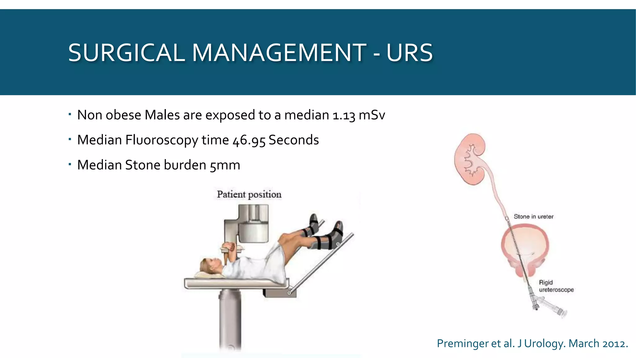
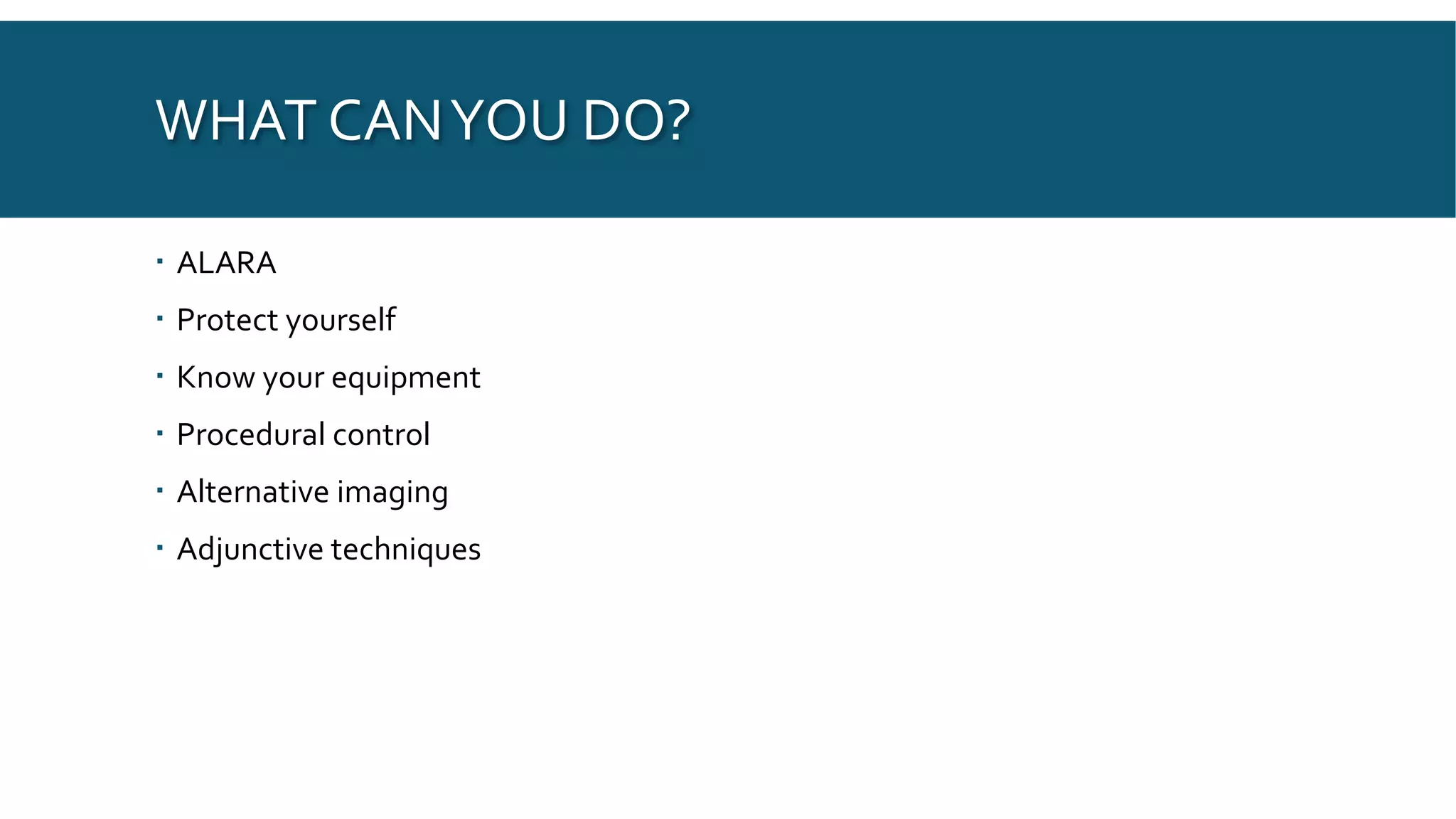
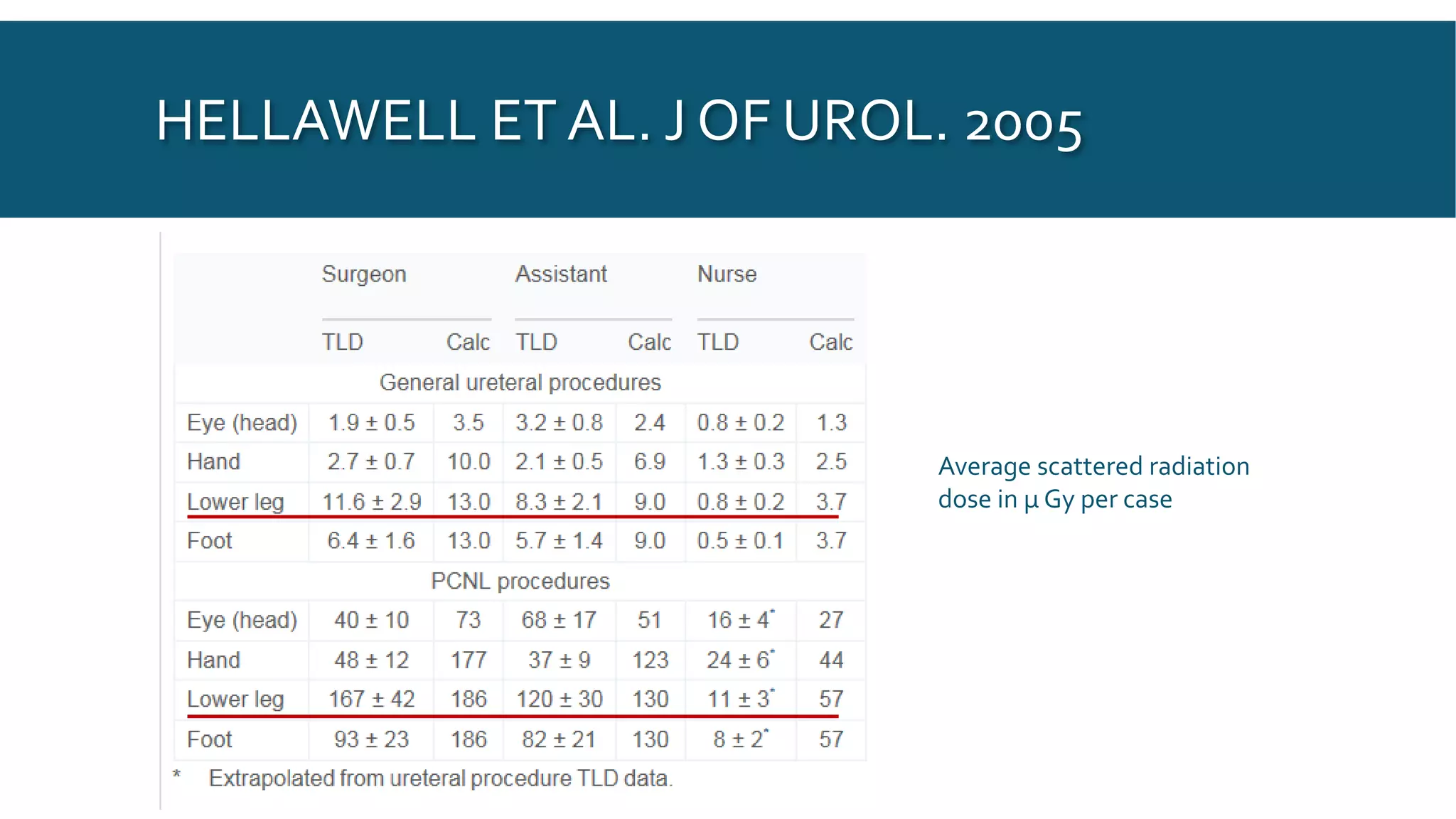
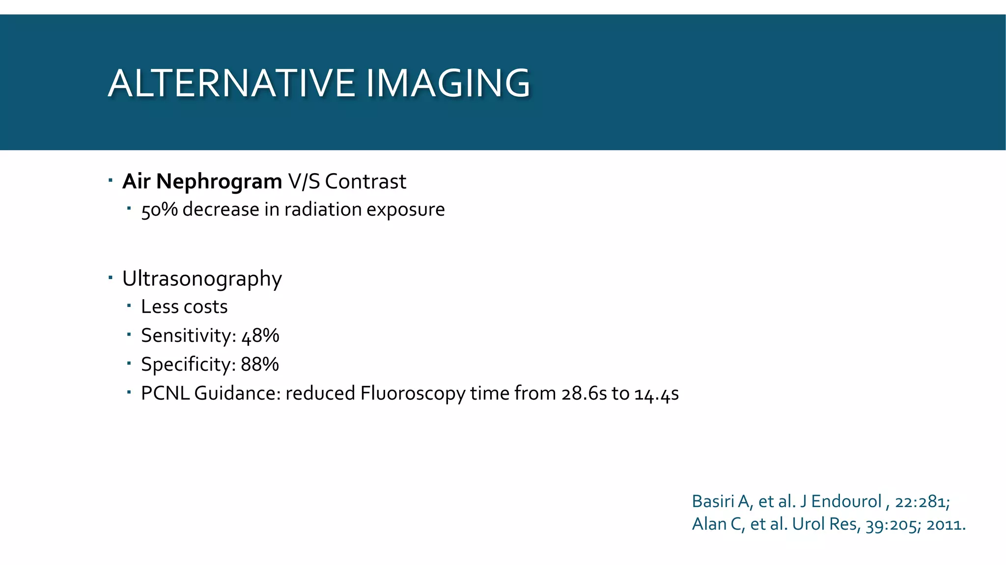
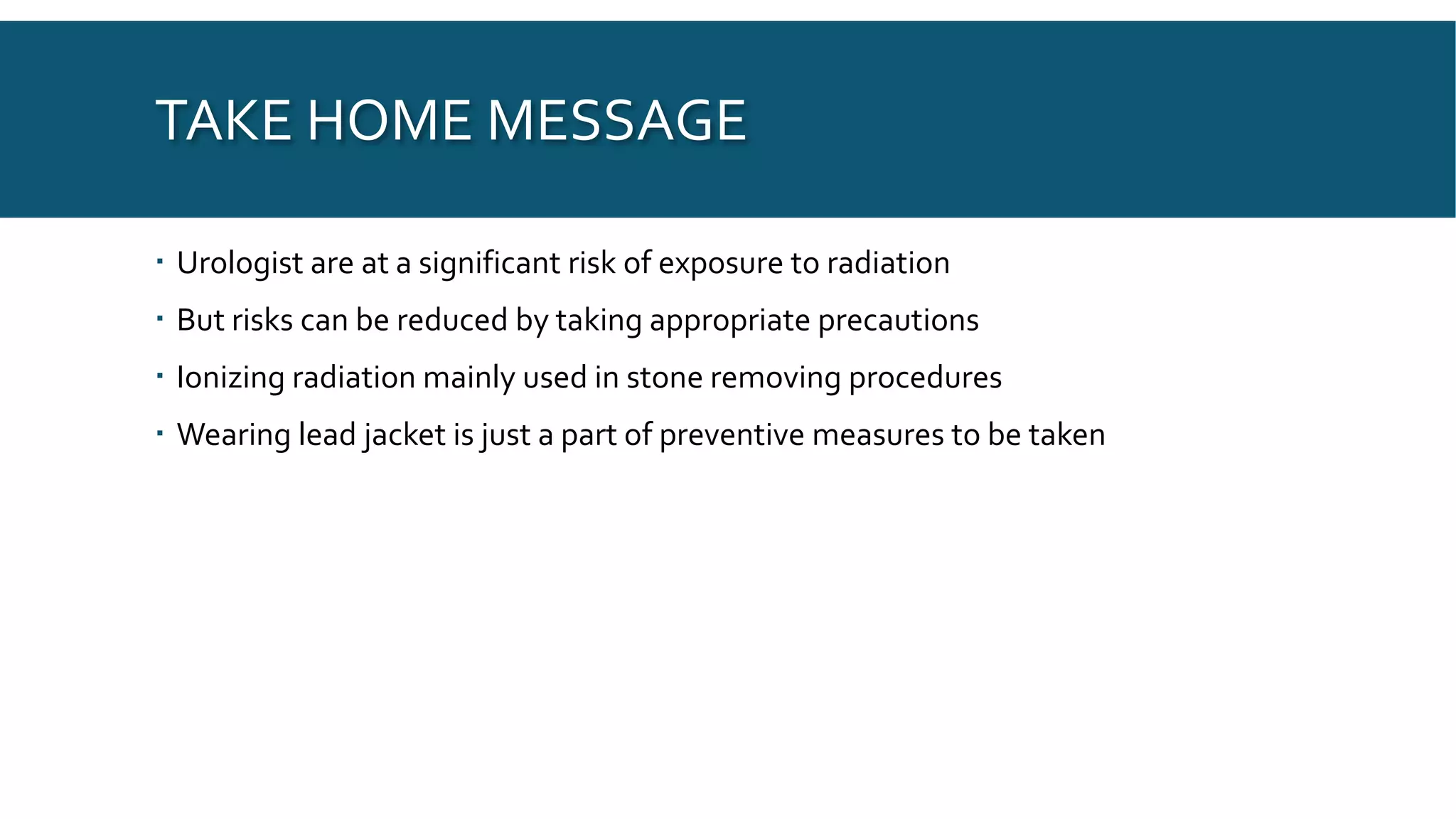
This document discusses radiation exposure in urology. It defines different types of radiation and their effects on the body. Radiation exposure can be a risk for both patients and surgeons in urology procedures like PCNL, URS, and ESWL that use fluoroscopy. The risks of radiation exposure include acute radiation syndrome and increased risk of cancer induction. The document provides recommendations for minimizing radiation exposure through principles like ALARA, protective equipment, procedural controls, alternative imaging techniques, and understanding radiology equipment settings.
9. SO,WHY AREWEWORRIED?
When exposed to Ionizing radiation
Causes changes in the cell division process of the body by affecting DNA structure
Damages protein structure of cells
Direct damage to tissues
The generic term for the detrimental effects of Radiation is termed as
ACUTE RADIATION SYNDROME
RADIATION SICKNESS
10. TERMINOLOGY
Ionizing Radiation:
Radiation from an source with enough energy to separate the subatomic particles and is
a known Carcinogen
Deterministic effect:
Describes the cause and effect relationship of Ionizing Radiation
Also known as Non-Stochastic Effect,
Dependent upon dose, dose rate, dose fractionation, irradiated volume and type of
radiation
Skin Erythema 2-5 Gy Irreversible Skin Damage 20-40Gy
Hair loss 2-5 Gy Sterility 2-3 Gy
Cataracts 0.5 -5Gy Lethality 3-5GyWhole body
Fetal Abnormality 0.1-0.5 Gy
11. TERMINOLOGY
Stochastic Effect:
Effect the occurs by chance
No threshold, not dependent upon dose
Caused by Mutational changes in cell DNA, Cell still remains viable after radiation effect
Increased level of exposure does not effect severity of disease but affect the probability
of development
Example:Cancer Induction
12. TERMINOLOGY
Absorbed Dose: Amount of radiation in theTissues
Measured in Gray (Gy)
Equivalent dose: Absorbed dose in a specific organ or tissue
Exposure: Number of ions produced by X-rays per Kilogram of air
Measured in Roentgen (R)
Effective Dose: Reflects whole body exposure/Radiation present in the
environment
Measured in rem or Sievert (Sv)
Dose area product (DAP): Radiation dose to air multipled by the are of X-ray field
Measured in Gy*cm2
13. THRESHOLD
European Union
Effective Dose limit is 100mSv over 5 years
No more than 50mSv in any one year
United States of America
Effective dose limit of 50mSv in one year
Lifetime limit of 10mSv x Age (Years)
International Commission on Radiological Protection
No more than 20mSv/yr over 5 years
No more than 50mSv in one year
14. BACKGROUND
X rays has been used to diagnosed diseases in the kidney and urinary tract for
about a century
However, use of Fluoroscopy is relatively new
Since the advent of Lithotripsy, use of CT has been increasing for mapping the
anatomy, diagnosis of diseases
A number of procedures, like PCNL, RIRS, Stent placement, ESWL,Tumor
ablation require fluoroscopy or other form of ionizing radiation.
In the United states; Per capita exposure to radiation has increased from
0.54mSv in 1980 TO 3.0mSv in 2006
600% increase
15.
16. RISKS IN UROLOGY - PATIENTS
Mainly in stone patients
Ranging from 1.18 to 37.66 mSv
Acute Stone episode patients undergo 1 – 4 radiographic studies in 1 year period
after stone event
Obesity increases mean Effective Dose by 177%
BMI 30-39.9 Kg/m2 = twofold increase in Effective Dose
BMI >40 kg/m2 =Threefold increase in Effective Dose
ChenTT, et al. J Urol, 194:878; 2015.
17. SURGICAL MANAGEMENT - URS
Non obese Males are exposed to a median 1.13 mSv
Median Fluoroscopy time 46.95 Seconds
Median Stone burden 5mm
Preminger et al. J Urology. March 2012.
18. SURGICAL MANAGEMENT - PCNL
Mean Effective Dose for Right PCNL: 7.63 mSv
Mean Effective Dose for Left PCNL: 8.11 mSv
Risks for increased Exposure:
High BMI,
Increased Stone Burden
Increased number of access tracts
Preminger et al. J Urology. March 2012.
19. SURGICAL MANAGEMENT - ESWL
Mean total effective dose: 1.71 mSv in males
Mean total effective dose: 1.82 mSv in females
Lipkin et al. J Urol 2015; 194: 878 885
21. RISKS IN UROLOGY - SURGEONS
Radiation exposure: arises due to scatter from the beam, patient and operating
table
Lens of the eye is most radiosensitive
>10mSv of exposure (cumulative) in 1 year increases lifetime risk of fatal cancer by
3%
>20mSV of exposure in 1 year increases risk of fatal cancer between 1:1000 and
1: 10000
Risk of fatal malignancy in general population is 1:500000
Hanna L, et al. J Endo, 29: 526; 2015.
22. WHAT CANYOU DO?
ALARA
Protect yourself
Know your equipment
Procedural control
Alternative imaging
Adjunctive techniques
23. ALARA
AS LOW AS REASONABLY ACHIEVABLE
Reduce Radiation exposure
Time: minimize “Beam-On”Time
Distance: Double the distance from the
source, exposure dose is 1/4th
Shielding: lightweight, radioprotective
clothing
Scatter
Affected by patient size, position, settings on
the machine, shielding, filtration, angulation.
28. HELLAWELL ET AL. J OF UROL. 2005
4 months period, 18 ureteral procedures
Average Fluoroscopy time: 78s
Mobile C-arm unit with under table X-rayTube
Dosimetry placed in 7 positions on body
Forehead
Little Finger B/L
Anterior Legs B/L
Upper aspect of foot/ankle B/L
29. HELLAWELL ET AL. J OF UROL. 2005
Average scattered radiation
dose in μ Gy per case
30. PROTECTIONS
Standard Lead jacket requires 0.35mm thickness
Does not prevent absolutely
But
Reduces transmission by up to 100 fold
0.25mm allow 10% radiation transmission
0.5mm allows 2% of radiation transmission
Leaded eyewear
Leaded Gloves
Wear Dosimeters
Hellawell G, et al. J Urol 174, 948; 2005.
31. KNOWYOUR EQUIPMENT
Low dose rate setting
Minimize use of Cine mode
Collimate the beam
Use magnification as little as possible
Image intensifier as close to patient as possible
Keep field clean of radiodense objects, such as metallic objects, as these increases
scatter by reflection.
32. Image Intensifier
X ray Source
Collimator
Filter
4 Axis Direction Control
Monitor
Exposure Indicator Light
X ray Generator
Control Panels
33.
34.
35. PROCEDURAL CONTROL
Mark Body surface forTargetting
Coning of radiation field
Radioprotective draping of patient
Laser guided C-arm
36. ALTERNATIVE IMAGING
Air Nephrogram V/S Contrast
50% decrease in radiation exposure
Ultrasonography
Less costs
Sensitivity: 48%
Specificity: 88%
PCNL Guidance: reduced Fluoroscopy time from 28.6s to 14.4s
BasiriA, et al. J Endourol , 22:281;
Alan C, et al. Urol Res, 39:205; 2011.
37. TAKE HOME MESSAGE
Urologist are at a significant risk of exposure to radiation
But risks can be reduced by taking appropriate precautions
Ionizing radiation mainly used in stone removing procedures
Wearing lead jacket is just a part of preventive measures to be taken
Editor's Notes
Gravitational radiation is generated, when a particle has enough energy be expelled form the source at the speed of light, and is able to change the fabric of time and space. This was previously though to be theoretical and was proposed by albert Einstein on 1916, and was only recently proven by the Hadron Collider and the discovery of the higgs boson particle. It is createdin small amounts by planets and star, and are largely created by black holes
Acoustic Radiation includes Sound wave released by objects because of their specific vibration. Also included sound emitted by speakers, ultrasounds and seismic waves and requires a medium for transmission
Particle radiation are formed when an object receives enough energy for breaking up of the atom and formation of subatomic particles. Includes Alpha waves, when 2 Neutron and 2 protons expelled from the atom. These are low energy radiation, which is even blocked by the cloth fabric, but may cause detrimental effects if it is inhaled or ingested. Beta waves are a type of particle radiation when an electron is expelled form an atom. These are high energy radiation, which is 8000 times higher than alpha particles, it is blocked by most solid objects, but travels through skin and may cause radiation sickness.
Electromagnetic radiation are energy radiation, in forms of waves and includes a variety of forms.
These are divided into ionizing and non ionizing radiations.
Particle radiation are a type of ionizing radiation, other than electromagnetic radiation formed when an object receives enough energy for breaking up of the atom and formation of subatomic particles. Includes Alpha waves, when 2 Neutron and 2 protons expelled from the atom. These are low energy radiation, which is even blocked by the cloth fabric, but may cause detrimental effects if it is inhaled or ingested. Beta waves are a type of particle radiation when an electron is expelled form an atom. These are high energy radiation, which is 8000 times higher than alpha particles, it is blocked by most solid objects, but travels through skin and may cause radiation sickness.
Gamma Rays are a type of electromagnetic radiation, which propel in form of waves of Photons and does not have any mass therefore, is able to pass through most solid structure including the human body.
So, to answer the Question, “Why are we worried?”
This was an experiment was done in the mayo clinic to measure the exposure in relation to the distance from the source.
At 50cm distance, exposure was found to be 220 milliroentgen per hour
At 100cm distance, exposure was found to be reduced to 55 milliroentgen per hour
At 200cm distance, exposure was found to be 12.8 milliroentgen per hour
All beams of electromagnetic waves, including visible light and and lionizing radiation likewise, has 3 methods of scattering.
Reflection: has the highest dose of ionizing radiation, which is present under the table
Refraction: and lower than those reflected doses, as part of the refracted dose is absorbed by the patient and the operating table.