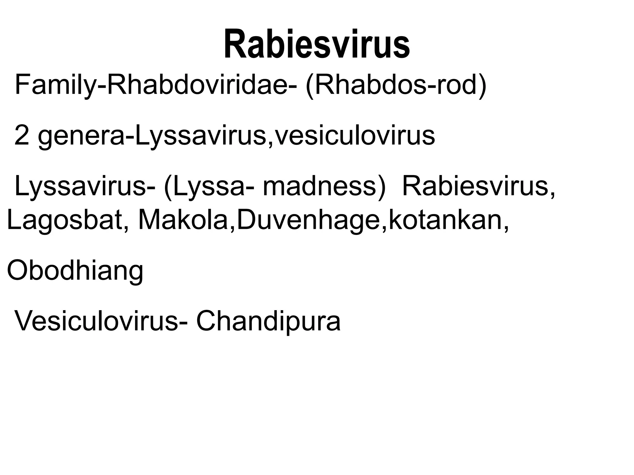
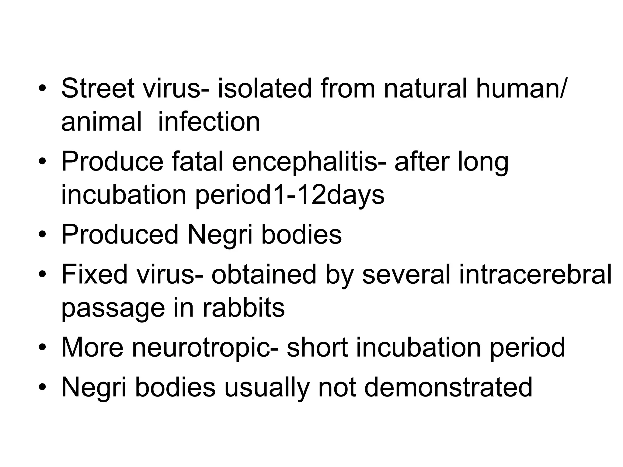
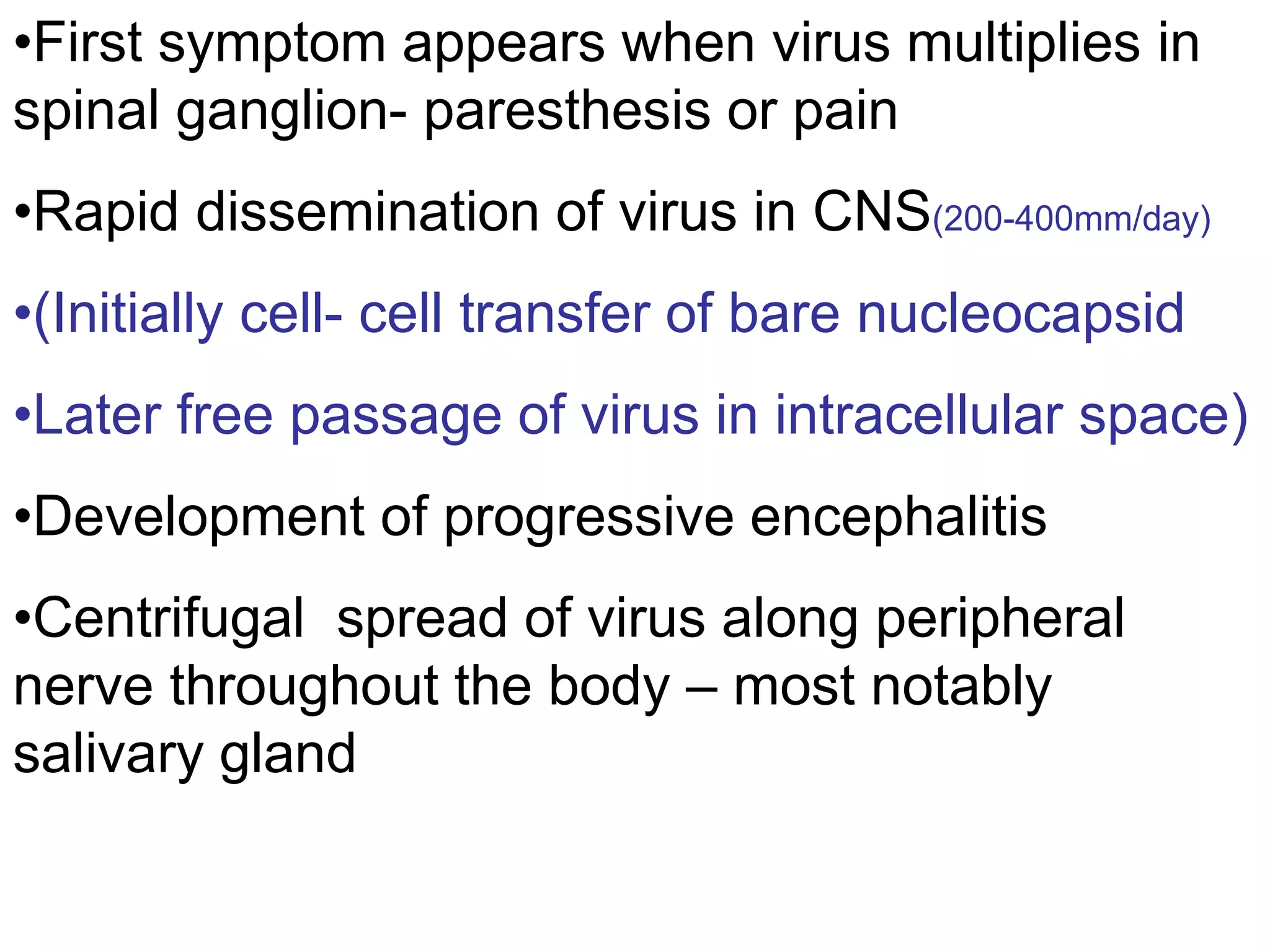
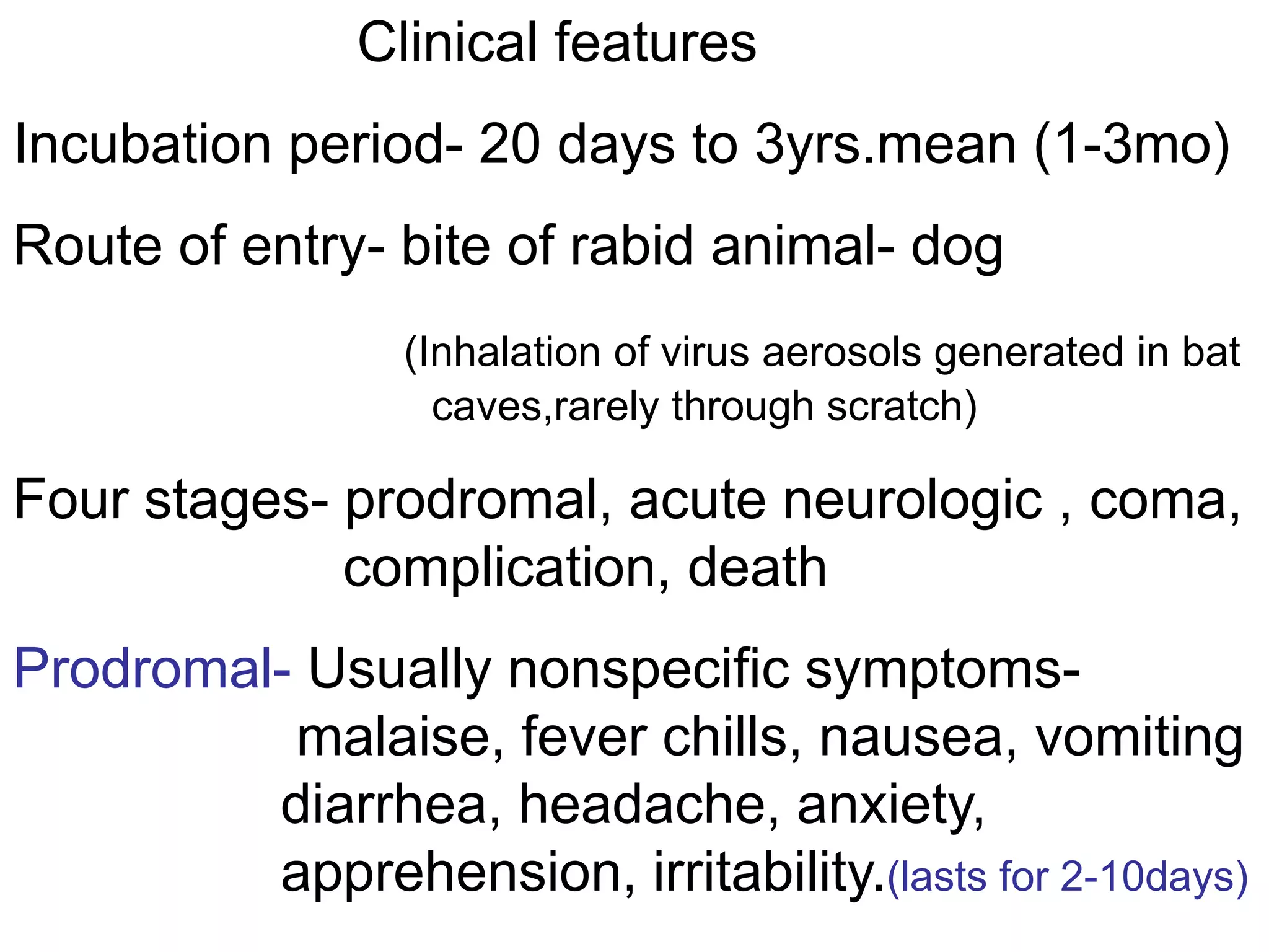
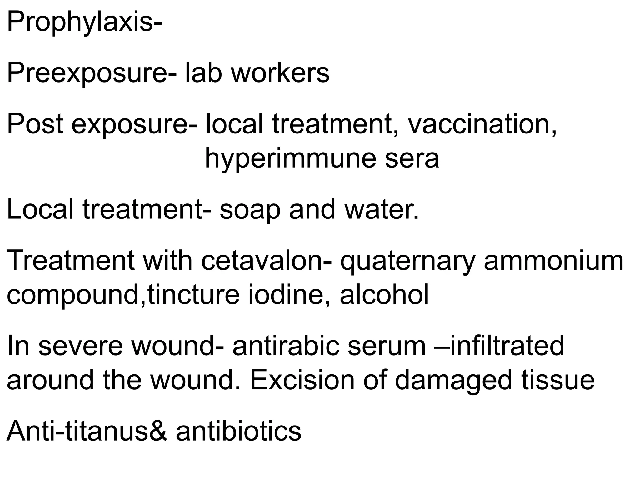
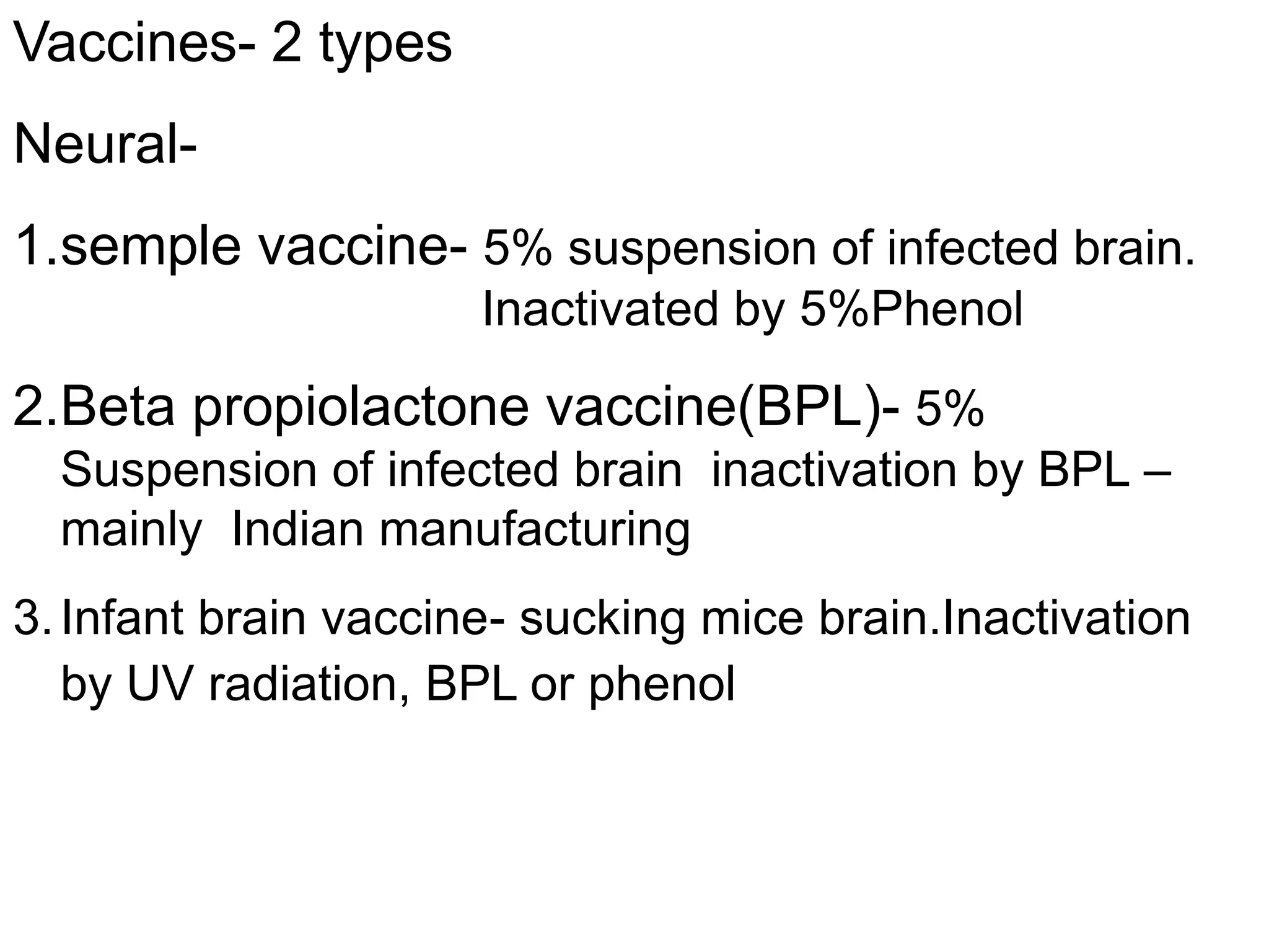
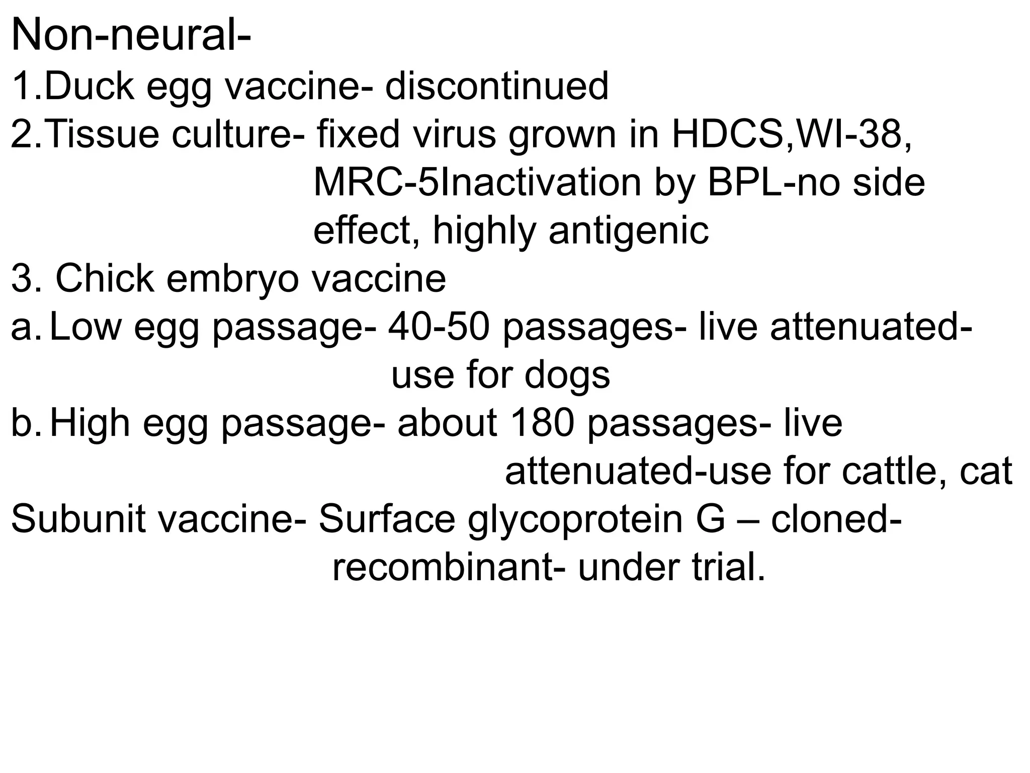
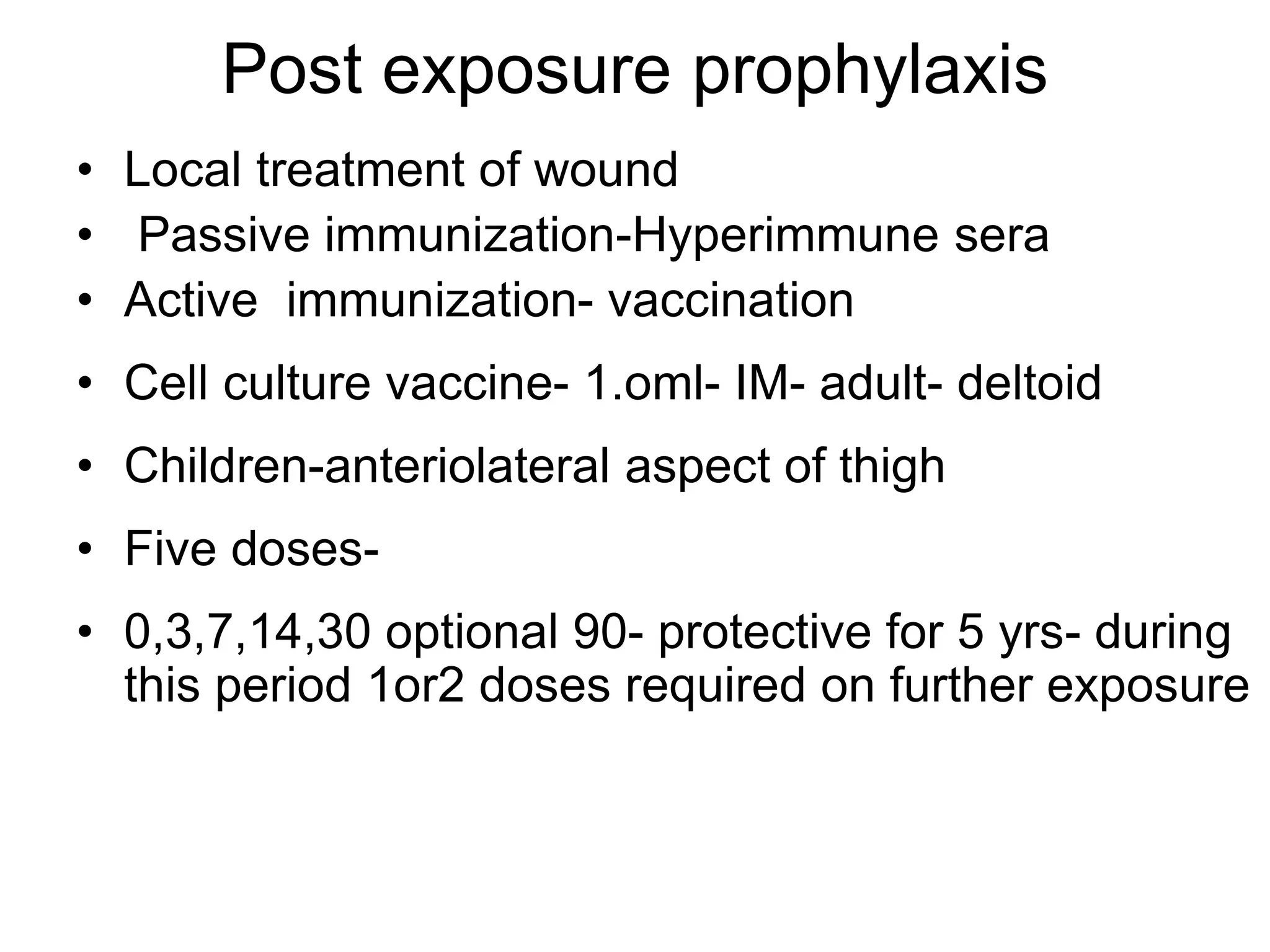
Rabies virus
Dr. Sshrutkirti Gupta
Rabiesvirus
Family-Rhabdoviridae- (Rhabdos-rod)
2 genera-Lyssavirus,vesiculovirus
Lyssavirus- (Lyssa- madness) Rabiesvirus,
Lagosbat, Makola,Duvenhage,kotankan,
Obodhiang
Vesiculovirus- Chandipura
Morphology
•Shape- Bullet shaped, one end round other end
concave or planer
Knob like surface projections all over the surface
except on concave end
Below the envelop has matrix (M) protein layer –
may project out as bleb from concave end
•Size- length varies
•Enveloped RNA Virus
• Genome –SSRNA-unsegmented-
• Codes for 5 proteins-
• Glycoprotein (G),
• Nucleocapsid (N),
• Viral polymerase (L),
• Two smaller nonstructural proteins (NS)
(P)-MW 38000 & (M)-MW 28000
Peplomer/spike- Present on surface,
Glycoprotein in nature,9 nm long
Haemagglutinating Property- optimal with goose
cells at 0-40cpH 6.2
Nucleocapsid-165x50nm,30-35 coils
Resistance-
highly resistant against dryness, cold
Infective for many wks in cadaver.
Sensitive to lipid solvent-chloroform, acetone,
ether. Ethanol, iodine,soap,phenol,formalin,beta
propiolactone, sunlight, UV
• Street virus- isolated from natural human/
animal infection
• Produce fatal encephalitis- after long
incubation period1-12days
• Produced Negri bodies
• Fixed virus- obtained by several intracerebral
passage in rabbits
• More neurotropic- short incubation period
• Negri bodies usually not demonstrated
Pathogenesis
Highly neurotropic
•Attachment of virus to host cell after inoculation
Via glycoprotein spike
Site of attachment- nicotinic acetylcholine binding
site of plasma mem. of muscle
cell (myotubule)
•Enters peripheral nerve
(time required depends upon conc. &site of bite)
After sufficient multiplication cross myoneural
junction- entry in CNS
(through myelinated sensory and motor axon
terminal)
•Once enter CNS infection cannot be halted by
vaccination
Passage to CNS occurs axonally through
axoplasmic flow (12-24mm/day)until it reaches
next neuron at the level of spinal cord
•First symptom appears when virus multiplies in
spinal ganglion- paresthesis or pain
•Rapid dissemination of virus in CNS(200-400mm/day)
•(Initially cell- cell transfer of bare nucleocapsid
•Later free passage of virus in intracellular space)
•Development of progressive encephalitis
•Centrifugal spread of virus along peripheral
nerve throughout the body – most notably
salivary gland
Clinical features
Incubation period- 20 days to 3yrs.mean (1-3mo)
Route of entry- bite of rabid animal- dog
(Inhalation of virus aerosols generated in bat
caves,rarely through scratch)
Four stages- prodromal, acute neurologic , coma,
complication, death
Prodromal- Usually nonspecific symptoms-
malaise, fever chills, nausea, vomiting
diarrhea, headache, anxiety,
apprehension, irritability.(lasts for 2-10days)
Acute neurologic phase-
Two forms- 1. furious encephalitis (>80%)
2. paralytic (dumb) form (20%)
Furious encephalitis- Hyperactivity, disorientation,
hallucination or bizarre behavior. Hyperactivity
becomes intermittent. Signs of autonomic
instability-often prominent (hyperthermia, tachycardia,
hypertension) Hydrophobia- Attempt of drinking
follows severe spasm of pharynx, larynx.
Aerophobia-due to exaggerated respiratory
irritant reflexes
•Fever, muscle fasciculation, hyperventilation, focal convulsion
Paralytic (dumb) rabies- initially patient is
mentally intact. No hyperactivity as in furious type
Fever, headache frequently present
Paralysis- maximal at bitten extremity,may be
diffuse& symmetric or may be ascending type
neck stiffness
Mental condition gradually deteriorate from
confusion to disorientation, stupor & finally coma
•Last for 2-7 days
•Abrupt death due to respiratory or cardiac arrest
Coma- develops in about 10 days after onset of
symptoms
Lasts for few hours to month
Complications- Neurologic, Pitutary, Pulmonary
Cardiovascular, other
Death due to respiratory or cardiac failure
Laboratory diagnosis
Specimens-
Antimortem- Saliva, CSF, Corneal impression
smear, biopsy from neck( above
hair line) , facial skin biopsy
Postmortem- CSF, brain biopsy.
1.Isolation of virus-
a. intracerebral inoculation in the mice
observe brain tissue for inclusion bodies on
28th day
b. Tissue culture- cell lines used-WI 38, BHK21 --
detection of virus by immunofluorescence-
Results available in 2-4days
2. Demonstration of viral antigen-
By immunofluorescence
3. Detection of inclusion bodies-(Negri bodies)
Intracytoplasmic, oval/ round, purple-pink
characteristic basophilic inner grannules
Size- 3-27 mm, eosinophilic
Stain- Seller’s technique, immunofluorescence
Common- hippocampus, horns of Ammon
Purkingie’s cells of cerebellum
20% of absent
Demonstration of Antibodies
Neutralising antibodies- high titre- in CSF
appear around 6th day
2- 25 times more than serum titre
Detection of Nucleic acid- RT PCR
Prophylaxis-
Preexposure- lab workers
Post exposure- local treatment, vaccination,
hyperimmune sera
Local treatment- soap and water.
Treatment with cetavalon- quaternary ammonium
compound,tincture iodine, alcohol
In severe wound- antirabic serum –infiltrated
around the wound. Excision of damaged tissue
Anti-titanus& antibiotics
Vaccines- 2 types
Neural-
1.semple vaccine- 5% suspension of infected brain.
Inactivated by 5%Phenol
2.Beta propiolactone vaccine(BPL)- 5%
Suspension of infected brain inactivation by BPL –
mainly Indian manufacturing
3.Infant brain vaccine- sucking mice brain.Inactivation
by UV radiation, BPL or phenol
Vaccination Schedule
Semple vaccine BPL vaccine
Class I 2ml x 7days 2ml x 7days
Class II 5ml x 14 days 3ml x 10days
Class III 10ml x 14 days 5ml x 10days
Booster required for BPL vaccine for Class II& III
Class II- 1 dose 3wks after last 10th injection
ClassIII- 2 doses- 1 7days after 10th injection 2nd 2wks
after 1st booster
Non-neural-
1.Duck egg vaccine- discontinued
2.Tissue culture- fixed virus grown in HDCS,WI-38,
MRC-5Inactivation by BPL-no side
effect, highly antigenic
3. Chick embryo vaccine
a.Low egg passage- 40-50 passages- live attenuated-
use for dogs
b.High egg passage- about 180 passages- live
attenuated-use for cattle, cat
Subunit vaccine- Surface glycoprotein G – cloned-
recombinant- under trial.
cell culture Vaccine schedule
•Same for children and adult
•Same for all three available vaccines
•Pre-exposure-
•Three doses- 0,7,21 dose-1ml Route- IM
•Booster after 1 yr & then every 5 yr.
Post exposure prophylaxis
• Local treatment of wound
• Passive immunization-Hyperimmune sera
• Active immunization- vaccination
• Cell culture vaccine- 1.oml- IM- adult- deltoid
• Children-anteriolateral aspect of thigh
• Five doses-
• 0,3,7,14,30 optional 90- protective for 5 yrs- during
this period 1or2 doses required on further exposure
Passive immunisation- Hyperimmune serum
1.Human antirabies immunoglobulin- (HRIG)
20 IU/kg body wt- half dose is infiltrated
locally in wound and other half administered- IM
•Should not be given to individual who had prior
active immunization
2.Equine/ horse hyperimmune serum-(ERIG)
40 IU /kg body wt
risk of hypersensitivity

Rabiesvirus.ppt

  • 1.
  • 2.
    Rabiesvirus Family-Rhabdoviridae- (Rhabdos-rod) 2 genera-Lyssavirus,vesiculovirus Lyssavirus-(Lyssa- madness) Rabiesvirus, Lagosbat, Makola,Duvenhage,kotankan, Obodhiang Vesiculovirus- Chandipura
  • 3.
    Morphology •Shape- Bullet shaped,one end round other end concave or planer Knob like surface projections all over the surface except on concave end Below the envelop has matrix (M) protein layer – may project out as bleb from concave end •Size- length varies •Enveloped RNA Virus
  • 4.
    • Genome –SSRNA-unsegmented- •Codes for 5 proteins- • Glycoprotein (G), • Nucleocapsid (N), • Viral polymerase (L), • Two smaller nonstructural proteins (NS) (P)-MW 38000 & (M)-MW 28000
  • 5.
    Peplomer/spike- Present onsurface, Glycoprotein in nature,9 nm long Haemagglutinating Property- optimal with goose cells at 0-40cpH 6.2 Nucleocapsid-165x50nm,30-35 coils
  • 7.
    Resistance- highly resistant againstdryness, cold Infective for many wks in cadaver. Sensitive to lipid solvent-chloroform, acetone, ether. Ethanol, iodine,soap,phenol,formalin,beta propiolactone, sunlight, UV
  • 9.
    • Street virus-isolated from natural human/ animal infection • Produce fatal encephalitis- after long incubation period1-12days • Produced Negri bodies • Fixed virus- obtained by several intracerebral passage in rabbits • More neurotropic- short incubation period • Negri bodies usually not demonstrated
  • 10.
    Pathogenesis Highly neurotropic •Attachment ofvirus to host cell after inoculation Via glycoprotein spike Site of attachment- nicotinic acetylcholine binding site of plasma mem. of muscle cell (myotubule) •Enters peripheral nerve (time required depends upon conc. &site of bite)
  • 11.
    After sufficient multiplicationcross myoneural junction- entry in CNS (through myelinated sensory and motor axon terminal) •Once enter CNS infection cannot be halted by vaccination Passage to CNS occurs axonally through axoplasmic flow (12-24mm/day)until it reaches next neuron at the level of spinal cord
  • 12.
    •First symptom appearswhen virus multiplies in spinal ganglion- paresthesis or pain •Rapid dissemination of virus in CNS(200-400mm/day) •(Initially cell- cell transfer of bare nucleocapsid •Later free passage of virus in intracellular space) •Development of progressive encephalitis •Centrifugal spread of virus along peripheral nerve throughout the body – most notably salivary gland
  • 13.
    Clinical features Incubation period-20 days to 3yrs.mean (1-3mo) Route of entry- bite of rabid animal- dog (Inhalation of virus aerosols generated in bat caves,rarely through scratch) Four stages- prodromal, acute neurologic , coma, complication, death Prodromal- Usually nonspecific symptoms- malaise, fever chills, nausea, vomiting diarrhea, headache, anxiety, apprehension, irritability.(lasts for 2-10days)
  • 14.
    Acute neurologic phase- Twoforms- 1. furious encephalitis (>80%) 2. paralytic (dumb) form (20%) Furious encephalitis- Hyperactivity, disorientation, hallucination or bizarre behavior. Hyperactivity becomes intermittent. Signs of autonomic instability-often prominent (hyperthermia, tachycardia, hypertension) Hydrophobia- Attempt of drinking follows severe spasm of pharynx, larynx. Aerophobia-due to exaggerated respiratory irritant reflexes •Fever, muscle fasciculation, hyperventilation, focal convulsion
  • 15.
    Paralytic (dumb) rabies-initially patient is mentally intact. No hyperactivity as in furious type Fever, headache frequently present Paralysis- maximal at bitten extremity,may be diffuse& symmetric or may be ascending type neck stiffness Mental condition gradually deteriorate from confusion to disorientation, stupor & finally coma •Last for 2-7 days •Abrupt death due to respiratory or cardiac arrest
  • 16.
    Coma- develops inabout 10 days after onset of symptoms Lasts for few hours to month Complications- Neurologic, Pitutary, Pulmonary Cardiovascular, other Death due to respiratory or cardiac failure
  • 17.
    Laboratory diagnosis Specimens- Antimortem- Saliva,CSF, Corneal impression smear, biopsy from neck( above hair line) , facial skin biopsy Postmortem- CSF, brain biopsy. 1.Isolation of virus- a. intracerebral inoculation in the mice observe brain tissue for inclusion bodies on 28th day
  • 18.
    b. Tissue culture-cell lines used-WI 38, BHK21 -- detection of virus by immunofluorescence- Results available in 2-4days 2. Demonstration of viral antigen- By immunofluorescence 3. Detection of inclusion bodies-(Negri bodies) Intracytoplasmic, oval/ round, purple-pink characteristic basophilic inner grannules Size- 3-27 mm, eosinophilic Stain- Seller’s technique, immunofluorescence
  • 19.
    Common- hippocampus, hornsof Ammon Purkingie’s cells of cerebellum 20% of absent Demonstration of Antibodies Neutralising antibodies- high titre- in CSF appear around 6th day 2- 25 times more than serum titre Detection of Nucleic acid- RT PCR
  • 20.
    Prophylaxis- Preexposure- lab workers Postexposure- local treatment, vaccination, hyperimmune sera Local treatment- soap and water. Treatment with cetavalon- quaternary ammonium compound,tincture iodine, alcohol In severe wound- antirabic serum –infiltrated around the wound. Excision of damaged tissue Anti-titanus& antibiotics
  • 21.
    Vaccines- 2 types Neural- 1.semplevaccine- 5% suspension of infected brain. Inactivated by 5%Phenol 2.Beta propiolactone vaccine(BPL)- 5% Suspension of infected brain inactivation by BPL – mainly Indian manufacturing 3.Infant brain vaccine- sucking mice brain.Inactivation by UV radiation, BPL or phenol
  • 22.
    Vaccination Schedule Semple vaccineBPL vaccine Class I 2ml x 7days 2ml x 7days Class II 5ml x 14 days 3ml x 10days Class III 10ml x 14 days 5ml x 10days Booster required for BPL vaccine for Class II& III Class II- 1 dose 3wks after last 10th injection ClassIII- 2 doses- 1 7days after 10th injection 2nd 2wks after 1st booster
  • 23.
    Non-neural- 1.Duck egg vaccine-discontinued 2.Tissue culture- fixed virus grown in HDCS,WI-38, MRC-5Inactivation by BPL-no side effect, highly antigenic 3. Chick embryo vaccine a.Low egg passage- 40-50 passages- live attenuated- use for dogs b.High egg passage- about 180 passages- live attenuated-use for cattle, cat Subunit vaccine- Surface glycoprotein G – cloned- recombinant- under trial.
  • 24.
    cell culture Vaccineschedule •Same for children and adult •Same for all three available vaccines •Pre-exposure- •Three doses- 0,7,21 dose-1ml Route- IM •Booster after 1 yr & then every 5 yr.
  • 25.
    Post exposure prophylaxis •Local treatment of wound • Passive immunization-Hyperimmune sera • Active immunization- vaccination • Cell culture vaccine- 1.oml- IM- adult- deltoid • Children-anteriolateral aspect of thigh • Five doses- • 0,3,7,14,30 optional 90- protective for 5 yrs- during this period 1or2 doses required on further exposure
  • 26.
    Passive immunisation- Hyperimmuneserum 1.Human antirabies immunoglobulin- (HRIG) 20 IU/kg body wt- half dose is infiltrated locally in wound and other half administered- IM •Should not be given to individual who had prior active immunization 2.Equine/ horse hyperimmune serum-(ERIG) 40 IU /kg body wt risk of hypersensitivity