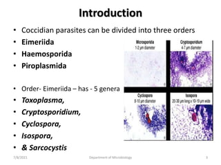
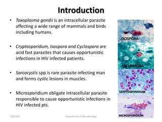
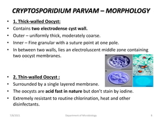
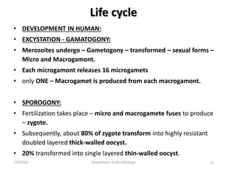
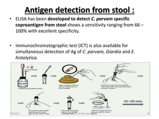
Coccidian Parasites
UNDER GARADUATE STUDENT’S LECTURE ON
BY
GUNJAL PN
ASSIST. PROF.
DEPT OF MICROBIOLOGY
DVVPF’S MEDICAL COLLEGE & HOSPITAL
AHMENDAGAR
7/8/2021 Department of Microbiology 1
Aims and Objectives
At the end of the session, the students will be able to
understand:
• Classification of Coccidian parasites
• Toxoplasma gondii
• Cryptosporidium parvam
• Cyclospora cayetanesis
• Isospora belli
• Pathogenicity and Clinical features of coccidian
parasites.
• Laboratory Diagnosis of coccidian parasites
• Treatment and prevention.
7/8/2021 Department of Microbiology 2
Introduction
• Coccidian parasites can be divided into three orders
• Eimeriida
• Haemosporida
• Piroplasmida
• Order- Eimeriida – has - 5 genera
• Toxoplasma,
• Cryptosporidium,
• Cyclospora,
• Isospora,
• & Sarcocystis
7/8/2021 Department of Microbiology 3
Introduction
• Toxoplasma gondii is an intracellular parasite
affecting a wide range of mammals and birds
including humans.
• Cryptosporidum, Isospora and Cyclospora are
acid fast parasites that causes opportunistic
infections in HIV infected patients.
• Sarcocystis spp is rare parasite infecting man
and forms cystic lesions in muscles.
• Microsporidium obligate intracellular parasite
responsible to cause opportunistic infections in
HIV infected pts.
ISOSPORA
CYCLOSPORA
CRYPTOSPORIDIUM
MICROSPORIDIUM
7/8/2021 Department of Microbiology 4
CRYPTOSPORIDIUM PARVAM
• It is an intestinal coccidian parasite affecting
many animals and humans.
• Can cause self limiting diarrhea in helathy
individuals.
• Can be an opportunistic pathogen in
immunocompromised patients (HIV) causing chronic
persistent life threatening diarrhea.
• Tyzzer (1907), was first to describe it in gastric crypts of
laboratory mice.
• The first human case was reported in 1976.
7/8/2021 Department of Microbiology 5
CRYPTOSPORIDIUM PARVAM
• Belongs to family Cryptosporidiidae.
• Different from other coccidian parasites- does not go into
deep host cells, but confined to an intracellular extra
cytoplasmic location.
• All sexual and asexual stages of development takes place
within a parasitophorous vaculoe
• That lies just below the cell membrane of the brush border
epithelium of the small intestine.
• Cryptosporidium parvam is the most common species
affecting humans.
• Other species infect wide range of mammals and reptiles.
7/8/2021 Department of Microbiology 6
CRYPTOSPORIDIUM PARVAM – MORPHOLOGY
• OOCYST:
• Infective form for humans.
• Diagnostic form.
• Excreted in the feces.
• Round, small, 4-6 um, surrounded by a cyst wall and bears four
sporozoites.
• Each sporozoite is crescentic shaped with pointed anterior end, blunt
posterior end and a nucleus located posteriorly .
• Two types of oocysts are demonstrated
• 1. thick walled
• 2. thin walled
Fig: Sporulated oocysts of A: Cryptosporidium B: Cyclospora C: Isospora
7/8/2021 Department of Microbiology 7
CRYPTOSPORIDIUM PARVAM – MORPHOLOGY
• 1. Thick-walled Oocyst:
• Contains two electrodense cyst wall.
• Outer – uniformly thick, moderately coarse.
• Inner – Fine granular with a suture point at one pole.
• In between two walls, lies an electroluscent middle zone containing
two oocyst membranes.
• 2. Thin-walled Oocyst :
• Surrounded by a single layered membrane.
• The oocysts are acid fast in nature but don’t stain by iodine.
• Extremely resistant to routine chlorination, heat and other
disinfectants.
7/8/2021 Department of Microbiology 8
Life cycle
• Host: Cryptosporidium parvam completes
its life cycle (both sexual and asexual stages)
in single host (human or animal).
• Infective stage: Sporulated oocyst.
• Thick-walled oocyst is infectious to other person.
• Thin-walled oocyst can cause autoinfection (through contaminated
fingers).
• Mode of transmission : Feco-oral route – ingestion of thick wall
oocyst.
Sporulated oocyst
7/8/2021 Department of Microbiology 9
Life cycle
• DEVELOPMENT IN HUMAN:
• EXCYSTATION:
• In small intestine, the suture present in the inner wall of the oocyst
gets dissolved and 4 slender crescent shaped sporozoites are
released from each oocyst.
• Various factors like pancreatic enzymes are bile salts help in
excystation.
• INVASION:
• Sporozoites invade the brush border epithelium of the small intestine
and lie inside a parasitophorous vaculoe near the microvilli surface,
within which all the stages of development take place.
7/8/2021 Department of Microbiology 10
Life cycle
• DEVELOPMENT IN HUMAN:
• EXCYSTATION - SCHIZOGONY:
• The sporozoites – differentiated into - trophozoites – which
undergo – asexual multiplication (Schizogony) to produce -
Type I Meronts.
• Each type I meront undergoes – schizogony – releases 8
merozoites – again invade the adjacent enterocytes and
undergo repeated schizogony to produce - Type II meronts.
7/8/2021 Department of Microbiology 11
Life cycle
• DEVELOPMENT IN HUMAN:
• EXCYSTATION - GAMATOGONY:
• Merozoites undergo – Gametogony – transformed – sexual forms –
Micro and Macrogamont.
• Each microgamont releases 16 microgamets
• only ONE – Macrogamet is produced from each macrogamont.
• SPOROGONY:
• Fertilization takes place – micro and macrogamete fuses to produce
– zygote.
• Subsequently, about 80% of zygote transform into highly resistant
doubled layered thick-walled oocyst.
• 20% transformed into single layered thin-walled oocyst.
7/8/2021 Department of Microbiology 12
Life cycle
• EXCYSTATION - SPOROGONY:
• Within host cell , oocyst undergo- Sporogony – sporozoites.
• Sporulated oocysts are excreted in feces.
• Thick-walled oocyst infects the new hosts where as the thin-
walled oocysts – infects – the same host (autoinfection).
• Prepatent period:
• Period from the time of ingestion of oocyst to completion of
the life cycle and release of new oocysts in environment
through human feces (4-22 days approx.).
7/8/2021 Department of Microbiology 13
7/8/2021 Department of Microbiology 14
7/8/2021 Department of Microbiology 15
Epidemiology
• Cryptosporidiosis is a zoonotic disease.
• PREVALENCE RATE OF CRYPTOSPORIDIOSIS:
• In immunocompetent people – prevalence – India (developing
countries) – 2.4 -15%, Whereas in Western Countries (1.4-6%).
• In immunocompromised hosts (HIV positive pts) – 12-46% in
developing countries, 7-21% in developed countries.
7/8/2021 Department of Microbiology 16
Epidemiology
• RISK FACTOR’S CONTRIBUTING TO DISEASE INCLUDE:
• Low infective dose of C. parvam (10-100 oocysts ) can initiate
infection.
• Large multiplication capacity (> 1010) in single host.
• Resistant to the available drugs and disinfectants.
• Large animals and humans are the reservoirs.
• Lack of appropriate immune response, poor sanitation, travel to
underdeveloped countries, zoonotic contact.
• Peak age of Infection: Infants and Children.
7/8/2021 Department of Microbiology 17
Pathogenesis and Clinical Features
• ATTACHMENT:
• Sporozoites attach to the brush border epithelium of the small
intestine with the help of a unique protein called as CP47
(47kDa C. parvam protein).
• PENETRATIO:
• Discharge from the apicomplex present in the anterior end of
the sporozoites help in invasion.
• Following penetration, parasite forms a parasitophorous
vacuole near microvilli surface of the host cells (intracellular
extra cytoplasmic location).
7/8/2021 Department of Microbiology 18
Pathogenesis and Clinical Features
• Then parasite activates host cell kinase signalling pathway that
liberates pro-inflammatory cytokines like tumor necrosis factor (TNF)
– α, Interleukin (Ic)- 8, prostaglandins, etc.
• Cytokines released from the inflammatory site can activate the
phagocytes; attract fresh leukocytes which in turn liberate soluble
factors.
• These factors increase intestinal secretion of chloride and water and
decreases the sodium absorption coupled to glucose transport . But
sodium-glutamine transport is not affected.
• So, glutamine transport based ORS (Oral Rehydration Solution) are
more effective in treatment.
7/8/2021 Department of Microbiology 19
Cryptosporidiosis in Immunocompetent Hosts
• Usually infection – asymptomatic.
• Sometimes, pts. develops self-limiting watery non-bloody
diarrhea.
• Other features like – abd. Pain, nausea, anorexia, fever, and/or
weight loss.
• Symptoms develop after an incubation period of 1 wk. and
subside within 1-2 wks.
• C. parvam accounts for 2-6% of cases of Traveler's Diarrhea.
7/8/2021 Department of Microbiology 20
Cryptosporidiosis in Immunocompromised Hosts
• More severe disease in IC pts with AIDS having CD4+ T cells
count less than 100/ul.
• Produces chronic, persistent remarkably profuse diarrhea (1-25
L/day), leading to significant fluid and electrolyte loss
(resembling cholera and diarrhea).
• Severe weight loss, wasting and abdominal pain may be seen.
• Autoinfection by thin-walled oocysts is key factor for the
chronic diarrhea which maintains the infection.
7/8/2021 Department of Microbiology 21
Cryptosporidiosis in Immunocompromised Hosts
• Involvement of sites other than small
intestine – like – pharynx, stomach,
large intestine and respiratory tract is
quite common in HIV positive pts.
• Involvement of biliary tract can cause
papillary stenosis - is a disturbance of
the sphincter of Oddi, a muscular
valve, that prevents the opening and
release of bile or pancreatic fluids
into the duodenum
• In response to food entering the
duodenum.
• Obstruction of the valve can cause:
pancreatic pain. jaundice - bile
leaking back into the blood stream.
7/8/2021 Department of Microbiology 22
• Sclerosing cholangitis - Along-term
progressive disease of the liver and
gallbladder characterized by
inflammation and scarring of the
bile ducts which normally allow
bile to drain from the gallbladder.
• OR Cholecystitis - is inflammation
of the gallbladder.
• Can cause mid-epigastric or right-
upper-quadrant pain.
Cryptosporidiosis in Immunocompromised Hosts
7/8/2021 Department of Microbiology 23
Laboratory Diagnosis
• Direct Microscopy:
• Stool Examination:
• 3 consecutive stool samples should be screened.
• Rarely (in HIV pts) –
• Sputum,
• Bronchial wash,
• Duodenal or
• Jejunal aspirates can be collected.
7/8/2021 Department of Microbiology 24
Laboratory Diagnosis
• DIRECT WET MOUNT:
• Direct mounting - From mucus plug of the stool sample is done.
• Can demonstrate highly refractile, round, double walled 4-6um sized
oocyst.
• Concentration Techniques:
• If oocyst load is less – conc. tech can be used –
• 1. Flotation technique –
• Sheather’s Sugar Floatation Technique – widely used for coccidian
parasites.
• Zinc Sulfate Flotation technique or Saturated Salt Flotation technique.
• 2. Sedimentation technique like –
• Formalin Ether or Formalin Ethyl Acetate Sedimentation technique.
7/8/2021 Department of Microbiology 25
Laboratory Diagnosis
• STAINING PROCEDURES:
• ACID FAST STAINING :
• Oocysts of C. parvam are acid fast to 1%
sulfuric acid or acid alcohol and appear as
round, 4-6 um red colored against blue
background.
• Sensitivity of AF staining is low and requires
minimum conc of more than 50,000
oocysts/mL of stool.
7/8/2021 Department of Microbiology 26
STAINING PROCEDURES:
• Commonly used modified ZN staining
methods: Kinyoun’s method (Cold AF
staining)
• Rapid Safranin Methylene blue method -
Oocysts of Cryptosporidium often (but not
always) stain a bright reddish-orange color.
• This method, advocated for Cyclospora, is not
widely used for Cryptosporidium because the
Cryptosporidium oocysts may not always
properly stained.
• Carbol Fuchsin negative staining method.
Kinyoun’s method
Rapid Safranin Methylene
blue method
7/8/2021 Department of Microbiology 27
STAINING PROCEDURES:
• Direct Fluorescent Antibody Staining –
• Done to detect C. parvam oocyst by using
fluorescent labelled monoclonal Abs
directed against cyst wall Ags.
• More sensitive (10 times) and specific than
Acid Fast staining.
• Useful to detect oocyst from water and
other environmental samples.
• Currently Gold Standard for
Cryptosporidiosis.
7/8/2021 Department of Microbiology 28
Antigen detection from stool :
• ELISA has been developed to detect C. parvam specific
coproantigen from stool shows a sensitivity ranging from 66 –
100% with excellent specificity.
• Immunochromatographic test (ICT) is also available for
simultaneous detection of Ag of C. parvam, Giardia and E.
histolytica.
7/8/2021 Department of Microbiology 29
Principal methods used for the detection of
Cryptosporidium in stool samples.
7/8/2021 Department of Microbiology 30
Antibody detection :
• ELISA has been developed to detect C. parvam specific antibodies
from serum (IgM and IgG) – seroepidemiological purpose.
• Indirect Immunofluorescent Antibody detection tests are also
available for detecting C. parvam specific antibodies against
oocyst antigens.
• MOLECULAR TECHNIQUES:
• PCR is available for detection of specific C. parvam genes from
both clinical and environmental specimens.
• HISTOPATHOLOGY-
• Parasites can be demonstrated in intestinal biopsy specimens.
7/8/2021 Department of Microbiology 31
Treatment
• Mild Cases: Self limited – Require – Fluid replacement – ORS,
lactose free glutamine supplemented diet.
• Severe Cases -
• Nitazoxanide – adult dose – 500 mg BD 3 days
• Paromomycin – alternative.
• Macrolide antibiotics – Spiramycin, Azithromycin and
Clarithromycin – have some activity against Cryptosporidium
• Prevention
• Requires minimizing exposure to infectious oocysts in human or
animal feces.
• Proper hand washing, using of submicron water filters, improved
personal hygiene are some of the efforts to prevent transmission.
7/8/2021 Department of Microbiology 32
CYCLOSPORA CAYETANENSIS
7/8/2021 Department of Microbiology 33
History
• Cyclospora cayetanensis is the most recently
described coccidian parasite as human
intestinal pathogen.
• Named by Schneider (1881).
• Human infection – described by Ashford in
1979.
7/8/2021 Department of Microbiology 34
Life cycle
• Host: Humans are only know host.
• Infective Form: Sporulated Oocyst
• Mode of Transmission: Feco-oral Route
• Life cycle is not fully understood but believed to be similar to
that of C. parvam except following points:
• Oocysts released in feces are not sporulated.
• Sporulation takes place in soil unlike C. parvam in which it
takes place in human intestine.
7/8/2021 Department of Microbiology 35
Life cycle of Cyclospora cayetanensis
7/8/2021 Department of Microbiology 36
Morphology of Sporulated Oocyst
• Mature oocyst is
round, 8-10um in
size.
• Contains 2
sporocysts .
• Each sporocysts
has 2 sporozoites.
7/8/2021 Department of Microbiology 37
Epidemiology
• Prevalent in Central America and South Asia.
• 11% of AIDS related diarrhea reported from Haiti.
• Children of Nepal 32%
• Traveler’s coming to India, Pakistan and Morocco.
• Less common in African countries.
7/8/2021 Department of Microbiology 38
Laboratory Diagnosis
• DIRECT DEMONSTRATION METHODS:
• STOOL EXAMINATION:
• 3 consecutive days sample should be screened
• Cyclospora oocysts are approx. twice the size of the
Cryptosporidium oocysts.
• It is round, 8-10um size and variably acid fast (i.e. 50%
of oocyst are acid fast, rest are non acid fast).
• Auto-fluorescence – The oocyst under ultraviolet
epifluorescence microscopy is both rapid and sensitive,
but not specific.
• Additional stains includes – Auramine, Safranin and
Lactophenol cotton blue stains.
7/8/2021 Department of Microbiology 39
Treatment
• Cotrimoxazole : Trimethoprim 160mg/ Sulphamethoxazole
800mg BD for 7 days.
• HIV pts may experience – relapse and may require long
term suppressive maintenance therapy.
• Pts who cannot tolerate cotrimoxazole may be treated with
Ciprofloxacin or Nitazoxanide.
7/8/2021 Department of Microbiology 40
ISOSPORA BELLI
7/8/2021 Department of Microbiology 41
Introduction
• Isospora more than 200 species are identified.
• Isospora belli is the only human pathogenic
species.
• No other animal reservoir known.
• It belongs to family – Sarcocystiidae.
• First described by Virchow in 1860 and was
named by Wenyon (1923).
7/8/2021 Department of Microbiology 42
Morphology
• OOCYST:
• Sporulated oocyst is oval/elliptical.
• Measuring 23-36um x 12-17um in
size.
• Contains 2 sporocysts.
• Each with 4 sporozoites.
• The oocyst is surrounded by a thin,
smooth, two layered cyst wall.
7/8/2021 Department of Microbiology 43
Life cycle – Isospora belli
• Host: Man.
• Infective form: Sporulated oocyst.
• Mode of Transmission: Feco-oral Route.
7/8/2021 Department of Microbiology 44
Life Cycle
• Man acquires infection after consumption of contaminated
food or water with the sporulated oocyst of Isospora belli.
• In the proximal small intestine – eight sporozoites are
released from each oocyst.
• Invade – duodenal and jejunal epithelium and transform into
trophozoites.
• Trophozoites multiply and transform into schizont that
undergoes asexual multiplication (Schizogony) to produce
merozoites.
7/8/2021 Department of Microbiology 45
Life Cycle
• Merozoites again attack fresh enterocytes to repeat the asexual cycle.
• Some of the merozoites transform into microgametocyte and
Macrogametocyte (Gametogony).
• Eventually, they form macrogametes and microgametes which fuse to
form the zygote (Fertilization).
• Zygote secrete the cyst wall and develops into immature oocysts,
excreted in the feces.
• In the soil, the sporulation occurs within 3-4 days and immature
oocyst transform into sporulated oocyst which bears two sporocysts
each containing four sporozoites.
7/8/2021 Department of Microbiology 46
Life Cycle – Isospora belli
7/8/2021 Department of Microbiology 47
7/8/2021 Department of Microbiology 48
Laboratory Diagnosis
• Lab diagnosis methods are similar to that of
Cryptosporidiosis.
• STOOL EXAMINATION:
• Detection and demonstration of
characteristic – Oocyst in pts stool
specimen.
• ACID FAST STAINING:
• The oocyst is uniformly acid fast,
• Oval/elliptical,
• 23-36X12-17um
• Surrounded by a thin, smooth, two layered
cyst wall.
• Other stains like- LPCB and Safranin can be
used.
7/8/2021 Department of Microbiology 49
Laboratory Diagnosis
• FLUORESCENT STAINED SMEARS:
• By Auramine Rhodamine stain.
• AUTO-FLUORESCENCE – can be seen under 330-380 nm ultraviolet
filer.
• However, this property is not consistent like Cyclospora.
• Phase contrast microscopy is also useful.
• If load of oocysts is less – stool samples are conc by Sheather’s Sugar
Flotation technique.
• Examination of small bowel specimens (e.g. duodenal aspirates) may
be helpful if stool examination is negative.
7/8/2021 Department of Microbiology 50
Laboratory Diagnosis
• OTHER METHODS:
• Peripheral Eosinophilia.
• Charcot-leyden crystals in stool.
• Low CD4-T cell count (in HIV infected)
• MOLECULAR METHODS:
• PCR using Isospora belli specific primers – highly sensitive and
specific but not used routinely.
• TREATMENT –
• Cotrimoxazole – 160mg (Trimethoprim) / 800mg
(Sulfamethoxazole) 4 times daily for 10 days.
7/8/2021 Department of Microbiology 51
Microsporidium
7/8/2021 Department of Microbiology 52
Microsporidium
• CLASSIFICATION:
• These are eukaryotic, spore forming obligate intracellular parasite
infecting a broad range of vertebrates and invertebrates.
• In human, they are opportunistic pathogens affecting HIV positive
pts.
• Microsporidia have a unique characteristics of entering into host
cell via a polar tube within a spore.
• Taxonomic classification –
• Previously under Sporozoa.
• Recent reports suggest – Fungus.
7/8/2021 Department of Microbiology 53
Taxonomic Classification
• Kingdom: Fungi
• Phylum : Microspora
• Class: Microsporea
• Order: Microsporidia
• Genus: Microsporidium
7/8/2021 Department of Microbiology 54
Morphology of Spores
• Highly resistant extracellular form (survives in the
environment) and also is the infective stage.
• Oval, variable in size ranging from 1.5-5um in diameter.
• Double layered cyst wall.
• Outer layer (exospore) is proteinaceous and electron-dense.
• Inner layer (endospore) is chitins and electron-luscent.
• Inner side to the cyst wall is lined by plasma memberane.
7/8/2021 Department of Microbiology 55
Morphology of Spores
• Cytoplasm contains various organelles like coiled polar tube,
polar sac, polaroplast, nucleus and a posterior vacuole
• Coiled polar tube has a spring like tubular extrusion mechanism
by which the infective material Sporoplasm is injected into the
host cell.
• The polar tube ends anteriorly into a Polar sac.
• Near the anterior pole, Polaroplast is situated on both the side
of the polar tube, which is a component of the extrusion
apparatus.
7/8/2021 Department of Microbiology 56
Life cycle
• Mode of transmission:
• Humans acquire infection by ingestion of
spores of microsporidia.
• Rarely inhalation.
• Or Ocular contact.
• Infective Form: Spore
7/8/2021 Department of Microbiology 57
Life Cycle – Microsporidia
• EXTRUSION OF SPOROPLASM :
• Infective material of spore (Sporoplasm) is injected into the host
cells (enterocytes).
• A long flexible cylindrical structure a polar tube comes out from
spore and with the extrusion mechanism Sporoplasm is injected in
the host cells.
• Helped by the polar sac, polaroplast, raised pH an calcium ion.
• By punching a hole in host cell plasma membrane for e. g.
Enterocytozoon.
• OR by expansion of host cell plasma membrane to cover the
emerging sporoplasm, for e.g. Encephalitozoon.
7/8/2021 Department of Microbiology 58
Life Cycle – Microsporidia
• ASEXUAL CYCLE :
• In host cell, sporoplasm multiplies to generate a number of
meronts.
• Multiplication –
• Binary fission (Merogony)
• or Multiple fission (Schizogony) or
• Division of cytoplasm without relation to nuclei produce
multinucleated offspring's (Plasmotomy).
• Meronts are round to elongated.
• Remain free in host cell cytoplasm OR lie inside
parasitophorous vacuole (Encephalitozoon).
7/8/2021 Department of Microbiology 59
Life Cycle – Microsporidia
• SEXUAL CYCLE :
• Finally meronts develop into sporonts;
• Eventually get surrounded by a double layered cyst wall and directly
transform into sporoblasts OR
• Multinucleated to form sporogonial plasmodia that later transform
into sporoblast.
• Sporoblast – undergo - Sporogony – develop into spores.
• Spores present – free in cytoplasm OR eclosed by sporophorous
vesicle as in Pleistophora & Trachipleistophora.
• Most of the spp. spores are released by lysis of the host cell
except in Encephalitozoon hellem, where the spores are germinate
with out lysing host cells. (Can be asked as MCQ) .
7/8/2021 Department of Microbiology 60
Life Cycle – Microsporidia
7/8/2021 Department of Microbiology 61
Pathogenesis and Clinical Features
• Microsporidium mainly cause:
• Opportunistic infections in AIDS pts.
• In pts with supressed immunity – Organ recipient of organ transplant :
• ENTERIC INFECTION:
• Immunocompromised individuals:
• Pts are usually co-infected with other coccidian parasites like Crypto,
Cyclospora, Isospora.
• Common features: Diarrhea, Malabsorption and wasting.
• Immunocompetent individuals:
• Traveler’s diarrhea
• Infections in chronically debilitated elderly people.
7/8/2021 Department of Microbiology 62
Pathogenesis and Clinical Features
• OCULAR INFECTION:
• Immunocompromised individuals:
• Involves conjunctival and corneal epithelium –
• Common features: Epithelial Keratopathy and Conjunctivitis.
• Immunocompetent individuals:
• Involves deep corneal stroma may lead to corneal scar and ulcer –
Stromal Keratitis.
• MUSCULOSKELETAL INFECTION:
• Facial myositis with muscle weakness
• Myalgia and fever
• DISSEMINATED INFECTION:
• Rhinitis, sinusitis, cholangitis, bronchitis, hepatitis, peritonitis, UTI,
smooth muscle infection.
7/8/2021 Department of Microbiology 63
Epidemiology
• Recognized as opportunistic infectious agent worldwide
since the advent of HIV AIDS.
• Prevalence ranging from 2 to 50% reported from America,
Europe and Australia in HIV pts.
• In India, few clusters of cases reported.
• First case reported in 2001 - Enteric microsporidiosis.
• Ocular microsporidiosis – 2003.
• In a study from PGI Chandigharh – Microsporidia were the
most common parasite detected 15% in the stool of HIV
pts.
7/8/2021 Department of Microbiology 64
Laboratory Diagnosis
• LIGHT MICROSCOPY:
• SAMPLES:
• Stool
• Small intestinal contents
• Corneal smear or small intestinal biopsies
• Sputum
• Urine
• Etc.
7/8/2021 Department of Microbiology 65
Laboratory Diagnosis
• MODIFIED TRICHROME STAIN
(MTS):
• The recommonded stain for
Microsporidium.
• Appears red oval refractile
spores against a blue
background.
• Various modifications of MTS
are Weber green MTS, Ryan
Blue MTS, Kokoskin Hot
Method of MTS.
7/8/2021 Department of Microbiology 66
• MODIFIED ACID FAST STAIN:
• Modified Acid Fast stain using 1% acid
alcohol – Spores of Microsporidia are acid
fast – red with darkly stained band at the
tip.
• GRAM’S STAIN (BROWN-BRENN
MODIFICATION):
• Spores stains – Gram positive.
• OTHER METHODS:
• Giemsa Stain, PAS, Gram Chromotrope stain
can be used.
Laboratory Diagnosis
Acid fast stain – Red
Mature spores and Blue
unstained immature or
degenerated spores
Duodenal Biopsy –
GM stain shows
spores inside
enterocyte
7/8/2021 Department of Microbiology 67
ELECTRON MICROSCOPY:
• Gold Standard Method for definitive
diagnosis of microsporidiosis.
• Highly specific, lacks sensitivity, time
consuming, labour-intensive and
expensive.
• Microsporidia can be identified upto
genus and species level based on ultra
structure of the spores –No of coils in
polar tubes.
• Method of division and nature of host
cell parasite interface – grow directly or
inside parasitophorous vocuoles.
7/8/2021 Department of Microbiology 68
Culture
• Can be successfully cultivated in
many mammalian cell lines:
• Monkey kidney cells
• Rabbit kidney cells
• Vero and RK131
• Human fetal lung fibroblast –
MRC-5
• Limited use in routine diagnosis.
• Useful for Antigen preparation
and drug susceptibility studies /
tests.
7/8/2021 Department of Microbiology 69
Serology
• ANTIBODY ASSAY:
• Various methods like:
• Immunofluorescence,
• Immunoperoxidase,
• ELISA
• Western Blot
• Can be used to detect Abs.
• Not very useful as lack specificity and give flase positive
• ANTIGEN DETECTION:
• Direct fluorescent Test (DFT): Using fluorescent tagged
monoclonal Abs – Ag on Microsporidia spores – detected.
7/8/2021 Department of Microbiology 70
MOLECULAR METHODS:
• Several PCR based methods are in use.
• Targeting different genes like: Small subunit and
Large subunit genes of Ribosomal Ribonucleic Acid
(rRNA).
• Intergenic spacer region (ISR) gene for diagnosis and
speciation of Microsporidia infecting humans.
7/8/2021 Department of Microbiology 71
TREATMENT
• Albendazole – effective – Enteric, Muscular, Ocular
microsporidiosis- 400mg BD 2-4 wks.
• Relapse may be seen in some cases.
• Other – Octreotide / Nitazoxanide /Fumagillin.
•
• Nutritional Therapy: to reduce malabsorption in enteric.
• Topical agents can be applied for corneal lesions –
Itraconazole, Metranidazole
• Contorl of AIDS by ART is essential to reconstitute
Immune system and to prevent reinfection
7/8/2021 Department of Microbiology 72
Expected Question
• Classify Coccidian parasites ? Describe the
lifecycle, pathogenesis and laboratory
diagnosis of Cryptosporidium.
• Enumerate the opportunistic parasitic
infections in AIDS pts. Describe the lifecycle,
pathogenesis and laboratory diagnosis of
Microsporidium.
7/8/2021 Department of Microbiology 73
Thank you all!
7/8/2021 Department of Microbiology 74

Opportunistic coccidian parasites

  • 1.
    Coccidian Parasites UNDER GARADUATESTUDENT’S LECTURE ON BY GUNJAL PN ASSIST. PROF. DEPT OF MICROBIOLOGY DVVPF’S MEDICAL COLLEGE & HOSPITAL AHMENDAGAR 7/8/2021 Department of Microbiology 1
  • 2.
    Aims and Objectives Atthe end of the session, the students will be able to understand: • Classification of Coccidian parasites • Toxoplasma gondii • Cryptosporidium parvam • Cyclospora cayetanesis • Isospora belli • Pathogenicity and Clinical features of coccidian parasites. • Laboratory Diagnosis of coccidian parasites • Treatment and prevention. 7/8/2021 Department of Microbiology 2
  • 3.
    Introduction • Coccidian parasitescan be divided into three orders • Eimeriida • Haemosporida • Piroplasmida • Order- Eimeriida – has - 5 genera • Toxoplasma, • Cryptosporidium, • Cyclospora, • Isospora, • & Sarcocystis 7/8/2021 Department of Microbiology 3
  • 4.
    Introduction • Toxoplasma gondiiis an intracellular parasite affecting a wide range of mammals and birds including humans. • Cryptosporidum, Isospora and Cyclospora are acid fast parasites that causes opportunistic infections in HIV infected patients. • Sarcocystis spp is rare parasite infecting man and forms cystic lesions in muscles. • Microsporidium obligate intracellular parasite responsible to cause opportunistic infections in HIV infected pts. ISOSPORA CYCLOSPORA CRYPTOSPORIDIUM MICROSPORIDIUM 7/8/2021 Department of Microbiology 4
  • 5.
    CRYPTOSPORIDIUM PARVAM • Itis an intestinal coccidian parasite affecting many animals and humans. • Can cause self limiting diarrhea in helathy individuals. • Can be an opportunistic pathogen in immunocompromised patients (HIV) causing chronic persistent life threatening diarrhea. • Tyzzer (1907), was first to describe it in gastric crypts of laboratory mice. • The first human case was reported in 1976. 7/8/2021 Department of Microbiology 5
  • 6.
    CRYPTOSPORIDIUM PARVAM • Belongsto family Cryptosporidiidae. • Different from other coccidian parasites- does not go into deep host cells, but confined to an intracellular extra cytoplasmic location. • All sexual and asexual stages of development takes place within a parasitophorous vaculoe • That lies just below the cell membrane of the brush border epithelium of the small intestine. • Cryptosporidium parvam is the most common species affecting humans. • Other species infect wide range of mammals and reptiles. 7/8/2021 Department of Microbiology 6
  • 7.
    CRYPTOSPORIDIUM PARVAM –MORPHOLOGY • OOCYST: • Infective form for humans. • Diagnostic form. • Excreted in the feces. • Round, small, 4-6 um, surrounded by a cyst wall and bears four sporozoites. • Each sporozoite is crescentic shaped with pointed anterior end, blunt posterior end and a nucleus located posteriorly . • Two types of oocysts are demonstrated • 1. thick walled • 2. thin walled Fig: Sporulated oocysts of A: Cryptosporidium B: Cyclospora C: Isospora 7/8/2021 Department of Microbiology 7
  • 8.
    CRYPTOSPORIDIUM PARVAM –MORPHOLOGY • 1. Thick-walled Oocyst: • Contains two electrodense cyst wall. • Outer – uniformly thick, moderately coarse. • Inner – Fine granular with a suture point at one pole. • In between two walls, lies an electroluscent middle zone containing two oocyst membranes. • 2. Thin-walled Oocyst : • Surrounded by a single layered membrane. • The oocysts are acid fast in nature but don’t stain by iodine. • Extremely resistant to routine chlorination, heat and other disinfectants. 7/8/2021 Department of Microbiology 8
  • 9.
    Life cycle • Host:Cryptosporidium parvam completes its life cycle (both sexual and asexual stages) in single host (human or animal). • Infective stage: Sporulated oocyst. • Thick-walled oocyst is infectious to other person. • Thin-walled oocyst can cause autoinfection (through contaminated fingers). • Mode of transmission : Feco-oral route – ingestion of thick wall oocyst. Sporulated oocyst 7/8/2021 Department of Microbiology 9
  • 10.
    Life cycle • DEVELOPMENTIN HUMAN: • EXCYSTATION: • In small intestine, the suture present in the inner wall of the oocyst gets dissolved and 4 slender crescent shaped sporozoites are released from each oocyst. • Various factors like pancreatic enzymes are bile salts help in excystation. • INVASION: • Sporozoites invade the brush border epithelium of the small intestine and lie inside a parasitophorous vaculoe near the microvilli surface, within which all the stages of development take place. 7/8/2021 Department of Microbiology 10
  • 11.
    Life cycle • DEVELOPMENTIN HUMAN: • EXCYSTATION - SCHIZOGONY: • The sporozoites – differentiated into - trophozoites – which undergo – asexual multiplication (Schizogony) to produce - Type I Meronts. • Each type I meront undergoes – schizogony – releases 8 merozoites – again invade the adjacent enterocytes and undergo repeated schizogony to produce - Type II meronts. 7/8/2021 Department of Microbiology 11
  • 12.
    Life cycle • DEVELOPMENTIN HUMAN: • EXCYSTATION - GAMATOGONY: • Merozoites undergo – Gametogony – transformed – sexual forms – Micro and Macrogamont. • Each microgamont releases 16 microgamets • only ONE – Macrogamet is produced from each macrogamont. • SPOROGONY: • Fertilization takes place – micro and macrogamete fuses to produce – zygote. • Subsequently, about 80% of zygote transform into highly resistant doubled layered thick-walled oocyst. • 20% transformed into single layered thin-walled oocyst. 7/8/2021 Department of Microbiology 12
  • 13.
    Life cycle • EXCYSTATION- SPOROGONY: • Within host cell , oocyst undergo- Sporogony – sporozoites. • Sporulated oocysts are excreted in feces. • Thick-walled oocyst infects the new hosts where as the thin- walled oocysts – infects – the same host (autoinfection). • Prepatent period: • Period from the time of ingestion of oocyst to completion of the life cycle and release of new oocysts in environment through human feces (4-22 days approx.). 7/8/2021 Department of Microbiology 13
  • 14.
    7/8/2021 Department ofMicrobiology 14
  • 15.
    7/8/2021 Department ofMicrobiology 15
  • 16.
    Epidemiology • Cryptosporidiosis isa zoonotic disease. • PREVALENCE RATE OF CRYPTOSPORIDIOSIS: • In immunocompetent people – prevalence – India (developing countries) – 2.4 -15%, Whereas in Western Countries (1.4-6%). • In immunocompromised hosts (HIV positive pts) – 12-46% in developing countries, 7-21% in developed countries. 7/8/2021 Department of Microbiology 16
  • 17.
    Epidemiology • RISK FACTOR’SCONTRIBUTING TO DISEASE INCLUDE: • Low infective dose of C. parvam (10-100 oocysts ) can initiate infection. • Large multiplication capacity (> 1010) in single host. • Resistant to the available drugs and disinfectants. • Large animals and humans are the reservoirs. • Lack of appropriate immune response, poor sanitation, travel to underdeveloped countries, zoonotic contact. • Peak age of Infection: Infants and Children. 7/8/2021 Department of Microbiology 17
  • 18.
    Pathogenesis and ClinicalFeatures • ATTACHMENT: • Sporozoites attach to the brush border epithelium of the small intestine with the help of a unique protein called as CP47 (47kDa C. parvam protein). • PENETRATIO: • Discharge from the apicomplex present in the anterior end of the sporozoites help in invasion. • Following penetration, parasite forms a parasitophorous vacuole near microvilli surface of the host cells (intracellular extra cytoplasmic location). 7/8/2021 Department of Microbiology 18
  • 19.
    Pathogenesis and ClinicalFeatures • Then parasite activates host cell kinase signalling pathway that liberates pro-inflammatory cytokines like tumor necrosis factor (TNF) – α, Interleukin (Ic)- 8, prostaglandins, etc. • Cytokines released from the inflammatory site can activate the phagocytes; attract fresh leukocytes which in turn liberate soluble factors. • These factors increase intestinal secretion of chloride and water and decreases the sodium absorption coupled to glucose transport . But sodium-glutamine transport is not affected. • So, glutamine transport based ORS (Oral Rehydration Solution) are more effective in treatment. 7/8/2021 Department of Microbiology 19
  • 20.
    Cryptosporidiosis in ImmunocompetentHosts • Usually infection – asymptomatic. • Sometimes, pts. develops self-limiting watery non-bloody diarrhea. • Other features like – abd. Pain, nausea, anorexia, fever, and/or weight loss. • Symptoms develop after an incubation period of 1 wk. and subside within 1-2 wks. • C. parvam accounts for 2-6% of cases of Traveler's Diarrhea. 7/8/2021 Department of Microbiology 20
  • 21.
    Cryptosporidiosis in ImmunocompromisedHosts • More severe disease in IC pts with AIDS having CD4+ T cells count less than 100/ul. • Produces chronic, persistent remarkably profuse diarrhea (1-25 L/day), leading to significant fluid and electrolyte loss (resembling cholera and diarrhea). • Severe weight loss, wasting and abdominal pain may be seen. • Autoinfection by thin-walled oocysts is key factor for the chronic diarrhea which maintains the infection. 7/8/2021 Department of Microbiology 21
  • 22.
    Cryptosporidiosis in ImmunocompromisedHosts • Involvement of sites other than small intestine – like – pharynx, stomach, large intestine and respiratory tract is quite common in HIV positive pts. • Involvement of biliary tract can cause papillary stenosis - is a disturbance of the sphincter of Oddi, a muscular valve, that prevents the opening and release of bile or pancreatic fluids into the duodenum • In response to food entering the duodenum. • Obstruction of the valve can cause: pancreatic pain. jaundice - bile leaking back into the blood stream. 7/8/2021 Department of Microbiology 22
  • 23.
    • Sclerosing cholangitis- Along-term progressive disease of the liver and gallbladder characterized by inflammation and scarring of the bile ducts which normally allow bile to drain from the gallbladder. • OR Cholecystitis - is inflammation of the gallbladder. • Can cause mid-epigastric or right- upper-quadrant pain. Cryptosporidiosis in Immunocompromised Hosts 7/8/2021 Department of Microbiology 23
  • 24.
    Laboratory Diagnosis • DirectMicroscopy: • Stool Examination: • 3 consecutive stool samples should be screened. • Rarely (in HIV pts) – • Sputum, • Bronchial wash, • Duodenal or • Jejunal aspirates can be collected. 7/8/2021 Department of Microbiology 24
  • 25.
    Laboratory Diagnosis • DIRECTWET MOUNT: • Direct mounting - From mucus plug of the stool sample is done. • Can demonstrate highly refractile, round, double walled 4-6um sized oocyst. • Concentration Techniques: • If oocyst load is less – conc. tech can be used – • 1. Flotation technique – • Sheather’s Sugar Floatation Technique – widely used for coccidian parasites. • Zinc Sulfate Flotation technique or Saturated Salt Flotation technique. • 2. Sedimentation technique like – • Formalin Ether or Formalin Ethyl Acetate Sedimentation technique. 7/8/2021 Department of Microbiology 25
  • 26.
    Laboratory Diagnosis • STAININGPROCEDURES: • ACID FAST STAINING : • Oocysts of C. parvam are acid fast to 1% sulfuric acid or acid alcohol and appear as round, 4-6 um red colored against blue background. • Sensitivity of AF staining is low and requires minimum conc of more than 50,000 oocysts/mL of stool. 7/8/2021 Department of Microbiology 26
  • 27.
    STAINING PROCEDURES: • Commonlyused modified ZN staining methods: Kinyoun’s method (Cold AF staining) • Rapid Safranin Methylene blue method - Oocysts of Cryptosporidium often (but not always) stain a bright reddish-orange color. • This method, advocated for Cyclospora, is not widely used for Cryptosporidium because the Cryptosporidium oocysts may not always properly stained. • Carbol Fuchsin negative staining method. Kinyoun’s method Rapid Safranin Methylene blue method 7/8/2021 Department of Microbiology 27
  • 28.
    STAINING PROCEDURES: • DirectFluorescent Antibody Staining – • Done to detect C. parvam oocyst by using fluorescent labelled monoclonal Abs directed against cyst wall Ags. • More sensitive (10 times) and specific than Acid Fast staining. • Useful to detect oocyst from water and other environmental samples. • Currently Gold Standard for Cryptosporidiosis. 7/8/2021 Department of Microbiology 28
  • 29.
    Antigen detection fromstool : • ELISA has been developed to detect C. parvam specific coproantigen from stool shows a sensitivity ranging from 66 – 100% with excellent specificity. • Immunochromatographic test (ICT) is also available for simultaneous detection of Ag of C. parvam, Giardia and E. histolytica. 7/8/2021 Department of Microbiology 29
  • 30.
    Principal methods usedfor the detection of Cryptosporidium in stool samples. 7/8/2021 Department of Microbiology 30
  • 31.
    Antibody detection : •ELISA has been developed to detect C. parvam specific antibodies from serum (IgM and IgG) – seroepidemiological purpose. • Indirect Immunofluorescent Antibody detection tests are also available for detecting C. parvam specific antibodies against oocyst antigens. • MOLECULAR TECHNIQUES: • PCR is available for detection of specific C. parvam genes from both clinical and environmental specimens. • HISTOPATHOLOGY- • Parasites can be demonstrated in intestinal biopsy specimens. 7/8/2021 Department of Microbiology 31
  • 32.
    Treatment • Mild Cases:Self limited – Require – Fluid replacement – ORS, lactose free glutamine supplemented diet. • Severe Cases - • Nitazoxanide – adult dose – 500 mg BD 3 days • Paromomycin – alternative. • Macrolide antibiotics – Spiramycin, Azithromycin and Clarithromycin – have some activity against Cryptosporidium • Prevention • Requires minimizing exposure to infectious oocysts in human or animal feces. • Proper hand washing, using of submicron water filters, improved personal hygiene are some of the efforts to prevent transmission. 7/8/2021 Department of Microbiology 32
  • 33.
  • 34.
    History • Cyclospora cayetanensisis the most recently described coccidian parasite as human intestinal pathogen. • Named by Schneider (1881). • Human infection – described by Ashford in 1979. 7/8/2021 Department of Microbiology 34
  • 35.
    Life cycle • Host:Humans are only know host. • Infective Form: Sporulated Oocyst • Mode of Transmission: Feco-oral Route • Life cycle is not fully understood but believed to be similar to that of C. parvam except following points: • Oocysts released in feces are not sporulated. • Sporulation takes place in soil unlike C. parvam in which it takes place in human intestine. 7/8/2021 Department of Microbiology 35
  • 36.
    Life cycle ofCyclospora cayetanensis 7/8/2021 Department of Microbiology 36
  • 37.
    Morphology of SporulatedOocyst • Mature oocyst is round, 8-10um in size. • Contains 2 sporocysts . • Each sporocysts has 2 sporozoites. 7/8/2021 Department of Microbiology 37
  • 38.
    Epidemiology • Prevalent inCentral America and South Asia. • 11% of AIDS related diarrhea reported from Haiti. • Children of Nepal 32% • Traveler’s coming to India, Pakistan and Morocco. • Less common in African countries. 7/8/2021 Department of Microbiology 38
  • 39.
    Laboratory Diagnosis • DIRECTDEMONSTRATION METHODS: • STOOL EXAMINATION: • 3 consecutive days sample should be screened • Cyclospora oocysts are approx. twice the size of the Cryptosporidium oocysts. • It is round, 8-10um size and variably acid fast (i.e. 50% of oocyst are acid fast, rest are non acid fast). • Auto-fluorescence – The oocyst under ultraviolet epifluorescence microscopy is both rapid and sensitive, but not specific. • Additional stains includes – Auramine, Safranin and Lactophenol cotton blue stains. 7/8/2021 Department of Microbiology 39
  • 40.
    Treatment • Cotrimoxazole :Trimethoprim 160mg/ Sulphamethoxazole 800mg BD for 7 days. • HIV pts may experience – relapse and may require long term suppressive maintenance therapy. • Pts who cannot tolerate cotrimoxazole may be treated with Ciprofloxacin or Nitazoxanide. 7/8/2021 Department of Microbiology 40
  • 41.
  • 42.
    Introduction • Isospora morethan 200 species are identified. • Isospora belli is the only human pathogenic species. • No other animal reservoir known. • It belongs to family – Sarcocystiidae. • First described by Virchow in 1860 and was named by Wenyon (1923). 7/8/2021 Department of Microbiology 42
  • 43.
    Morphology • OOCYST: • Sporulatedoocyst is oval/elliptical. • Measuring 23-36um x 12-17um in size. • Contains 2 sporocysts. • Each with 4 sporozoites. • The oocyst is surrounded by a thin, smooth, two layered cyst wall. 7/8/2021 Department of Microbiology 43
  • 44.
    Life cycle –Isospora belli • Host: Man. • Infective form: Sporulated oocyst. • Mode of Transmission: Feco-oral Route. 7/8/2021 Department of Microbiology 44
  • 45.
    Life Cycle • Manacquires infection after consumption of contaminated food or water with the sporulated oocyst of Isospora belli. • In the proximal small intestine – eight sporozoites are released from each oocyst. • Invade – duodenal and jejunal epithelium and transform into trophozoites. • Trophozoites multiply and transform into schizont that undergoes asexual multiplication (Schizogony) to produce merozoites. 7/8/2021 Department of Microbiology 45
  • 46.
    Life Cycle • Merozoitesagain attack fresh enterocytes to repeat the asexual cycle. • Some of the merozoites transform into microgametocyte and Macrogametocyte (Gametogony). • Eventually, they form macrogametes and microgametes which fuse to form the zygote (Fertilization). • Zygote secrete the cyst wall and develops into immature oocysts, excreted in the feces. • In the soil, the sporulation occurs within 3-4 days and immature oocyst transform into sporulated oocyst which bears two sporocysts each containing four sporozoites. 7/8/2021 Department of Microbiology 46
  • 47.
    Life Cycle –Isospora belli 7/8/2021 Department of Microbiology 47
  • 48.
    7/8/2021 Department ofMicrobiology 48
  • 49.
    Laboratory Diagnosis • Labdiagnosis methods are similar to that of Cryptosporidiosis. • STOOL EXAMINATION: • Detection and demonstration of characteristic – Oocyst in pts stool specimen. • ACID FAST STAINING: • The oocyst is uniformly acid fast, • Oval/elliptical, • 23-36X12-17um • Surrounded by a thin, smooth, two layered cyst wall. • Other stains like- LPCB and Safranin can be used. 7/8/2021 Department of Microbiology 49
  • 50.
    Laboratory Diagnosis • FLUORESCENTSTAINED SMEARS: • By Auramine Rhodamine stain. • AUTO-FLUORESCENCE – can be seen under 330-380 nm ultraviolet filer. • However, this property is not consistent like Cyclospora. • Phase contrast microscopy is also useful. • If load of oocysts is less – stool samples are conc by Sheather’s Sugar Flotation technique. • Examination of small bowel specimens (e.g. duodenal aspirates) may be helpful if stool examination is negative. 7/8/2021 Department of Microbiology 50
  • 51.
    Laboratory Diagnosis • OTHERMETHODS: • Peripheral Eosinophilia. • Charcot-leyden crystals in stool. • Low CD4-T cell count (in HIV infected) • MOLECULAR METHODS: • PCR using Isospora belli specific primers – highly sensitive and specific but not used routinely. • TREATMENT – • Cotrimoxazole – 160mg (Trimethoprim) / 800mg (Sulfamethoxazole) 4 times daily for 10 days. 7/8/2021 Department of Microbiology 51
  • 52.
  • 53.
    Microsporidium • CLASSIFICATION: • Theseare eukaryotic, spore forming obligate intracellular parasite infecting a broad range of vertebrates and invertebrates. • In human, they are opportunistic pathogens affecting HIV positive pts. • Microsporidia have a unique characteristics of entering into host cell via a polar tube within a spore. • Taxonomic classification – • Previously under Sporozoa. • Recent reports suggest – Fungus. 7/8/2021 Department of Microbiology 53
  • 54.
    Taxonomic Classification • Kingdom:Fungi • Phylum : Microspora • Class: Microsporea • Order: Microsporidia • Genus: Microsporidium 7/8/2021 Department of Microbiology 54
  • 55.
    Morphology of Spores •Highly resistant extracellular form (survives in the environment) and also is the infective stage. • Oval, variable in size ranging from 1.5-5um in diameter. • Double layered cyst wall. • Outer layer (exospore) is proteinaceous and electron-dense. • Inner layer (endospore) is chitins and electron-luscent. • Inner side to the cyst wall is lined by plasma memberane. 7/8/2021 Department of Microbiology 55
  • 56.
    Morphology of Spores •Cytoplasm contains various organelles like coiled polar tube, polar sac, polaroplast, nucleus and a posterior vacuole • Coiled polar tube has a spring like tubular extrusion mechanism by which the infective material Sporoplasm is injected into the host cell. • The polar tube ends anteriorly into a Polar sac. • Near the anterior pole, Polaroplast is situated on both the side of the polar tube, which is a component of the extrusion apparatus. 7/8/2021 Department of Microbiology 56
  • 57.
    Life cycle • Modeof transmission: • Humans acquire infection by ingestion of spores of microsporidia. • Rarely inhalation. • Or Ocular contact. • Infective Form: Spore 7/8/2021 Department of Microbiology 57
  • 58.
    Life Cycle –Microsporidia • EXTRUSION OF SPOROPLASM : • Infective material of spore (Sporoplasm) is injected into the host cells (enterocytes). • A long flexible cylindrical structure a polar tube comes out from spore and with the extrusion mechanism Sporoplasm is injected in the host cells. • Helped by the polar sac, polaroplast, raised pH an calcium ion. • By punching a hole in host cell plasma membrane for e. g. Enterocytozoon. • OR by expansion of host cell plasma membrane to cover the emerging sporoplasm, for e.g. Encephalitozoon. 7/8/2021 Department of Microbiology 58
  • 59.
    Life Cycle –Microsporidia • ASEXUAL CYCLE : • In host cell, sporoplasm multiplies to generate a number of meronts. • Multiplication – • Binary fission (Merogony) • or Multiple fission (Schizogony) or • Division of cytoplasm without relation to nuclei produce multinucleated offspring's (Plasmotomy). • Meronts are round to elongated. • Remain free in host cell cytoplasm OR lie inside parasitophorous vacuole (Encephalitozoon). 7/8/2021 Department of Microbiology 59
  • 60.
    Life Cycle –Microsporidia • SEXUAL CYCLE : • Finally meronts develop into sporonts; • Eventually get surrounded by a double layered cyst wall and directly transform into sporoblasts OR • Multinucleated to form sporogonial plasmodia that later transform into sporoblast. • Sporoblast – undergo - Sporogony – develop into spores. • Spores present – free in cytoplasm OR eclosed by sporophorous vesicle as in Pleistophora & Trachipleistophora. • Most of the spp. spores are released by lysis of the host cell except in Encephalitozoon hellem, where the spores are germinate with out lysing host cells. (Can be asked as MCQ) . 7/8/2021 Department of Microbiology 60
  • 61.
    Life Cycle –Microsporidia 7/8/2021 Department of Microbiology 61
  • 62.
    Pathogenesis and ClinicalFeatures • Microsporidium mainly cause: • Opportunistic infections in AIDS pts. • In pts with supressed immunity – Organ recipient of organ transplant : • ENTERIC INFECTION: • Immunocompromised individuals: • Pts are usually co-infected with other coccidian parasites like Crypto, Cyclospora, Isospora. • Common features: Diarrhea, Malabsorption and wasting. • Immunocompetent individuals: • Traveler’s diarrhea • Infections in chronically debilitated elderly people. 7/8/2021 Department of Microbiology 62
  • 63.
    Pathogenesis and ClinicalFeatures • OCULAR INFECTION: • Immunocompromised individuals: • Involves conjunctival and corneal epithelium – • Common features: Epithelial Keratopathy and Conjunctivitis. • Immunocompetent individuals: • Involves deep corneal stroma may lead to corneal scar and ulcer – Stromal Keratitis. • MUSCULOSKELETAL INFECTION: • Facial myositis with muscle weakness • Myalgia and fever • DISSEMINATED INFECTION: • Rhinitis, sinusitis, cholangitis, bronchitis, hepatitis, peritonitis, UTI, smooth muscle infection. 7/8/2021 Department of Microbiology 63
  • 64.
    Epidemiology • Recognized asopportunistic infectious agent worldwide since the advent of HIV AIDS. • Prevalence ranging from 2 to 50% reported from America, Europe and Australia in HIV pts. • In India, few clusters of cases reported. • First case reported in 2001 - Enteric microsporidiosis. • Ocular microsporidiosis – 2003. • In a study from PGI Chandigharh – Microsporidia were the most common parasite detected 15% in the stool of HIV pts. 7/8/2021 Department of Microbiology 64
  • 65.
    Laboratory Diagnosis • LIGHTMICROSCOPY: • SAMPLES: • Stool • Small intestinal contents • Corneal smear or small intestinal biopsies • Sputum • Urine • Etc. 7/8/2021 Department of Microbiology 65
  • 66.
    Laboratory Diagnosis • MODIFIEDTRICHROME STAIN (MTS): • The recommonded stain for Microsporidium. • Appears red oval refractile spores against a blue background. • Various modifications of MTS are Weber green MTS, Ryan Blue MTS, Kokoskin Hot Method of MTS. 7/8/2021 Department of Microbiology 66
  • 67.
    • MODIFIED ACIDFAST STAIN: • Modified Acid Fast stain using 1% acid alcohol – Spores of Microsporidia are acid fast – red with darkly stained band at the tip. • GRAM’S STAIN (BROWN-BRENN MODIFICATION): • Spores stains – Gram positive. • OTHER METHODS: • Giemsa Stain, PAS, Gram Chromotrope stain can be used. Laboratory Diagnosis Acid fast stain – Red Mature spores and Blue unstained immature or degenerated spores Duodenal Biopsy – GM stain shows spores inside enterocyte 7/8/2021 Department of Microbiology 67
  • 68.
    ELECTRON MICROSCOPY: • GoldStandard Method for definitive diagnosis of microsporidiosis. • Highly specific, lacks sensitivity, time consuming, labour-intensive and expensive. • Microsporidia can be identified upto genus and species level based on ultra structure of the spores –No of coils in polar tubes. • Method of division and nature of host cell parasite interface – grow directly or inside parasitophorous vocuoles. 7/8/2021 Department of Microbiology 68
  • 69.
    Culture • Can besuccessfully cultivated in many mammalian cell lines: • Monkey kidney cells • Rabbit kidney cells • Vero and RK131 • Human fetal lung fibroblast – MRC-5 • Limited use in routine diagnosis. • Useful for Antigen preparation and drug susceptibility studies / tests. 7/8/2021 Department of Microbiology 69
  • 70.
    Serology • ANTIBODY ASSAY: •Various methods like: • Immunofluorescence, • Immunoperoxidase, • ELISA • Western Blot • Can be used to detect Abs. • Not very useful as lack specificity and give flase positive • ANTIGEN DETECTION: • Direct fluorescent Test (DFT): Using fluorescent tagged monoclonal Abs – Ag on Microsporidia spores – detected. 7/8/2021 Department of Microbiology 70
  • 71.
    MOLECULAR METHODS: • SeveralPCR based methods are in use. • Targeting different genes like: Small subunit and Large subunit genes of Ribosomal Ribonucleic Acid (rRNA). • Intergenic spacer region (ISR) gene for diagnosis and speciation of Microsporidia infecting humans. 7/8/2021 Department of Microbiology 71
  • 72.
    TREATMENT • Albendazole –effective – Enteric, Muscular, Ocular microsporidiosis- 400mg BD 2-4 wks. • Relapse may be seen in some cases. • Other – Octreotide / Nitazoxanide /Fumagillin. • • Nutritional Therapy: to reduce malabsorption in enteric. • Topical agents can be applied for corneal lesions – Itraconazole, Metranidazole • Contorl of AIDS by ART is essential to reconstitute Immune system and to prevent reinfection 7/8/2021 Department of Microbiology 72
  • 73.
    Expected Question • ClassifyCoccidian parasites ? Describe the lifecycle, pathogenesis and laboratory diagnosis of Cryptosporidium. • Enumerate the opportunistic parasitic infections in AIDS pts. Describe the lifecycle, pathogenesis and laboratory diagnosis of Microsporidium. 7/8/2021 Department of Microbiology 73
  • 74.
    Thank you all! 7/8/2021Department of Microbiology 74