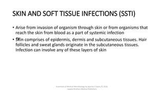
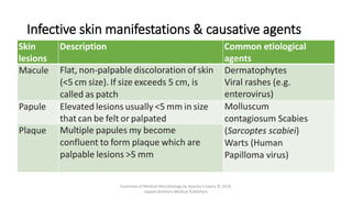
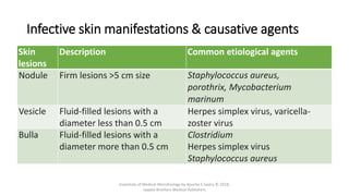
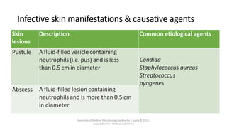
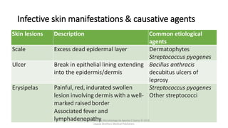
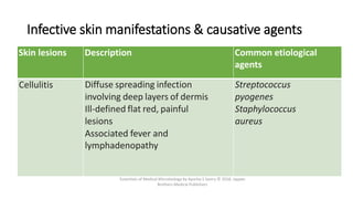
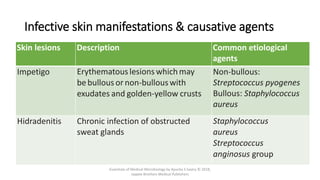
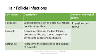
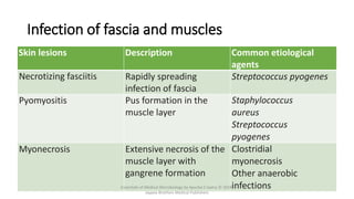
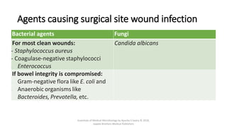
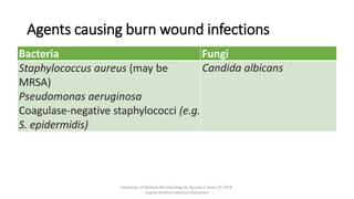
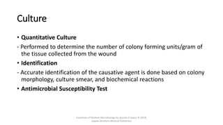
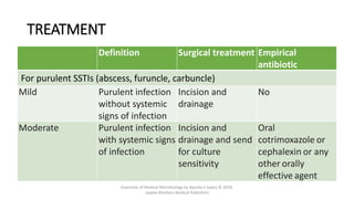
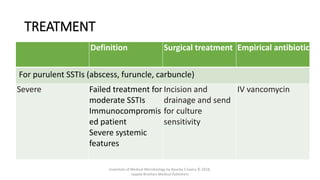
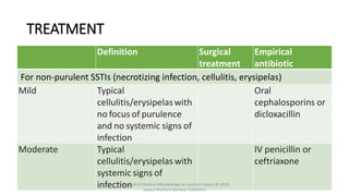
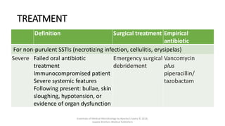
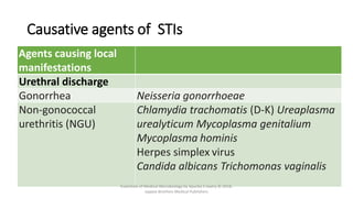
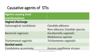
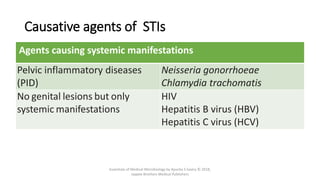
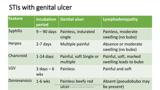
Miscellaneous
Infectious
Syndromes
SKIN AND SOFT TISSUE INFECTIONS (SSTI)
• Arise from invasion of organism through skin or from organisms that
reach the skin from blood as a part of systemic infection
• ™
Skin comprises of epidermis, dermis and subcutaneous tissues. Hair
follicles and sweat glands originate in the subcutaneous tissues.
Infection can involve any of these layers of skin
Essentials of Medical Microbiology by Apurba S Sastry © 2018,
Jaypee Brothers Medical Publishers
Infective skin manifestations & causative agents
Skin
lesions
Description Common etiological
agents
Macule Flat, non-palpable discoloration of skin
(<5 cm size). If size exceeds 5 cm, is
called as patch
Dermatophytes
Viral rashes (e.g.
enterovirus)
Papule Elevated lesions usually <5 mm in size
that can be felt or palpated
Molluscum
contagiosum Scabies
(Sarcoptes scabiei)
Warts (Human
Papilloma virus)
Plaque Multiple papules my become
confluent to form plaque which are
palpable lesions >5 mm
Essentials of Medical Microbiology by Apurba S Sastry © 2018,
Jaypee Brothers Medical Publishers
Infective skin manifestations & causative agents
Skin
lesions
Description Common etiological agents
Nodule Firm lesions >5 cm size Staphylococcus aureus,
porothrix, Mycobacterium
marinum
Vesicle Fluid-filled lesions with a
diameter less than 0.5 cm
Herpes simplex virus, varicella-
zoster virus
Bulla Fluid-filled lesions with a
diameter more than 0.5 cm
Clostridium
Herpes simplex virus
Staphylococcus aureus
Essentials of Medical Microbiology by Apurba S Sastry © 2018,
Jaypee Brothers Medical Publishers
Infective skin manifestations & causative agents
Skin
lesions
Description Common etiological agents
Pustule A fluid-filled vesicle containing
neutrophils (i.e. pus) and is less
than 0.5 cm in diameter
Candida
Staphylococcus aureus
Streptococcus
pyogenes
Abscess A fluid-filled lesion containing
neutrophils and is more than 0.5 cm
in diameter
Essentials of Medical Microbiology by Apurba S Sastry © 2018,
Jaypee Brothers Medical Publishers
Infective skin manifestations & causative agents
Skin lesions Description Common etiological
agents
Scale Excess dead epidermal layer Dermatophytes
Streptococcus pyogenes
Ulcer Break in epithelial lining extending
into the epidermis/dermis
Bacillus anthracis
decubitus ulcers of
leprosy
Erysipelas Painful, red, indurated swollen
lesion involving dermis with a well-
marked raised border
Associated fever and
lymphadenopathy
Streptococcus pyogenes
Other streptococci
Essentials of Medical Microbiology by Apurba S Sastry © 2018,
Jaypee Brothers Medical Publishers
Infective skin manifestations & causative agents
Skin lesions Description Common etiological
agents
Cellulitis Diffuse spreading infection
involving deep layers of dermis
Ill-defined flat red, painful
lesions
Associated fever and
lymphadenopathy
Streptococcus
pyogenes
Staphylococcus
aureus
Essentials of Medical Microbiology by Apurba S Sastry © 2018, Jaypee
Brothers Medical Publishers
Infective skin manifestations & causative agents
Skin lesions Description Common etiological
agents
Impetigo Erythematouslesions which may
be bullous or non-bullouswith
exudates and golden-yellow crusts
Non-bullous:
Streptococcus pyogenes
Bullous: Staphylococcus
aureus
Hidradenitis Chronic infection of obstructed
sweat glands
Staphylococcus
aureus
Streptococcus
anginosus group
Essentials of Medical Microbiology by Apurba S Sastry © 2018,
Jaypee Brothers Medical Publishers
Hair Follicle Infections
Skin lesions Description Common etiological
agents
Folliculitis Superficial infection of single hair follicle,
presents as pustule
Staphylococcus
aureus
Furuncle Deeper infections of the hair follicles,
presents as abscess, spread deeply into
dermis and subcutaneous tissues
Carbuncle Represents the coalescence of a number
of furuncles
Essentials of Medical Microbiology by Apurba S Sastry © 2018,
Jaypee Brothers Medical Publishers
Infection of fascia and muscles
Skin lesions Description Common etiological
agents
Necrotizing fasciitis Rapidly spreading
infection of fascia
Streptococcus pyogenes
Pyomyositis Pus formation in the
muscle layer
Staphylococcus
aureus
Streptococcus
pyogenes
Myonecrosis Extensive necrosis of the
muscle layer with
gangrene formation
Clostridial
myonecrosis
Other anaerobic
infections
Essentials of Medical Microbiology by Apurba S Sastry © 2018,
Jaypee Brothers Medical Publishers
Agents causing surgical site wound infection
Bacterial agents Fungi
For most clean wounds:
- Staphylococcus aureus
- Coagulase-negative staphylococci
- Enterococcus
Candida albicans
If bowel integrity is compromised:
- Gram-negative flora like E. coli and
- Anaerobic organisms like
Bacteroides, Prevotella, etc.
Essentials of Medical Microbiology by Apurba S Sastry © 2018,
Jaypee Brothers Medical Publishers
Agents causing burn wound infections
Bacteria Fungi
Staphylococcus aureus (may be
MRSA)
Pseudomonas aeruginosa
Coagulase-negative staphylococci (e.g.
S. epidermidis)
Candida albicans
Essentials of Medical Microbiology by Apurba S Sastry © 2018,
Jaypee Brothers Medical Publishers
Clinical types of SSTIs
• Primary lesion: An area of tissue with impaired structure/function
due to damage by trauma or disease
• ™
Secondary lesion: A lesion arising as a consequence of any primary
infection
Essentials of Medical Microbiology by Apurba S Sastry © 2018,
Jaypee Brothers Medical Publishers
Laboratory Diagnosis
• Specimen Collection
- Pus from wound collected by sterile swab
- Pus from abscess collected by incision and drainage or needle
aspiration
- Vesicle or bulla fluid, collected by needle aspiration or sterile swab
- Subcutaneous infections: from the base of the lesion or biopsy of the
deep tissues
- Skin scrapings, plucked hair or nail clippings in suspected fungal
infections
Essentials of Medical Microbiology by Apurba S Sastry © 2018,
Jaypee Brothers Medical Publishers
Microscopy
• ™
Gram staining
• ™
KOH mount for suspected fungal infections (e.g.dermatophyte)
• ™
Tzanck smear of the vesicle fluid suspected of herpes simplex or
varicella virus infections
Essentials of Medical Microbiology by Apurba S Sastry © 2018,
Jaypee Brothers Medical Publishers
Culture
• ™
Aerobic culture - inoculated onto blood agar and MacConkey agar and
incubated overnight at 37°C
• ™
Atypical Mycobacterium: Lowenstein Jensen medium
• ™
Dermatophytes: Sabouraud’s dextrose agar
• ™
Anaerobic organisms: Robertson’s cooked meat broth and BHIS (brain
heart infusion agar with supplements)
Essentials of Medical Microbiology by Apurba S Sastry © 2018,
Jaypee Brothers Medical Publishers
Culture
• Quantitative Culture
- Performed to determine the number of colony forming units/gram of
the tissue collected from the wound
• Identification
- Accurate identification of the causative agent is done based on colony
morphology, culture smear, and biochemical reactions
• Antimicrobial Susceptibility Test
Essentials of Medical Microbiology by Apurba S Sastry © 2018,
Jaypee Brothers Medical Publishers
TREATMENT
Definition Surgical treatment Empirical
antibiotic
For purulent SSTIs (abscess, furuncle, carbuncle)
Mild Purulent infection
without systemic
signs of infection
Incision and
drainage
No
Moderate Purulent infection
with systemic signs
of infection
Incision and
drainage and send
for culture
sensitivity
Oral
cotrimoxazole or
cephalexin or any
other orally
effective agent
Essentials of Medical Microbiology by Apurba S Sastry © 2018,
Jaypee Brothers Medical Publishers
TREATMENT
Definition Surgical treatment Empirical antibiotic
For purulent SSTIs (abscess, furuncle, carbuncle)
Severe Failed treatment for
moderate SSTIs
Immunocompromis
ed patient
Severe systemic
features
Incision and
drainage and send
for culture
sensitivity
IV vancomycin
Essentials of Medical Microbiology by Apurba S Sastry © 2018,
Jaypee Brothers Medical Publishers
TREATMENT
Definition Surgical
treatment
Empirical
antibiotic
For non-purulent SSTIs (necrotizing infection, cellulitis, erysipelas)
Mild Typical
cellulitis/erysipelaswith
no focus of purulence
and no systemic signs of
infection
Oral
cephalosporins or
dicloxacillin
Moderate Typical
cellulitis/erysipelas with
systemic signs of
infection
IV penicillin or
ceftriaxone
Essentials of Medical Microbiology by Apurba S Sastry © 2018,
Jaypee Brothers Medical Publishers
TREATMENT
Definition Surgical treatment Empirical
antibiotic
For non-purulent SSTIs (necrotizing infection, cellulitis, erysipelas)
Severe Failed oral antibiotic
treatment
Immunocompromised patient
Severe systemic features
Following present: bullae, skin
sloughing, hypotension, or
evidence of organ dysfunction
Emergency surgical
debridement
Vancomycin
plus
piperacillin/
tazobactam
Essentials of Medical Microbiology by Apurba S Sastry © 2018,
Jaypee Brothers Medical Publishers
SEXUALLY TRANSmITTED
INFECTIONS
Essentials of Medical Microbiology by Apurba S Sastry © 2018,
Jaypee Brothers Medical Publishers
SEXUALLY TRANSMITTED INFECTIONS
• Agents causing local manifestations such as:
• Genital ulcers
• Urethral discharge
• Vaginal discharge
• Genital warts
• Pelvic inflammatory diseases.
• Agents transmitted by sexual route, producing only systemic manifestations
and do not cause local manifestations (e.g. HIV)
Essentials of Medical Microbiology by Apurba S Sastry © 2018,
Jaypee Brothers Medical Publishers
Causative agents of STIs
Agents causing local
manifestations
Genital ulcers
Syphilis Treponema pallidum
Herpes genitalis Herpes simplex viruses
Chancroid Haemophilus ducreyi
Lymphogranuloma venereum Chlamydia trachomatis
Donovanosis Klebsiella granulomatis
Essentials of Medical Microbiology by Apurba S Sastry © 2018,
Jaypee Brothers Medical Publishers
Causative agents of STIs
Agents causing local
manifestations
Urethral discharge
Gonorrhea Neisseria gonorrhoeae
Non-gonococcal
urethritis (NGU)
Chlamydia trachomatis (D-K) Ureaplasma
urealyticum Mycoplasma genitalium
Mycoplasma hominis
Herpes simplex virus
Candida albicans Trichomonas vaginalis
Essentials of Medical Microbiology by Apurba S Sastry © 2018,
Jaypee Brothers Medical Publishers
Causative agents of STIs
Agents causing local
manifestations
Vaginal discharge
Vulvovaginal candidiasis Candida albicans
Non-albicans Candida species
Bacterial vaginosis Gardnerella vaginalis
Mobiluncus species
Trichomonal vaginitis Trichomonas vaginalis
Genital warts
Condyloma acuminata Human papilloma viruses
Essentials of Medical Microbiology by Apurba S Sastry © 2018,
Jaypee Brothers Medical Publishers
Causative agents of STIs
Agents causing systemic manifestations
Pelvic inflammatory diseases
(PID)
Neisseria gonorrhoeae
Chlamydia trachomatis
No genital lesions but only
systemic manifestations
HIV
Hepatitis B virus (HBV)
Hepatitis C virus (HCV)
Essentials of Medical Microbiology by Apurba S Sastry © 2018,
Jaypee Brothers Medical Publishers
STIs with genital ulcer
Feature Incubation
period
Genital ulcer Lymphadenopathy
Syphilis 9 – 90 days Painless, indurated
single
Painless, moderate
swelling (no bubo)
Herpes 2-7 days Multiple painful Absence or moderate
swelling (no bubo)
Chancroid 1-14 days Painful, soft Single or
multiple
Painful, soft, marked
swelling leads to bubo
LGV 3 days – 6
wks
Painless Painful and soft
Donovanosis 1-6 wks Painless beefy red
ulcer
Absent (pseudobubo may
be present)
Essentials of Medical Microbiology by Apurba S Sastry © 2018,
Jaypee Brothers Medical Publishers
Laboratory Diagnosis of STIs
• Specimen Collection
• Discharge from the infected area - vaginal or urethral discharge in a
sterile container
• Sterile swabs may be used to collect the discharge: Charcoal
impregnated swabs are used for suspected gonococcal infection
• Fluid from the vesicles (genital herpes)
Essentials of Medical Microbiology by Apurba S Sastry © 2018,
Jaypee Brothers Medical Publishers
Microscopy
• Wet mount examination: vaginal discharge
- Trichomoniasis: Pus cells along with motile trophozoites
- Candidiasis: Yeast cells along with pseudohyphae
• Gram-stained smear
- Bacterial vaginosis—clue cells (vaginal epithelial cells studded with
gram variable pleomorphic coccobacilli)  Gardnerella vaginalis
- Gonorrhea—intracellular kidney-shaped diplococci
- Candidiasis—gram-positive budding yeast cells along with
pseudohyphae
Essentials of Medical Microbiology by Apurba S Sastry © 2018,
Jaypee Brothers Medical Publishers
Microscopy
• Giemsa stain
- Klebsiella granulomatis - Donovan’s bodies
- Chlamydia trachomatis - inclusion bodies
• Dark field microscopy and silver impregnation - in syphilis - spirally
coiled bacilli
Essentials of Medical Microbiology by Apurba S Sastry © 2018,
Jaypee Brothers Medical Publishers
Culture
• Specimens are inoculated onto the appropriate culture media/cell
line:
• Thayer-Martin medium—for N. gonorrhoeae
• Chocolate agar added with isovitalex and vancomycin— for H. ducreyi
• McCoy cell line—for Chlamydia trachomatis
• Sabouraud’s dextrose agar (SDA)—for Candida species
• Vero cells, monkey kidney cell line - herpes simplex virus.
Essentials of Medical Microbiology by Apurba S Sastry © 2018,
Jaypee Brothers Medical Publishers
Serology
• VDRL or RPR test -syphilis
• Molecular Test
• Multiplex PCR and real-time PCR
- C. trachomatis (opacity protein gene or 16s or 23s rRNA)
- Gonorrhoeae (16s or 23s rRNA gene)
- T. pallidum (47 kDa tpp gene or polA gene)
- H. ducreyi (16s rRNA) and HSV (TSK3 gene)
Essentials of Medical Microbiology by Apurba S Sastry © 2018,
Jaypee Brothers Medical Publishers
Treatment - Urethritis
• Ceftriaxone + Azithromycin - ensure cure and prevent further
development of resistance
• Ceftriaxone - act against gonococcus
• Azithromycin - C. trachomatis
• Treatment to both the sexual partners
Essentials of Medical Microbiology by Apurba S Sastry © 2018,
Jaypee Brothers Medical Publishers
Congenital infections
Essentials of Medical Microbiology by Apurba S Sastry © 2018,
Jaypee Brothers Medical Publishers
Congenital infections
• Infection that crosses placenta to infect the fetus
• Often lead to defects in fetal development or even death
• TORCH
- Toxoplasmosis
- Other infections (congenital syphilis, hepatitis B, Coxsackie virus, Epstein-
Barr virus, varicella-zoster virus, Plasmodium falciparum and human
parvovirus)
- Rubella
- Cytomegalovirus (CMV)
- Herpes simplex virus
Essentials of Medical Microbiology by Apurba S Sastry © 2018,
Jaypee Brothers Medical Publishers
Perinatal Infections (During Delivery)
• Occur while the baby moves through an infected birth canal
• Usually caused by the agents of STIs or fecal contamination
- Cytomegalovirus
- Neisseria gonorrhoeae
- Chlamydia species
- Herpes simplex virus
- Human papilloma virus (genital warts)
- Group B streptococci
Essentials of Medical Microbiology by Apurba S Sastry © 2018,
Jaypee Brothers Medical Publishers
Postnatal Infections (After Delivery)
• Spread from mother to baby following delivery, usually during
breastfeeding
- CMV
- HIV
- Group B streptococci
Essentials of Medical Microbiology by Apurba S Sastry © 2018,
Jaypee Brothers Medical Publishers
Eye infections
Essentials of Medical Microbiology by Apurba S Sastry © 2018,
Jaypee Brothers Medical Publishers
Eye infections
• Infections involving external structures of the eyes: eyelid
(blepharitis), conjunctiva (conjunctivitis), cornea (keratitis) & sclera
(scleritis)
• ™
Infections involving internal structures: Retina (retinitis), uvea
(uveitis) and aqueous humor or vitreous humor (endophthalmitis)
Essentials of Medical Microbiology by Apurba S Sastry © 2018,
Jaypee Brothers Medical Publishers
Causative agents of Ocular infections
Infections Organisms
Blepharitis (Infection of eyelids) Staphylococcus aureus
Conjunctivitis
(Infection of conjunctiva )
Haemophilus influenzae
Staphylococcus aureus
Chlamydia trachomatis
Neisseria gonorrhoeae
Moraxella lacunata (angular
conjunctivitis)
Adenovirus, Herpes simplex virus
Keratitis
(Infection of cornea)
Staphylococcus aureus
Streptococcus pneumoniae
Fusarium, Candida
Acanthamoeba
Essentials of Medical Microbiology by Apurba S Sastry © 2018,
Jaypee Brothers Medical Publishers
Causative agents of Ocular infections
Infections Organisms
Scleritis (Infection of sclera ) Staphylococcus aureus
Chorioretinitis and uveitis
(Infection of choroid, retina,
and uvea )
Mycobacterium tuberculosis
Treponema pallidum
Borrelia burgdorferi
Cytomegalovirus
Toxoplasma gondii
Endophthalmitis
(Infection of aqueous humor or
vitreous humor )
Staphylococcus aureus
Streptococcus pneumoniae
Pseudomonas aeruginosa
Other gram-negative bacilli
Herpes simplex virus, Candida
Essentials of Medical Microbiology by Apurba S Sastry © 2018,
Jaypee Brothers Medical Publishers
EAR INFECTIONS
Essentials of Medical Microbiology by Apurba S Sastry © 2018,
Jaypee Brothers Medical Publishers
EAR INFECTIONS
• ™
Otitis externa: Inflammation, irritation, or infection of
the outer ear and ear canal
• „
Symptoms –
- Itchy ear canal
- Discharge/ pus in ear canal
- Earache that is aggravated when the ear lobe is pulled
Essentials of Medical Microbiology by Apurba S Sastry © 2018,
Jaypee Brothers Medical Publishers
Agents causing otitis externa
• Acute otitis externa
- Staphylococcus aureus (MC), Streptococcus pyogenes
- Pseudomonas (malignant otitis externa), Other GNB
- Aspergillus species, Candida species
• Chronic otitis externa: Anaerobes (most common), Pseudomonas
Essentials of Medical Microbiology by Apurba S Sastry © 2018,
Jaypee Brothers Medical Publishers
Otitis media
• ™
Infections of middle ear
• Earache and ear discharge
• Usually begins as sore throat, cold or respiratory problem  spread
to the middle ear
• „
Symptoms : Intense earache, headache, fever and nausea
- Leaking of discharge from ear  rupture of tympanic membrane
Essentials of Medical Microbiology by Apurba S Sastry © 2018,
Jaypee Brothers Medical Publishers
Organisms causing otitis media
• Acute otitis media
- Streptococcus pneumoniae: MC, (33%, in children)
- Haemophilus influenzae type b (second MC)
- Moraxella catarrhalis
- Streptococcus pyogenes
- Respiratory syncytial virus
- Influenza virus
• Chronic otitis media - Anaerobes (MC)
Essentials of Medical Microbiology by Apurba S Sastry © 2018,
Jaypee Brothers Medical Publishers
Quick Assessment
Essentials of Medical Microbiology by Apurba S Sastry © 2018,
Jaypee Brothers Medical Publishers
MCQs
• Which of the following sexually
transmitted infection produces
painful genital ulcers and
painful lymph nodes?
a. Syphilis
b. Chancroid
c. Herpes
d. Donovanosis
• The agent of malignant otitis
externa is:
a. Staphylococcus aureus
b. Pseudomonas species
c. Streptococcus pyogenes
d. Candida species
Essentials of Medical Microbiology by Apurba S Sastry © 2018,
Jaypee Brothers Medical Publishers
Thank you...!
Essentials of Medical Microbiology by Apurba S Sastry © 2018,
Jaypee Brothers Medical Publishers

Miscellaneous Infectious.pptx

  • 1.
  • 2.
    SKIN AND SOFTTISSUE INFECTIONS (SSTI) • Arise from invasion of organism through skin or from organisms that reach the skin from blood as a part of systemic infection • ™ Skin comprises of epidermis, dermis and subcutaneous tissues. Hair follicles and sweat glands originate in the subcutaneous tissues. Infection can involve any of these layers of skin Essentials of Medical Microbiology by Apurba S Sastry © 2018, Jaypee Brothers Medical Publishers
  • 3.
    Infective skin manifestations& causative agents Skin lesions Description Common etiological agents Macule Flat, non-palpable discoloration of skin (<5 cm size). If size exceeds 5 cm, is called as patch Dermatophytes Viral rashes (e.g. enterovirus) Papule Elevated lesions usually <5 mm in size that can be felt or palpated Molluscum contagiosum Scabies (Sarcoptes scabiei) Warts (Human Papilloma virus) Plaque Multiple papules my become confluent to form plaque which are palpable lesions >5 mm Essentials of Medical Microbiology by Apurba S Sastry © 2018, Jaypee Brothers Medical Publishers
  • 4.
    Infective skin manifestations& causative agents Skin lesions Description Common etiological agents Nodule Firm lesions >5 cm size Staphylococcus aureus, porothrix, Mycobacterium marinum Vesicle Fluid-filled lesions with a diameter less than 0.5 cm Herpes simplex virus, varicella- zoster virus Bulla Fluid-filled lesions with a diameter more than 0.5 cm Clostridium Herpes simplex virus Staphylococcus aureus Essentials of Medical Microbiology by Apurba S Sastry © 2018, Jaypee Brothers Medical Publishers
  • 5.
    Infective skin manifestations& causative agents Skin lesions Description Common etiological agents Pustule A fluid-filled vesicle containing neutrophils (i.e. pus) and is less than 0.5 cm in diameter Candida Staphylococcus aureus Streptococcus pyogenes Abscess A fluid-filled lesion containing neutrophils and is more than 0.5 cm in diameter Essentials of Medical Microbiology by Apurba S Sastry © 2018, Jaypee Brothers Medical Publishers
  • 6.
    Infective skin manifestations& causative agents Skin lesions Description Common etiological agents Scale Excess dead epidermal layer Dermatophytes Streptococcus pyogenes Ulcer Break in epithelial lining extending into the epidermis/dermis Bacillus anthracis decubitus ulcers of leprosy Erysipelas Painful, red, indurated swollen lesion involving dermis with a well- marked raised border Associated fever and lymphadenopathy Streptococcus pyogenes Other streptococci Essentials of Medical Microbiology by Apurba S Sastry © 2018, Jaypee Brothers Medical Publishers
  • 7.
    Infective skin manifestations& causative agents Skin lesions Description Common etiological agents Cellulitis Diffuse spreading infection involving deep layers of dermis Ill-defined flat red, painful lesions Associated fever and lymphadenopathy Streptococcus pyogenes Staphylococcus aureus Essentials of Medical Microbiology by Apurba S Sastry © 2018, Jaypee Brothers Medical Publishers
  • 8.
    Infective skin manifestations& causative agents Skin lesions Description Common etiological agents Impetigo Erythematouslesions which may be bullous or non-bullouswith exudates and golden-yellow crusts Non-bullous: Streptococcus pyogenes Bullous: Staphylococcus aureus Hidradenitis Chronic infection of obstructed sweat glands Staphylococcus aureus Streptococcus anginosus group Essentials of Medical Microbiology by Apurba S Sastry © 2018, Jaypee Brothers Medical Publishers
  • 9.
    Hair Follicle Infections Skinlesions Description Common etiological agents Folliculitis Superficial infection of single hair follicle, presents as pustule Staphylococcus aureus Furuncle Deeper infections of the hair follicles, presents as abscess, spread deeply into dermis and subcutaneous tissues Carbuncle Represents the coalescence of a number of furuncles Essentials of Medical Microbiology by Apurba S Sastry © 2018, Jaypee Brothers Medical Publishers
  • 10.
    Infection of fasciaand muscles Skin lesions Description Common etiological agents Necrotizing fasciitis Rapidly spreading infection of fascia Streptococcus pyogenes Pyomyositis Pus formation in the muscle layer Staphylococcus aureus Streptococcus pyogenes Myonecrosis Extensive necrosis of the muscle layer with gangrene formation Clostridial myonecrosis Other anaerobic infections Essentials of Medical Microbiology by Apurba S Sastry © 2018, Jaypee Brothers Medical Publishers
  • 11.
    Agents causing surgicalsite wound infection Bacterial agents Fungi For most clean wounds: - Staphylococcus aureus - Coagulase-negative staphylococci - Enterococcus Candida albicans If bowel integrity is compromised: - Gram-negative flora like E. coli and - Anaerobic organisms like Bacteroides, Prevotella, etc. Essentials of Medical Microbiology by Apurba S Sastry © 2018, Jaypee Brothers Medical Publishers
  • 12.
    Agents causing burnwound infections Bacteria Fungi Staphylococcus aureus (may be MRSA) Pseudomonas aeruginosa Coagulase-negative staphylococci (e.g. S. epidermidis) Candida albicans Essentials of Medical Microbiology by Apurba S Sastry © 2018, Jaypee Brothers Medical Publishers
  • 13.
    Clinical types ofSSTIs • Primary lesion: An area of tissue with impaired structure/function due to damage by trauma or disease • ™ Secondary lesion: A lesion arising as a consequence of any primary infection Essentials of Medical Microbiology by Apurba S Sastry © 2018, Jaypee Brothers Medical Publishers
  • 14.
    Laboratory Diagnosis • SpecimenCollection - Pus from wound collected by sterile swab - Pus from abscess collected by incision and drainage or needle aspiration - Vesicle or bulla fluid, collected by needle aspiration or sterile swab - Subcutaneous infections: from the base of the lesion or biopsy of the deep tissues - Skin scrapings, plucked hair or nail clippings in suspected fungal infections Essentials of Medical Microbiology by Apurba S Sastry © 2018, Jaypee Brothers Medical Publishers
  • 15.
    Microscopy • ™ Gram staining •™ KOH mount for suspected fungal infections (e.g.dermatophyte) • ™ Tzanck smear of the vesicle fluid suspected of herpes simplex or varicella virus infections Essentials of Medical Microbiology by Apurba S Sastry © 2018, Jaypee Brothers Medical Publishers
  • 16.
    Culture • ™ Aerobic culture- inoculated onto blood agar and MacConkey agar and incubated overnight at 37°C • ™ Atypical Mycobacterium: Lowenstein Jensen medium • ™ Dermatophytes: Sabouraud’s dextrose agar • ™ Anaerobic organisms: Robertson’s cooked meat broth and BHIS (brain heart infusion agar with supplements) Essentials of Medical Microbiology by Apurba S Sastry © 2018, Jaypee Brothers Medical Publishers
  • 17.
    Culture • Quantitative Culture -Performed to determine the number of colony forming units/gram of the tissue collected from the wound • Identification - Accurate identification of the causative agent is done based on colony morphology, culture smear, and biochemical reactions • Antimicrobial Susceptibility Test Essentials of Medical Microbiology by Apurba S Sastry © 2018, Jaypee Brothers Medical Publishers
  • 18.
    TREATMENT Definition Surgical treatmentEmpirical antibiotic For purulent SSTIs (abscess, furuncle, carbuncle) Mild Purulent infection without systemic signs of infection Incision and drainage No Moderate Purulent infection with systemic signs of infection Incision and drainage and send for culture sensitivity Oral cotrimoxazole or cephalexin or any other orally effective agent Essentials of Medical Microbiology by Apurba S Sastry © 2018, Jaypee Brothers Medical Publishers
  • 19.
    TREATMENT Definition Surgical treatmentEmpirical antibiotic For purulent SSTIs (abscess, furuncle, carbuncle) Severe Failed treatment for moderate SSTIs Immunocompromis ed patient Severe systemic features Incision and drainage and send for culture sensitivity IV vancomycin Essentials of Medical Microbiology by Apurba S Sastry © 2018, Jaypee Brothers Medical Publishers
  • 20.
    TREATMENT Definition Surgical treatment Empirical antibiotic For non-purulentSSTIs (necrotizing infection, cellulitis, erysipelas) Mild Typical cellulitis/erysipelaswith no focus of purulence and no systemic signs of infection Oral cephalosporins or dicloxacillin Moderate Typical cellulitis/erysipelas with systemic signs of infection IV penicillin or ceftriaxone Essentials of Medical Microbiology by Apurba S Sastry © 2018, Jaypee Brothers Medical Publishers
  • 21.
    TREATMENT Definition Surgical treatmentEmpirical antibiotic For non-purulent SSTIs (necrotizing infection, cellulitis, erysipelas) Severe Failed oral antibiotic treatment Immunocompromised patient Severe systemic features Following present: bullae, skin sloughing, hypotension, or evidence of organ dysfunction Emergency surgical debridement Vancomycin plus piperacillin/ tazobactam Essentials of Medical Microbiology by Apurba S Sastry © 2018, Jaypee Brothers Medical Publishers
  • 22.
    SEXUALLY TRANSmITTED INFECTIONS Essentials ofMedical Microbiology by Apurba S Sastry © 2018, Jaypee Brothers Medical Publishers
  • 23.
    SEXUALLY TRANSMITTED INFECTIONS •Agents causing local manifestations such as: • Genital ulcers • Urethral discharge • Vaginal discharge • Genital warts • Pelvic inflammatory diseases. • Agents transmitted by sexual route, producing only systemic manifestations and do not cause local manifestations (e.g. HIV) Essentials of Medical Microbiology by Apurba S Sastry © 2018, Jaypee Brothers Medical Publishers
  • 24.
    Causative agents ofSTIs Agents causing local manifestations Genital ulcers Syphilis Treponema pallidum Herpes genitalis Herpes simplex viruses Chancroid Haemophilus ducreyi Lymphogranuloma venereum Chlamydia trachomatis Donovanosis Klebsiella granulomatis Essentials of Medical Microbiology by Apurba S Sastry © 2018, Jaypee Brothers Medical Publishers
  • 25.
    Causative agents ofSTIs Agents causing local manifestations Urethral discharge Gonorrhea Neisseria gonorrhoeae Non-gonococcal urethritis (NGU) Chlamydia trachomatis (D-K) Ureaplasma urealyticum Mycoplasma genitalium Mycoplasma hominis Herpes simplex virus Candida albicans Trichomonas vaginalis Essentials of Medical Microbiology by Apurba S Sastry © 2018, Jaypee Brothers Medical Publishers
  • 26.
    Causative agents ofSTIs Agents causing local manifestations Vaginal discharge Vulvovaginal candidiasis Candida albicans Non-albicans Candida species Bacterial vaginosis Gardnerella vaginalis Mobiluncus species Trichomonal vaginitis Trichomonas vaginalis Genital warts Condyloma acuminata Human papilloma viruses Essentials of Medical Microbiology by Apurba S Sastry © 2018, Jaypee Brothers Medical Publishers
  • 27.
    Causative agents ofSTIs Agents causing systemic manifestations Pelvic inflammatory diseases (PID) Neisseria gonorrhoeae Chlamydia trachomatis No genital lesions but only systemic manifestations HIV Hepatitis B virus (HBV) Hepatitis C virus (HCV) Essentials of Medical Microbiology by Apurba S Sastry © 2018, Jaypee Brothers Medical Publishers
  • 28.
    STIs with genitalulcer Feature Incubation period Genital ulcer Lymphadenopathy Syphilis 9 – 90 days Painless, indurated single Painless, moderate swelling (no bubo) Herpes 2-7 days Multiple painful Absence or moderate swelling (no bubo) Chancroid 1-14 days Painful, soft Single or multiple Painful, soft, marked swelling leads to bubo LGV 3 days – 6 wks Painless Painful and soft Donovanosis 1-6 wks Painless beefy red ulcer Absent (pseudobubo may be present) Essentials of Medical Microbiology by Apurba S Sastry © 2018, Jaypee Brothers Medical Publishers
  • 29.
    Laboratory Diagnosis ofSTIs • Specimen Collection • Discharge from the infected area - vaginal or urethral discharge in a sterile container • Sterile swabs may be used to collect the discharge: Charcoal impregnated swabs are used for suspected gonococcal infection • Fluid from the vesicles (genital herpes) Essentials of Medical Microbiology by Apurba S Sastry © 2018, Jaypee Brothers Medical Publishers
  • 30.
    Microscopy • Wet mountexamination: vaginal discharge - Trichomoniasis: Pus cells along with motile trophozoites - Candidiasis: Yeast cells along with pseudohyphae • Gram-stained smear - Bacterial vaginosis—clue cells (vaginal epithelial cells studded with gram variable pleomorphic coccobacilli)  Gardnerella vaginalis - Gonorrhea—intracellular kidney-shaped diplococci - Candidiasis—gram-positive budding yeast cells along with pseudohyphae Essentials of Medical Microbiology by Apurba S Sastry © 2018, Jaypee Brothers Medical Publishers
  • 31.
    Microscopy • Giemsa stain -Klebsiella granulomatis - Donovan’s bodies - Chlamydia trachomatis - inclusion bodies • Dark field microscopy and silver impregnation - in syphilis - spirally coiled bacilli Essentials of Medical Microbiology by Apurba S Sastry © 2018, Jaypee Brothers Medical Publishers
  • 32.
    Culture • Specimens areinoculated onto the appropriate culture media/cell line: • Thayer-Martin medium—for N. gonorrhoeae • Chocolate agar added with isovitalex and vancomycin— for H. ducreyi • McCoy cell line—for Chlamydia trachomatis • Sabouraud’s dextrose agar (SDA)—for Candida species • Vero cells, monkey kidney cell line - herpes simplex virus. Essentials of Medical Microbiology by Apurba S Sastry © 2018, Jaypee Brothers Medical Publishers
  • 33.
    Serology • VDRL orRPR test -syphilis • Molecular Test • Multiplex PCR and real-time PCR - C. trachomatis (opacity protein gene or 16s or 23s rRNA) - Gonorrhoeae (16s or 23s rRNA gene) - T. pallidum (47 kDa tpp gene or polA gene) - H. ducreyi (16s rRNA) and HSV (TSK3 gene) Essentials of Medical Microbiology by Apurba S Sastry © 2018, Jaypee Brothers Medical Publishers
  • 34.
    Treatment - Urethritis •Ceftriaxone + Azithromycin - ensure cure and prevent further development of resistance • Ceftriaxone - act against gonococcus • Azithromycin - C. trachomatis • Treatment to both the sexual partners Essentials of Medical Microbiology by Apurba S Sastry © 2018, Jaypee Brothers Medical Publishers
  • 35.
    Congenital infections Essentials ofMedical Microbiology by Apurba S Sastry © 2018, Jaypee Brothers Medical Publishers
  • 36.
    Congenital infections • Infectionthat crosses placenta to infect the fetus • Often lead to defects in fetal development or even death • TORCH - Toxoplasmosis - Other infections (congenital syphilis, hepatitis B, Coxsackie virus, Epstein- Barr virus, varicella-zoster virus, Plasmodium falciparum and human parvovirus) - Rubella - Cytomegalovirus (CMV) - Herpes simplex virus Essentials of Medical Microbiology by Apurba S Sastry © 2018, Jaypee Brothers Medical Publishers
  • 37.
    Perinatal Infections (DuringDelivery) • Occur while the baby moves through an infected birth canal • Usually caused by the agents of STIs or fecal contamination - Cytomegalovirus - Neisseria gonorrhoeae - Chlamydia species - Herpes simplex virus - Human papilloma virus (genital warts) - Group B streptococci Essentials of Medical Microbiology by Apurba S Sastry © 2018, Jaypee Brothers Medical Publishers
  • 38.
    Postnatal Infections (AfterDelivery) • Spread from mother to baby following delivery, usually during breastfeeding - CMV - HIV - Group B streptococci Essentials of Medical Microbiology by Apurba S Sastry © 2018, Jaypee Brothers Medical Publishers
  • 39.
    Eye infections Essentials ofMedical Microbiology by Apurba S Sastry © 2018, Jaypee Brothers Medical Publishers
  • 40.
    Eye infections • Infectionsinvolving external structures of the eyes: eyelid (blepharitis), conjunctiva (conjunctivitis), cornea (keratitis) & sclera (scleritis) • ™ Infections involving internal structures: Retina (retinitis), uvea (uveitis) and aqueous humor or vitreous humor (endophthalmitis) Essentials of Medical Microbiology by Apurba S Sastry © 2018, Jaypee Brothers Medical Publishers
  • 41.
    Causative agents ofOcular infections Infections Organisms Blepharitis (Infection of eyelids) Staphylococcus aureus Conjunctivitis (Infection of conjunctiva ) Haemophilus influenzae Staphylococcus aureus Chlamydia trachomatis Neisseria gonorrhoeae Moraxella lacunata (angular conjunctivitis) Adenovirus, Herpes simplex virus Keratitis (Infection of cornea) Staphylococcus aureus Streptococcus pneumoniae Fusarium, Candida Acanthamoeba Essentials of Medical Microbiology by Apurba S Sastry © 2018, Jaypee Brothers Medical Publishers
  • 42.
    Causative agents ofOcular infections Infections Organisms Scleritis (Infection of sclera ) Staphylococcus aureus Chorioretinitis and uveitis (Infection of choroid, retina, and uvea ) Mycobacterium tuberculosis Treponema pallidum Borrelia burgdorferi Cytomegalovirus Toxoplasma gondii Endophthalmitis (Infection of aqueous humor or vitreous humor ) Staphylococcus aureus Streptococcus pneumoniae Pseudomonas aeruginosa Other gram-negative bacilli Herpes simplex virus, Candida Essentials of Medical Microbiology by Apurba S Sastry © 2018, Jaypee Brothers Medical Publishers
  • 43.
    EAR INFECTIONS Essentials ofMedical Microbiology by Apurba S Sastry © 2018, Jaypee Brothers Medical Publishers
  • 44.
    EAR INFECTIONS • ™ Otitisexterna: Inflammation, irritation, or infection of the outer ear and ear canal • „ Symptoms – - Itchy ear canal - Discharge/ pus in ear canal - Earache that is aggravated when the ear lobe is pulled Essentials of Medical Microbiology by Apurba S Sastry © 2018, Jaypee Brothers Medical Publishers
  • 45.
    Agents causing otitisexterna • Acute otitis externa - Staphylococcus aureus (MC), Streptococcus pyogenes - Pseudomonas (malignant otitis externa), Other GNB - Aspergillus species, Candida species • Chronic otitis externa: Anaerobes (most common), Pseudomonas Essentials of Medical Microbiology by Apurba S Sastry © 2018, Jaypee Brothers Medical Publishers
  • 46.
    Otitis media • ™ Infectionsof middle ear • Earache and ear discharge • Usually begins as sore throat, cold or respiratory problem  spread to the middle ear • „ Symptoms : Intense earache, headache, fever and nausea - Leaking of discharge from ear  rupture of tympanic membrane Essentials of Medical Microbiology by Apurba S Sastry © 2018, Jaypee Brothers Medical Publishers
  • 47.
    Organisms causing otitismedia • Acute otitis media - Streptococcus pneumoniae: MC, (33%, in children) - Haemophilus influenzae type b (second MC) - Moraxella catarrhalis - Streptococcus pyogenes - Respiratory syncytial virus - Influenza virus • Chronic otitis media - Anaerobes (MC) Essentials of Medical Microbiology by Apurba S Sastry © 2018, Jaypee Brothers Medical Publishers
  • 48.
    Quick Assessment Essentials ofMedical Microbiology by Apurba S Sastry © 2018, Jaypee Brothers Medical Publishers
  • 49.
    MCQs • Which ofthe following sexually transmitted infection produces painful genital ulcers and painful lymph nodes? a. Syphilis b. Chancroid c. Herpes d. Donovanosis • The agent of malignant otitis externa is: a. Staphylococcus aureus b. Pseudomonas species c. Streptococcus pyogenes d. Candida species Essentials of Medical Microbiology by Apurba S Sastry © 2018, Jaypee Brothers Medical Publishers
  • 50.
    Thank you...! Essentials ofMedical Microbiology by Apurba S Sastry © 2018, Jaypee Brothers Medical Publishers