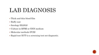
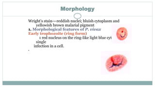
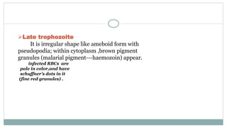
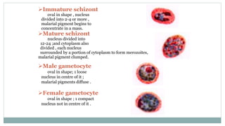
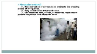
Mosab Nouraldein Mohammed Hamad
Musab.noor13@gmail.com
Haemamoeba vivax Grassi and Feletti, 1890
Mostly occur in Asia , Latin America and
some parts of Africa
P.vivax originate from Asia
P.vivax cause benign tertian malaria
P. vivax (as well as P. ovale) has dormant
liver stages (“hypnozoites”) that can activate
and invade the blood (“relapse”) several
months or years after the infecting mosquito
bite.
Pathogenesis results from rupture of infected
red blood cells, leading to fever. Infected red
blood cells may also stick to each other and to
walls of capillaries. Vessels plug up and
deprive tissues of oxygen. Infection may also
cause the spleen to enlarge
P. vivax can populate the bloodstream (all
sexual and asexual stages present in
peripheral blood)
The parasite can go dormant in the liver for
days to years, causing no symptoms and
remaining undetectable in blood tests (form
hypnozoites)
Cardinal signs : fever, sweat and cold stages
Abdominal pain
Nausea
Vomiting
 Thick and thin blood film
 Buffy coat
 Serology (ELISA)
 Culture in RPMI or NNN medium
 Molecular methods (PCR)
 Rapid test (ICT) is a screening test not diagnostic.
Morphology
Wright’s stain---reddish nuclei; bluish cytoplasm and
yellowish brown malarial pigment
1. Morphological features of P. vivax
Early trophozoite (ring form)
1 red nucleus on the ring-like light blue cytoplasm ;
single
infection in a cell.
.
Late trophozoite
It is irregular shape like ameboid form with
pseudopodia; within cytoplasm ,brown pigment
granules (malarial pigment---haemozoin) appear.
infected RBCs are
pale in color,and have
schuffner’s dots in it
(fine red granules) .
Immature schizont
oval in shape , nucleus
divided into 2-4 or more ,
malarial pigment begins to
concentrate in a mass.
Mature schizont
nucleus divided into
12-24 ;and cytoplasm also
divided , each nucleus
surrounded by a portion of cytoplasm to form merozoites,
malarial pigment clumped.
Male gametocyte
oval in shape; 1 loose
nucleus in centre of it ;
malarial pigments diffuse .
Female gametocyte
oval in shape ; 1 compact
nucleus not in centre of it .
 Thrombocytopenia as occur in Falciparum malaria
 Anemia
 Elevated LDH
Treatment
Chlorquine and quinine----anti-erythrocytic stage drugs.
(question: Which stage of plasmodium can these drugs kill?)
Primaquine and pyrimethamine ----anti-exoerythrocytic
stage drugs.
Prevention
Chemoprophylaxis
-----Chloroquine / pyrimethamine
used for
prophylaxis of malaria
-----Chemotherapy: 1 week before entry into the endemic area ; for 4 weeks
after returning from the endemic area.
Mosquito control
(1). Reconstruction of environment: eradicate the breeding
places of mosquitoes.
(2). Spry insecticides: DDVP and so on.
(3). Use mosquito nets, screen, or mosquito repellents to
protect the person from mosquito bites.
Plasmodium vivax m

Plasmodium vivax m

  • 1.
    Mosab Nouraldein MohammedHamad Musab.noor13@gmail.com
  • 2.
    Haemamoeba vivax Grassiand Feletti, 1890
  • 3.
    Mostly occur inAsia , Latin America and some parts of Africa P.vivax originate from Asia P.vivax cause benign tertian malaria
  • 4.
    P. vivax (aswell as P. ovale) has dormant liver stages (“hypnozoites”) that can activate and invade the blood (“relapse”) several months or years after the infecting mosquito bite.
  • 6.
    Pathogenesis results fromrupture of infected red blood cells, leading to fever. Infected red blood cells may also stick to each other and to walls of capillaries. Vessels plug up and deprive tissues of oxygen. Infection may also cause the spleen to enlarge
  • 7.
    P. vivax canpopulate the bloodstream (all sexual and asexual stages present in peripheral blood)
  • 8.
    The parasite cango dormant in the liver for days to years, causing no symptoms and remaining undetectable in blood tests (form hypnozoites)
  • 9.
    Cardinal signs :fever, sweat and cold stages Abdominal pain Nausea Vomiting
  • 10.
     Thick andthin blood film  Buffy coat  Serology (ELISA)  Culture in RPMI or NNN medium  Molecular methods (PCR)  Rapid test (ICT) is a screening test not diagnostic.
  • 11.
    Morphology Wright’s stain---reddish nuclei;bluish cytoplasm and yellowish brown malarial pigment 1. Morphological features of P. vivax Early trophozoite (ring form) 1 red nucleus on the ring-like light blue cytoplasm ; single infection in a cell. .
  • 12.
    Late trophozoite It isirregular shape like ameboid form with pseudopodia; within cytoplasm ,brown pigment granules (malarial pigment---haemozoin) appear. infected RBCs are pale in color,and have schuffner’s dots in it (fine red granules) .
  • 13.
    Immature schizont oval inshape , nucleus divided into 2-4 or more , malarial pigment begins to concentrate in a mass. Mature schizont nucleus divided into 12-24 ;and cytoplasm also divided , each nucleus surrounded by a portion of cytoplasm to form merozoites, malarial pigment clumped. Male gametocyte oval in shape; 1 loose nucleus in centre of it ; malarial pigments diffuse . Female gametocyte oval in shape ; 1 compact nucleus not in centre of it .
  • 14.
     Thrombocytopenia asoccur in Falciparum malaria  Anemia  Elevated LDH
  • 15.
    Treatment Chlorquine and quinine----anti-erythrocyticstage drugs. (question: Which stage of plasmodium can these drugs kill?) Primaquine and pyrimethamine ----anti-exoerythrocytic stage drugs. Prevention Chemoprophylaxis -----Chloroquine / pyrimethamine used for prophylaxis of malaria -----Chemotherapy: 1 week before entry into the endemic area ; for 4 weeks after returning from the endemic area.
  • 16.
    Mosquito control (1). Reconstructionof environment: eradicate the breeding places of mosquitoes. (2). Spry insecticides: DDVP and so on. (3). Use mosquito nets, screen, or mosquito repellents to protect the person from mosquito bites.