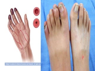
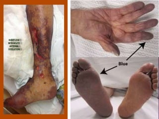
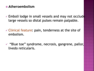
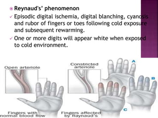
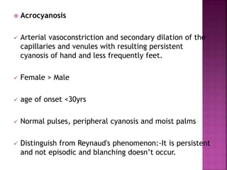
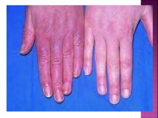
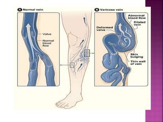
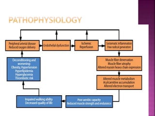
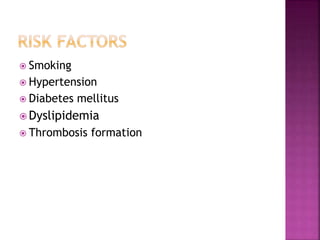
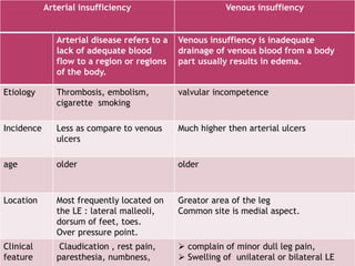
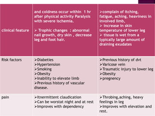
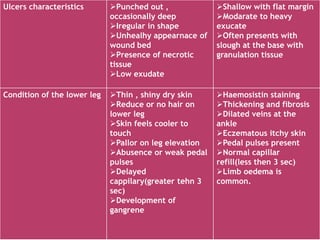
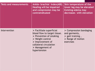
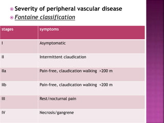
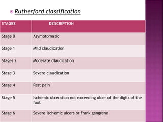
SlideShare a Scribd company logo
PERIPHERAL VASCULAR DISEASE
 Any disorder that interferes with arterial or venous
blood flow of the extremities.
 Peripheral vascular disease includes a group of
diseases in which blood vessels become restricted
or blocked
 Arterial disorder
 Venous disorder
 The chance of having PAD increases as you
get older.
 People over age 50 have a higher risk for PAD
and the majority of patients will fall in this
demographic.
 One in 20 adults over age 50 and one in five
over age 70 will have PAD.
Arterial disorder
 Fibro muscular dysplasia-
Hyperplasic disorder affecting medium and small
sized arteries.
 More in females and involves renal and carotid
arteries but can affect iliac and subclavian arteries.
 Symptoms: atherosclerosis including claudication
and rest pain.
 “string of beads” appearance.
 Thromboanginitis obliterance(Burger’s disease)
 Inflammatory occlusive disorder involving small and
medium sized arteries and veins in distal upper and
lower extremities.
 Cerebral, visceral and coronary vessels may also be
involved.
 More in men under age 40.
 Cause is unknown, but shows a definite relationship
with cigarette smoking.
 Acute arterial occlusion
 Sudden cessation of blood flow to an extremity.
 Severity of ischemia depend on location and extent
of the occlusion and presence of collateral blood
vessels.
 causes:- embolism and thrombus
 Embolism causes- Cardiac disorders include atrial
fibrillation, Myocardial infarction, ventricular
aneurysm.
 Emboli may lodge in femoral , iliac , aorta ,
popliteal and tibioperoneal arteries.
 Thrombosis causes- trauma, thoracic outlet
compression syndrome and entrapment of popliteal
artery.
 Thrombosis most frequently occurs in
atherosclerotic vessels.
 Clinical fearture- pain, paresthesia,
numbness, and coldness occur within 1 hr.
Paralysis with severe ischemia. loss of pulses
distal to occlusion, cyanosis, decreases skin
temp, mottling, muscle stiffening, loss of
sensation, weakness, and absent deep
tendon reflexes.
 Atheroembolism
 Emboli lodge in small vessels and may not occlude
large vessels so distal pulses remain palpable.
 Clinical feature: pain, tenderness at the site of
embolism.
 “Blue toe” syndrome, necrosis, gangrene, pallor,
livedo reticularis.
 Reynaud's’ phenomenon
 Episodic digital ischemia, digital blanching, cyanosis
and rubor of fingers or toes following cold exposure
and subsequent rewarming.
 One or more digits will appear white when exposed
to cold environment.
 Acrocyanosis
 Arterial vasoconstriction and secondary dilation of the
capillaries and venules with resulting persistent
cyanosis of hand and less frequently feet.
 Female > Male
 age of onset <30yrs
 Normal pulses, peripheral cyanosis and moist palms
 Distinguish from Reynaud's phenomenon:-It is persistent
and not episodic and blanching doesn’t occur.
Venous disorder
 Venous thrombosis(thrombophlebitis)
 Presence of thrombus within superficial or deep
vein and that accompany inflammatory response in
the vessel wall.
 Prevalence- 50% pts with orthopedic surgical
procedure, 10-40% pts with abdominal or thoracic
surgeries.
 Deep vein thrombosis
 Most imp consequences of this disorder are
pulmonary embolism and chronic venous
insufficiency.
 More in lower extremity than upper extremity.
 DVT of iliac, femoral and popliteal veins is
suggested by leg swelling, warmth,
erythema.
 Examination:- post calf tenderness, warmth,
increased tissue turger or modest swelling
and Homans’s sign(unreliable).
 Superficial vein thrombosis
 Associated with i.v catheters and infusion, occures
in varicose veins, thromboangitis obliterans.
 Clinical feature: pain at localized to the site of
thrombus.
 Varicose vein
 Dilated, tortuous superficial veins results from
defective structure and function of the valves of
the saphenous veins, from weakness of the vein
wall, from high luminal pressure
 Clinical feature:- dull ache, mild ankle oedema
which is relieved with leg elevation.
 Chronic venous insufficiency
 May result from deep vein thrombosis and/or
valvular incompetence.
 Valves become thickened and contracted, veins
become rigid and thick-walled.
 Clinical feature: dull ache on long standing,
relieved by leg elevation.
 Erythema, dermatitis and hyper
pigmentation develops along distal aspect of
leg, skin ulceration occur near the medial
and lateral malleoli
 Smoking
 Hypertension
 Diabetes mellitus
 Dyslipidemia
 Thrombosis formation
Arterial ulcerations Venous ulcerations
Arterial disease refers to a
lack of adequate blood
flow to a region or regions
of the body.
Venous insuffiency is inadequate
drainage of venous blood from a body
part usually results in edema.
Etiology Thrombosis, embolism,
cigarette smoking
valvular incompetence
Incidence Less as compare to venous
ulcers
Much higher then arterial ulcers
age older older
Location Most frequently located on
the LE : lateral malleoli,
dorsum of feet, toes.
Over pressure point.
Greator area of the leg
Common site is medial aspect.
Clinical
feature
Claudication , rest pain,
paresthesia, numbness,
 complain of minor dull leg pain,
 Swelling of unilateral or bilateral LE
Arterial insufficiency Venous insuffiency
clinical feature
and coldness occur within 1 hr
after physical activity Paralysis
with severe ischemia.
 Trophic changes : abnormal
nail growth, dry skin , decrease
leg and foot hair.
complain of itching,
fatigue, aching, heaviness in
involved limb,
 increase in skin
temperature of lower leg
 tissue is wet from a
typically large amount of
draining exudates
Risk factors Diabeties
Hypertension
Smoking
Obesity
Inability to elevate limb
Previous history of vascular
disease.
Previous history of dvt
Varicose vein
Traumatic injury to lower leg
Obesity
pregnency
pain Itnermittent claudication
Can be worstat night and at rest
Improves with dependency
Throbing,aching, heavy
feelings in leg
Improves with elevation and
rest.
Ulcers characteristics Punched out ,
occasionally deep
Iregular in shape
Unhealhy appearnace of
wound bed
Presence of necrotic
tissue
Low exudate
Shallow with flat margin
Modarate to heavy
exucate
Often presents with
slough at the base with
granulation tissue
Condition of the lower leg Thin , shiny dry skin
Reduce or no hair on
lower leg
Skin feels cooler to
touch
Pallor on leg elevation
Abusence or weak pedal
pulses
Delayed
cappilary(greater tehn 3
sec)
Development of
gangrene
Haemosistin staining
Thickening and fibrosis
Dilated veins at the
ankle
Eczematous itchy skin
Pedal pulses present
Normal capillar
refill(less then 3 sec)
Limb oedema is
common.
Tests and measurements Ankle- brachial Index(ABI)
Healing will be impaired
and compression may be
contraindicated
Skin temperature of the
lower leg may be elevated
Existing edema may
decreases with elevation
Intervention  Facilitate superficial
blood flow to target tissue.
 Prevention of smoking
 Weight control
 Improvement of
collateral circulation
 Management of
hypertension
 Compression bandaging
and garments,
 gait training ,
 Range of motion
exercises
 Severity of peripheral vascular disease
 Fontaine classification
stages symptoms
I Asymptomatic
II Intermittent claudication
IIa Pain-free, claudication walking >200 m
IIb Pain-free, claudication walking <200 m
III Rest/nocturnal pain
IV Necrosis/gangrene
 Rutherford classification
STAGES DESCRIPTION
Stage 0 Asymptomatic
Stage 1 Mild claudication
Stages 2 Moderate claudication
Stage 3 Severe claudication
Stage 4 Rest pain
Stage 5 Ischemic ulceration not exceeding ulcer of the digits of the
foot
Stage 6 Severe ischemic ulcers or frank gangrene
 Ankle brachial index
 Pulse examination.
 Treadmill exercise testing
 CT angiography
 MR angiography
 Used to examine the vascular system.
 Provide information about the potential loss
of perfusion in L.Es
 Performed with DOPPLER ultrasound.
 Brachial ,radial, femoral, popliteal, dorsalis
pedis, and posterior tibialis.
In following conditions ABI should be used:-
Exertional leg pain
Non-healing wound
Patients with 50 yrs and older with a
history of smoking or diabetes
It provide information about possible vascular
system involvement.
Pulse scale:-
0 =no pulse
1+ =weak pulse, difficult to palpate
2+ =palpable but not normal, diminished
3+ =normal, easy to palpate
4+ =bounding, very strong
 primary objectives are to obtain reliable
measures of:
1) the rate of claudication pain development,
2) The ABI response to exercise,
3) the presence of coexisting coronary heart
disease.
 The test should be progressive with gradual
increments in grade
 A highly reliable treadmill test for patients
with PAD uses a constant walking speed of 2
mph and gradual increases in grade of 2%
every 2 minutes beginning at 0% grade
 Functional claudication distance: a reliable and
valid measurement to assess functional
limitation in patients with intermittent
claudication Lotte M Kruidenier1, Saskia PA
Nicolaï1, BMC Cardiovascular Disorders 2009,
9:9
 Disease severity and functional impairment in
patients with intermittent claudication is usually
quantified by the measurement of pain-free
walking distance (intermittent claudication
distance, ICD) and maximal walking distance
(absolute claudication distance, ACD).
 However, the distance at which a patient would
prefer to stop because of claudication pain
seems a definition that is more correspondent
with the actual daily life walking distance.
 A study is conducted in which the distance a
patient prefers to stop was defined as the
functional claudication distance (FCD), and
estimated the reliability and validity of this
measurement.
 FCD is a reliable and valid measurement for
determining functional capacity in trained
patients with intermittent claudication.
Furthermore it seems that FCD better reflects
the actual functional impairment.
 By using this treadmill protocol, typical
distances to the onset of pain and the
maximal claudication pain are approximately
170 m (3 minutes) and 360 m (6.5 minutes),
respectively.
 Measurement of the ABI immediately after a
treadmill exercise stress test can help diagnose
PAD in difficult cases and determine the extent
of impairment of the peripheral circulation.
 Exercise increases systemic blood pressure (ie,
the brachial pressure), whereas pressure distal
to an arterial lesion in the lower extremity falls
with exercise as a consequence of dilation of
secondary arterioles.
 As a result, ABI typically drops from a resting
value of 0.7 to approximately 0.3 immediately
following the treadmill test.
 The sensitivity of ABI measured after treadmill
walking is more than 95%.
 CT Angiography is done when MRA is
contraindicated to patients.
 Done in patient with endovascular
intervention, surgical bypass and to select
sites of surgical anastomosis.
 Visual analogue scale(VAS)
 Walking impairment questionnaire(WIQ)
 Claudication pain scale(CLAU-S)
 Short form 36(SF-36)
 Claudication distances and the Walking
Impairment Questionnaire best describe
the ambulatory limitations in patients
with symptomatic peripheral arterial
disease Sara A. Myers, ( J Vasc Surg
2008;47:550-5.)
 Qualitative measurements
 Quantitative measurements
 Qualitative measurements included
• Walking Impairment Questionnaire (WIQ) and
• Medical Outcomes Study Short Form-36 (SF-36)
Health Survey
 Quantitative evaluation outcome measures
included
• Ankle-brachial index,
• Iinitial claudication distance,
• Absolute claudication distance, and
• Self-selected treadmill pace.
 The WIQ is a disease-specific
questionnaire validated in patients with
intermittent claudication.
 It consists of four subcategories:
• pain,
• distance,
• walking speed, and
• stair climbing.
 Eight health domains are assessed with
the SF-36:
 Physical Function,
 limitation due to Physical Health,
 limitation due to Emotional Problems,
 Energy,
 Mental Health,
 Bodily Pain,
 General Health, and
 Social Function.
 The WIQ and SF-36 both use a scale from 0 to
100,with a score of 0 representing the low score
and 100 being the high score.
 data demonstrated that absolute and initial
claudication distances, and WIQ pain, speed, and
distance subscales are the measures with the
best ability to describe the ambulatory
limitation of claudication patients and also
closely relate to their QOL
Claudication-Scale is a
disease-specific quality
of life (QoL) scale for
patients with
intermittent claudication
due to peripheral arterial
disease of the lower
limbs.
 RUBOR OF DEPENDENCY
non invasive test to examine the LE for
the presence of ischemia.
Following elevation of limb,lowering of
limb should return the skin of the limb to
a pink colour. If the colour is dark red and
takes more then 30seconds to appear—
the test is +ve arterial insufficiency.
TRANSCUTANEOUS OXYGEN- non-invasive
method
 Measurement of O2 at skin level gives
information about what is happening at
cellular level.
 Result predictive for healing of ulcers and
amputation wounds.
 Used primarily for research purpose
because time consuming.
 Air plethysmography
A non invasive test of both arterial and venous
circulation. Changes in leg volume are measured
using a pressure cuff that quantifies volume
changes during rest, standing, and light walking,
venous obstruction and arterial inflow can be
observed with this test.
o Venous filling time
the extremity is elevated and then lowered in
to a dependent position. The time it takes for
the veins on top of the foot to refill is recorded.
Normal filling time is 15 sec. greater then 15 sec
indicates arterial disease while less then 5 sec
indicates venous disease.
Neurological
 Spinal canal stenosis
 >50 yrs
 Proximal location
 Initially lumber and
buttocks then radiates
distally
 Weakness, burning,
numbness and tingling
 Worse walking up or
downhill, or prolonged
standing relief by lying
down or flexing supine
takes 20 to 30 mins
 Bowel and bladder can be
affected
Vascular
 Aortoilliac arterial occlusive
disease
 >50yrs
 Distal location, calf ,thigh
 Radiates proximally
 Cramping aching squeezing
 Walking a set distance each
time, especially uphill
 Relief with standing still-
fast
 Slow walking decreases
severity
 Associated impotence
 Pharmacological therapy
 Smoking cessation
 Exercise therapy
Algorithm for the evaluation and management of patients with peripheral arterial
disease. (MWD = maximal walking distance; PFWD = pain-free walking distance; SF-
36 = medical outcomes short form 36 questionnaire; WIQ = walking impairment
questionnaire; A1c = hemoglobin A1c; JNC-VI = Sixth report of the Joint National
Committee on Prevention, Detection, Evaluation, and Treatment of High Blood
Pressure; LDL = low-density lipoprotein; MRA = magnetic resonance angiography)
Drugs Dosage comments
Cilostazol (Pletal) 100 mg twice per day
orally
Correct dosing is
critical; avoid in
patients with heart
failure; may cause loose
stools and gastric upset
Pentoxifylline (Trental) 400 mg 3times per day May have a small effect
on walking ability,
Aspirin 75to 325 mg per day
orally
reduce risk of MI,
stroke, vascular death
Clopidogrel (Plavix) 75 mg per day orally Fewer side effects than
aspirin .
Pharmacological Therapy
 Smoking cessation reduces severity of
claudication .In some studies it shows in an
increase in walking time.
 Smoking cessation reduces progression of this
disease.
 It also reduces risk of MI and death due to other
vascular disease.
 Two approaches have strong evidence of efficacy
for smoking cessation
i. Counselling
ii. Pharmacotherapy
Both above significantly increased the rate of
smoking cessation at 6 month compared with
verbal advice to quit smoking.
 exercise program is the most effective
treatment of PAD. Exercise increased the
distance to onset of claudication.
 Improvements in walking ability occur when
• Each exercise session lasts longer than 30
minutes,
• sessions take place at least three times per
week,
• when the patient walks until near-maximal
pain is reached in each session, and
• when the program lasts at least six months.
 The American Heart Association and American
College of Cardiology (AHA/ACC) Guidelines for
the Management of Patients With Lower
Extremity Peripheral Arterial Disease (Physical
Therapy July 2011 vol. 91 no. 7 997-1002 )
recommend the following key elements of an
exercise program for patients with IC
• Supervised treadmill or track walking at an
intensity that elicits claudication symptoms
within 3 to 5 minutes (a score of 1 on the
Claudication Pain Rating Scale)
• Walking until the claudication pain is rated as
moderate (a score of 2 on the Claudication Pain
Rating Scale), followed by standing or sitting rest
to permit symptoms to resolve.
• Repeating these exercise and rest cycles for 35
minutes of intermittent walking.
• Increasing the exercise program by 5 minutes
per session to 50 minutes, 3 to 5 times per week,
for a minimum of 12 weeks.
Exercise
component
Comment
Frequency Three exercise sessions per wk
Intensity Initially, 50% of peak exercise capacity,
with gradual progression to 80% by
the end of the program
duration Initially, 15 min of exercise per session,
with gradual progression to 40 to 50
min by the end of the program
mode Weight bearing (eg, walking, stair
climbing); non–weight-bearing tasks
(eg, bicycling) may be used for
warming up and cooling down
Type of
exercise
Intermittent walking to a claudication
pain score of 3 using a 4-point pain
scale
Programme
length
Approximately 6 months
 Mediators and Readiness to Exercise in Patients
at Risk of Symptomatic Peripheral Arterial
Disease.(angiology 2010)
 Adults aged 65 years and older are the fastest
growing group in the United States and have the
lowest level of activity. While time is a major
barrier to exercise for younger adults, older cite
poor health as the most common barrier, along
with Pain.
Walking is a well-accepted form of exercise.
 Efficacy of arm-ergometry versus treadmill
exercise training to improve walking distance
in patients with claudication. (Vasc Med 2009)
 The efficacy of treadmill walking training to
improve pain-free (PFWD) and maximal (MWD)
walking distance in patients with claudication is
well documented.
 The data from this study demonstrate for the
first time that dynamic arm exercise training
can improve walking capacity in people with
PAD induced claudication compared to
participants receiving usual care, and
improvement was not different from that
seen in response to treadmill walking
exercise training.
 Effects of a long-term exercise program on
lower limb mobility, physiological responses,
walking performance, and physical activity
levels in patients with peripheral arterial
disease(J Vasc Surg 2008)
 The results of this study confirm that a 12-
month supervised exercise program will
result in improved walking performance, but
does not have an impact on lower limb
mobility, peak physiological responses, or
physical activity levels of PAD-IC patients.
 Patients With Peripheral Artery Disease Who
Complete 12-Week Supervised Exercise
Training Program Show Reduced Cardiovascular
Mortality and Morbidity(Circ J 2009)
 Supervised exercise training improved cardiovascular
mortality and morbidity in patients with PAD, which
suggests that exercise training should be considered
as a secondary prevention strategy for these patients
 The development and implementation of a
regional network of physiotherapists for
exercise therapy in patients with peripheral
arterial disease (12 July 2005 BMC Health
Services Research 2005,)
 This preliminary report describes the
development and implementation of the
Network Exercise Therapy Parkstad. The main
goal of this regional network was to improve the
access to care, and the communication between
the different care providers.
 The Importance of Conservative Measures
in Peripheral Arterial DISEASE(AngiologyVolume
59 Number 5 October/November 2008)
 Conservative measures (smoking cessation,
adoption of a regular exercise program,
management of dyslipidemia, strict
glycemic control, antihypertensive, and
antiplatelet treatment) comprise the
mainstay of the therapeutic approach of
patients with PAD.
 Cost–effectiveness of exercise training to
improve claudication symptoms in patients
with peripheral arterial disease. vascular
medicine 2004;9:279-285
 Exercise rehabilitation at 6mths is more
effective and cost less than
PTA(percutaneous transluminal angioplasty),
and its cost saving.
Optimizing supervised exercise therapy for
patients with intermittent claudication Journal of
Vascular Surgery 2010
A SET programs consisting of at least 2 training
sessions a week, each lasting over 30 minutes, should
be offered during the first 3 months of the SET
program to optimize improvement in terms of
maximum walking distance.
Benefits of low intensity pain free treadmill
exercise (LIPFE) on functional capacity of
individuals presenting with intermittent
claudication due to peripheral arterial
disease.(Angiology 2009;60:477 486
LIPFE training is an effective intervention for
individuals presenting with intermittent claudication
in peripheral arterial disease
 Physical Therapy Improves Venous
Hemodynamic in Cases of Primary Varicosity:
Results of a Controlled Study Angiology1997
Feb;48(2):157-62
 The combined physical therapy was shown to
be of long-term therapeutic value, improving
venous function and reducing patients'
symptoms. These findings indicate that for
the further development of this combined
treatment regimen it would be useful to
identify the individual factors contributing to
its efficacy and evaluate them separately.
 Structured exercise improves calf muscle
pump function in chronic venous
insufficiency: A randomized trial Journal of
Vascular Surgery 2004
 Calf muscle pump function and dynamic calf
muscle strength were improved after a 6-
month program of structured exercise.
Directed physical conditioning of the calf
musculature may prove beneficial for
patients with or without alternative
management options for severe CVI
 Comparison of reduction of edema after rest
and after muscle exercises in treatment of
chronic venous insufficiency International
Archives of Medicine 2009
 exercises are more efficient to reduce the
edema of lower limbs than resting in the
Trendelenburg position.

More Related Content

What's hot

Atherosclerosis - Physiotherapy Assessment and Management
Atherosclerosis - Physiotherapy Assessment and ManagementAtherosclerosis - Physiotherapy Assessment and Management
Atherosclerosis - Physiotherapy Assessment and Management
UPASANA AGARWAL
 
physiotherapy management for chronic obstructive pulmonary disease
physiotherapy management  for chronic obstructive pulmonary disease physiotherapy management  for chronic obstructive pulmonary disease
physiotherapy management for chronic obstructive pulmonary disease
Sunil kumar
 
Exercise Tolerance Test
Exercise Tolerance TestExercise Tolerance Test
Exercise Tolerance Test
Dr.Sayeedur Rumi
 
Exercise testing
Exercise testingExercise testing
Exercise testing
Anshu Yadav
 
PT in thoracic surgery
PT in thoracic surgeryPT in thoracic surgery
PT in thoracic surgery
BPT4thyearJamiaMilli
 
6 minute walk test
6 minute walk test6 minute walk test
6 minute walk test
DrSmita Kanase
 
ABG analysis
ABG analysisABG analysis
Varicose vein
Varicose veinVaricose vein
Varicose vein
Simran Shaw
 
Humidification & nebulization
Humidification & nebulizationHumidification & nebulization
Humidification & nebulization
Meghan Phutane
 
Exercise tolerance testing
Exercise tolerance testingExercise tolerance testing
Exercise tolerance testing
Physioaadhar Physiotherapy Services
 
PNEUMONIA PT ASSESSMENT AND MANAGEMENT
PNEUMONIA PT ASSESSMENT AND MANAGEMENTPNEUMONIA PT ASSESSMENT AND MANAGEMENT
PNEUMONIA PT ASSESSMENT AND MANAGEMENT
Dr Samir Jadav
 
FLUTTER DEVICE.ppt
FLUTTER DEVICE.pptFLUTTER DEVICE.ppt
FLUTTER DEVICE.ppt
BlessyThomas39
 
Mannual hyperinflation
Mannual hyperinflationMannual hyperinflation
Mannual hyperinflation
Sunil kumar
 
Physiotherapy Management in Peripheral arterial disease
Physiotherapy Management in Peripheral arterial diseasePhysiotherapy Management in Peripheral arterial disease
Physiotherapy Management in Peripheral arterial disease
AkhilaNatesan
 
6 mwt
6 mwt6 mwt
Pre & post operative physiotherapy in abdominal surgeries
Pre & post operative physiotherapy in abdominal surgeriesPre & post operative physiotherapy in abdominal surgeries
Pre & post operative physiotherapy in abdominal surgeries
kajal sansoya
 
Coughing and huffing
Coughing and huffingCoughing and huffing
Coughing and huffing
Hina Vaish
 
Lobectomy
LobectomyLobectomy
Cardiac rehabilitation
Cardiac rehabilitationCardiac rehabilitation
Cardiac rehabilitation
Alireza Pishgahi
 

What's hot (20)

Acbt
AcbtAcbt
Acbt
 
Atherosclerosis - Physiotherapy Assessment and Management
Atherosclerosis - Physiotherapy Assessment and ManagementAtherosclerosis - Physiotherapy Assessment and Management
Atherosclerosis - Physiotherapy Assessment and Management
 
physiotherapy management for chronic obstructive pulmonary disease
physiotherapy management  for chronic obstructive pulmonary disease physiotherapy management  for chronic obstructive pulmonary disease
physiotherapy management for chronic obstructive pulmonary disease
 
Exercise Tolerance Test
Exercise Tolerance TestExercise Tolerance Test
Exercise Tolerance Test
 
Exercise testing
Exercise testingExercise testing
Exercise testing
 
PT in thoracic surgery
PT in thoracic surgeryPT in thoracic surgery
PT in thoracic surgery
 
6 minute walk test
6 minute walk test6 minute walk test
6 minute walk test
 
ABG analysis
ABG analysisABG analysis
ABG analysis
 
Varicose vein
Varicose veinVaricose vein
Varicose vein
 
Humidification & nebulization
Humidification & nebulizationHumidification & nebulization
Humidification & nebulization
 
Exercise tolerance testing
Exercise tolerance testingExercise tolerance testing
Exercise tolerance testing
 
PNEUMONIA PT ASSESSMENT AND MANAGEMENT
PNEUMONIA PT ASSESSMENT AND MANAGEMENTPNEUMONIA PT ASSESSMENT AND MANAGEMENT
PNEUMONIA PT ASSESSMENT AND MANAGEMENT
 
FLUTTER DEVICE.ppt
FLUTTER DEVICE.pptFLUTTER DEVICE.ppt
FLUTTER DEVICE.ppt
 
Mannual hyperinflation
Mannual hyperinflationMannual hyperinflation
Mannual hyperinflation
 
Physiotherapy Management in Peripheral arterial disease
Physiotherapy Management in Peripheral arterial diseasePhysiotherapy Management in Peripheral arterial disease
Physiotherapy Management in Peripheral arterial disease
 
6 mwt
6 mwt6 mwt
6 mwt
 
Pre & post operative physiotherapy in abdominal surgeries
Pre & post operative physiotherapy in abdominal surgeriesPre & post operative physiotherapy in abdominal surgeries
Pre & post operative physiotherapy in abdominal surgeries
 
Coughing and huffing
Coughing and huffingCoughing and huffing
Coughing and huffing
 
Lobectomy
LobectomyLobectomy
Lobectomy
 
Cardiac rehabilitation
Cardiac rehabilitationCardiac rehabilitation
Cardiac rehabilitation
 

Similar to pvd ppt for mpt.pptx

Peripheral Vascular Diseases
Peripheral Vascular DiseasesPeripheral Vascular Diseases
Peripheral Vascular Diseases
Jack Frost
 
Peripheral vascular diseases
Peripheral vascular diseasesPeripheral vascular diseases
Peripheral vascular diseases
Dr. Armaan Singh
 
Peripheral Vascular Diseases
Peripheral Vascular DiseasesPeripheral Vascular Diseases
Peripheral Vascular DiseasesJessie Madz
 
peripheralvasculardiseases PVHD (1).pptx
peripheralvasculardiseases PVHD (1).pptxperipheralvasculardiseases PVHD (1).pptx
peripheralvasculardiseases PVHD (1).pptx
emmanuelnareng
 
Peripheral vascular disease
Peripheral vascular diseasePeripheral vascular disease
Peripheral vascular disease
AndrewCrofton
 
peripheral vascular disease.pptx
peripheral vascular disease.pptxperipheral vascular disease.pptx
peripheral vascular disease.pptx
vaibhavpaul9
 
Buerger’s disease
Buerger’s diseaseBuerger’s disease
Buerger’s disease
Sai Sashãnk
 
Peripheral vascular disease
Peripheral vascular diseasePeripheral vascular disease
Peripheral vascular disease
Madhur Anand
 
Chronic limb ischemia
Chronic limb ischemia Chronic limb ischemia
Chronic limb ischemia
Kundan Singh
 
Circulatory diseases and disorders
Circulatory diseases and disordersCirculatory diseases and disorders
Circulatory diseases and disordersRonnie Z. Valenciano
 
Peripheral vascular examination (2).pptx
Peripheral vascular examination (2).pptxPeripheral vascular examination (2).pptx
Peripheral vascular examination (2).pptx
cvkrishnapriya575
 
Amol gulhane -peripheral vascular disease
Amol gulhane -peripheral vascular diseaseAmol gulhane -peripheral vascular disease
Amol gulhane -peripheral vascular disease
Amol Gulhane
 
peripheral-vascular-diseases-1223958778280146-9 (1).pptx
peripheral-vascular-diseases-1223958778280146-9 (1).pptxperipheral-vascular-diseases-1223958778280146-9 (1).pptx
peripheral-vascular-diseases-1223958778280146-9 (1).pptx
RAKSHITHMS11
 
Peripheral arterial diseases
Peripheral arterial diseasesPeripheral arterial diseases
Peripheral arterial diseases
Selvaraj Balasubramani
 
Peripheral Arterial Disease.pptx
Peripheral Arterial Disease.pptxPeripheral Arterial Disease.pptx
Peripheral Arterial Disease.pptx
Shilpasree Saha
 
vasculardisorders-160920130338_8_PdfToPowerPoint.pptx
vasculardisorders-160920130338_8_PdfToPowerPoint.pptxvasculardisorders-160920130338_8_PdfToPowerPoint.pptx
vasculardisorders-160920130338_8_PdfToPowerPoint.pptx
mousaderhem1
 
Vascular disorders
Vascular disordersVascular disorders
Vascular disorders
Thangamani Ramalingam
 
Presentation1.pptx, radiological imaging of upper limb ischemia.
Presentation1.pptx, radiological imaging of upper limb ischemia.Presentation1.pptx, radiological imaging of upper limb ischemia.
Presentation1.pptx, radiological imaging of upper limb ischemia.Abdellah Nazeer
 
Atherosclerosis
AtherosclerosisAtherosclerosis
AtherosclerosisDeep Deep
 
Peripheral vascular disease pvd
Peripheral vascular disease pvdPeripheral vascular disease pvd
Peripheral vascular disease pvd
Jignesh Vora
 

Similar to pvd ppt for mpt.pptx (20)

Peripheral Vascular Diseases
Peripheral Vascular DiseasesPeripheral Vascular Diseases
Peripheral Vascular Diseases
 
Peripheral vascular diseases
Peripheral vascular diseasesPeripheral vascular diseases
Peripheral vascular diseases
 
Peripheral Vascular Diseases
Peripheral Vascular DiseasesPeripheral Vascular Diseases
Peripheral Vascular Diseases
 
peripheralvasculardiseases PVHD (1).pptx
peripheralvasculardiseases PVHD (1).pptxperipheralvasculardiseases PVHD (1).pptx
peripheralvasculardiseases PVHD (1).pptx
 
Peripheral vascular disease
Peripheral vascular diseasePeripheral vascular disease
Peripheral vascular disease
 
peripheral vascular disease.pptx
peripheral vascular disease.pptxperipheral vascular disease.pptx
peripheral vascular disease.pptx
 
Buerger’s disease
Buerger’s diseaseBuerger’s disease
Buerger’s disease
 
Peripheral vascular disease
Peripheral vascular diseasePeripheral vascular disease
Peripheral vascular disease
 
Chronic limb ischemia
Chronic limb ischemia Chronic limb ischemia
Chronic limb ischemia
 
Circulatory diseases and disorders
Circulatory diseases and disordersCirculatory diseases and disorders
Circulatory diseases and disorders
 
Peripheral vascular examination (2).pptx
Peripheral vascular examination (2).pptxPeripheral vascular examination (2).pptx
Peripheral vascular examination (2).pptx
 
Amol gulhane -peripheral vascular disease
Amol gulhane -peripheral vascular diseaseAmol gulhane -peripheral vascular disease
Amol gulhane -peripheral vascular disease
 
peripheral-vascular-diseases-1223958778280146-9 (1).pptx
peripheral-vascular-diseases-1223958778280146-9 (1).pptxperipheral-vascular-diseases-1223958778280146-9 (1).pptx
peripheral-vascular-diseases-1223958778280146-9 (1).pptx
 
Peripheral arterial diseases
Peripheral arterial diseasesPeripheral arterial diseases
Peripheral arterial diseases
 
Peripheral Arterial Disease.pptx
Peripheral Arterial Disease.pptxPeripheral Arterial Disease.pptx
Peripheral Arterial Disease.pptx
 
vasculardisorders-160920130338_8_PdfToPowerPoint.pptx
vasculardisorders-160920130338_8_PdfToPowerPoint.pptxvasculardisorders-160920130338_8_PdfToPowerPoint.pptx
vasculardisorders-160920130338_8_PdfToPowerPoint.pptx
 
Vascular disorders
Vascular disordersVascular disorders
Vascular disorders
 
Presentation1.pptx, radiological imaging of upper limb ischemia.
Presentation1.pptx, radiological imaging of upper limb ischemia.Presentation1.pptx, radiological imaging of upper limb ischemia.
Presentation1.pptx, radiological imaging of upper limb ischemia.
 
Atherosclerosis
AtherosclerosisAtherosclerosis
Atherosclerosis
 
Peripheral vascular disease pvd
Peripheral vascular disease pvdPeripheral vascular disease pvd
Peripheral vascular disease pvd
 

More from ssuserdf29f0

4. SUHU UDARA, TEKANAN UDARA, DAN ANGIN.pptx
4. SUHU UDARA, TEKANAN UDARA, DAN ANGIN.pptx4. SUHU UDARA, TEKANAN UDARA, DAN ANGIN.pptx
4. SUHU UDARA, TEKANAN UDARA, DAN ANGIN.pptx
ssuserdf29f0
 
2. ATMOSFER.pptx
2. ATMOSFER.pptx2. ATMOSFER.pptx
2. ATMOSFER.pptx
ssuserdf29f0
 
Zeolites.pptx
Zeolites.pptxZeolites.pptx
Zeolites.pptx
ssuserdf29f0
 
UnitHydrograph.pptx
UnitHydrograph.pptxUnitHydrograph.pptx
UnitHydrograph.pptx
ssuserdf29f0
 
Lecture3 - Streams Sediments.ppt
Lecture3 - Streams Sediments.pptLecture3 - Streams Sediments.ppt
Lecture3 - Streams Sediments.ppt
ssuserdf29f0
 
Ore deposit classification (1).ppt
Ore deposit classification (1).pptOre deposit classification (1).ppt
Ore deposit classification (1).ppt
ssuserdf29f0
 
geological_features.ppt
geological_features.pptgeological_features.ppt
geological_features.ppt
ssuserdf29f0
 
Earthquakes.pptx
Earthquakes.pptxEarthquakes.pptx
Earthquakes.pptx
ssuserdf29f0
 
Problems_of_Urbanization.pptx
Problems_of_Urbanization.pptxProblems_of_Urbanization.pptx
Problems_of_Urbanization.pptx
ssuserdf29f0
 
BM_FI.ppt
BM_FI.pptBM_FI.ppt
BM_FI.ppt
ssuserdf29f0
 
porphyry Cu-Mo.ppt
porphyry Cu-Mo.pptporphyry Cu-Mo.ppt
porphyry Cu-Mo.ppt
ssuserdf29f0
 
Dfghcont
DfghcontDfghcont
Dfghcont
ssuserdf29f0
 
Ipr goods
Ipr goods Ipr goods
Ipr goods
ssuserdf29f0
 
7 TRIPS and Public Health.pptx
7 TRIPS and Public Health.pptx7 TRIPS and Public Health.pptx
7 TRIPS and Public Health.pptx
ssuserdf29f0
 
11 Biotechnology Patents_Subject Matter Exclusions.pptx
11 Biotechnology Patents_Subject Matter Exclusions.pptx11 Biotechnology Patents_Subject Matter Exclusions.pptx
11 Biotechnology Patents_Subject Matter Exclusions.pptx
ssuserdf29f0
 
concillation.pptx
concillation.pptxconcillation.pptx
concillation.pptx
ssuserdf29f0
 
techniques of ADR- With Polls.pptx
techniques of ADR- With Polls.pptxtechniques of ADR- With Polls.pptx
techniques of ADR- With Polls.pptx
ssuserdf29f0
 
negotiation.pptx
negotiation.pptxnegotiation.pptx
negotiation.pptx
ssuserdf29f0
 
PPT on Arbitration.pptx
PPT on Arbitration.pptxPPT on Arbitration.pptx
PPT on Arbitration.pptx
ssuserdf29f0
 

More from ssuserdf29f0 (19)

4. SUHU UDARA, TEKANAN UDARA, DAN ANGIN.pptx
4. SUHU UDARA, TEKANAN UDARA, DAN ANGIN.pptx4. SUHU UDARA, TEKANAN UDARA, DAN ANGIN.pptx
4. SUHU UDARA, TEKANAN UDARA, DAN ANGIN.pptx
 
2. ATMOSFER.pptx
2. ATMOSFER.pptx2. ATMOSFER.pptx
2. ATMOSFER.pptx
 
Zeolites.pptx
Zeolites.pptxZeolites.pptx
Zeolites.pptx
 
UnitHydrograph.pptx
UnitHydrograph.pptxUnitHydrograph.pptx
UnitHydrograph.pptx
 
Lecture3 - Streams Sediments.ppt
Lecture3 - Streams Sediments.pptLecture3 - Streams Sediments.ppt
Lecture3 - Streams Sediments.ppt
 
Ore deposit classification (1).ppt
Ore deposit classification (1).pptOre deposit classification (1).ppt
Ore deposit classification (1).ppt
 
geological_features.ppt
geological_features.pptgeological_features.ppt
geological_features.ppt
 
Earthquakes.pptx
Earthquakes.pptxEarthquakes.pptx
Earthquakes.pptx
 
Problems_of_Urbanization.pptx
Problems_of_Urbanization.pptxProblems_of_Urbanization.pptx
Problems_of_Urbanization.pptx
 
BM_FI.ppt
BM_FI.pptBM_FI.ppt
BM_FI.ppt
 
porphyry Cu-Mo.ppt
porphyry Cu-Mo.pptporphyry Cu-Mo.ppt
porphyry Cu-Mo.ppt
 
Dfghcont
DfghcontDfghcont
Dfghcont
 
Ipr goods
Ipr goods Ipr goods
Ipr goods
 
7 TRIPS and Public Health.pptx
7 TRIPS and Public Health.pptx7 TRIPS and Public Health.pptx
7 TRIPS and Public Health.pptx
 
11 Biotechnology Patents_Subject Matter Exclusions.pptx
11 Biotechnology Patents_Subject Matter Exclusions.pptx11 Biotechnology Patents_Subject Matter Exclusions.pptx
11 Biotechnology Patents_Subject Matter Exclusions.pptx
 
concillation.pptx
concillation.pptxconcillation.pptx
concillation.pptx
 
techniques of ADR- With Polls.pptx
techniques of ADR- With Polls.pptxtechniques of ADR- With Polls.pptx
techniques of ADR- With Polls.pptx
 
negotiation.pptx
negotiation.pptxnegotiation.pptx
negotiation.pptx
 
PPT on Arbitration.pptx
PPT on Arbitration.pptxPPT on Arbitration.pptx
PPT on Arbitration.pptx
 

Recently uploaded

Orion Air Quality Monitoring Systems - CWS
Orion Air Quality Monitoring Systems - CWSOrion Air Quality Monitoring Systems - CWS
Orion Air Quality Monitoring Systems - CWS
Columbia Weather Systems
 
GBSN- Microbiology (Lab 3) Gram Staining
GBSN- Microbiology (Lab 3) Gram StainingGBSN- Microbiology (Lab 3) Gram Staining
GBSN- Microbiology (Lab 3) Gram Staining
Areesha Ahmad
 
Seminar of U.V. Spectroscopy by SAMIR PANDA
 Seminar of U.V. Spectroscopy by SAMIR PANDA Seminar of U.V. Spectroscopy by SAMIR PANDA
Seminar of U.V. Spectroscopy by SAMIR PANDA
SAMIR PANDA
 
What is greenhouse gasses and how many gasses are there to affect the Earth.
What is greenhouse gasses and how many gasses are there to affect the Earth.What is greenhouse gasses and how many gasses are there to affect the Earth.
What is greenhouse gasses and how many gasses are there to affect the Earth.
moosaasad1975
 
Hemostasis_importance& clinical significance.pptx
Hemostasis_importance& clinical significance.pptxHemostasis_importance& clinical significance.pptx
Hemostasis_importance& clinical significance.pptx
muralinath2
 
in vitro propagation of plants lecture note.pptx
in vitro propagation of plants lecture note.pptxin vitro propagation of plants lecture note.pptx
in vitro propagation of plants lecture note.pptx
yusufzako14
 
GBSN - Biochemistry (Unit 5) Chemistry of Lipids
GBSN - Biochemistry (Unit 5) Chemistry of LipidsGBSN - Biochemistry (Unit 5) Chemistry of Lipids
GBSN - Biochemistry (Unit 5) Chemistry of Lipids
Areesha Ahmad
 
general properties of oerganologametal.ppt
general properties of oerganologametal.pptgeneral properties of oerganologametal.ppt
general properties of oerganologametal.ppt
IqrimaNabilatulhusni
 
NuGOweek 2024 Ghent - programme - final version
NuGOweek 2024 Ghent - programme - final versionNuGOweek 2024 Ghent - programme - final version
NuGOweek 2024 Ghent - programme - final version
pablovgd
 
Unveiling the Energy Potential of Marshmallow Deposits.pdf
Unveiling the Energy Potential of Marshmallow Deposits.pdfUnveiling the Energy Potential of Marshmallow Deposits.pdf
Unveiling the Energy Potential of Marshmallow Deposits.pdf
Erdal Coalmaker
 
erythropoiesis-I_mechanism& clinical significance.pptx
erythropoiesis-I_mechanism& clinical significance.pptxerythropoiesis-I_mechanism& clinical significance.pptx
erythropoiesis-I_mechanism& clinical significance.pptx
muralinath2
 
Deep Behavioral Phenotyping in Systems Neuroscience for Functional Atlasing a...
Deep Behavioral Phenotyping in Systems Neuroscience for Functional Atlasing a...Deep Behavioral Phenotyping in Systems Neuroscience for Functional Atlasing a...
Deep Behavioral Phenotyping in Systems Neuroscience for Functional Atlasing a...
Ana Luísa Pinho
 
Comparative structure of adrenal gland in vertebrates
Comparative structure of adrenal gland in vertebratesComparative structure of adrenal gland in vertebrates
Comparative structure of adrenal gland in vertebrates
sachin783648
 
extra-chromosomal-inheritance[1].pptx.pdfpdf
extra-chromosomal-inheritance[1].pptx.pdfpdfextra-chromosomal-inheritance[1].pptx.pdfpdf
extra-chromosomal-inheritance[1].pptx.pdfpdf
DiyaBiswas10
 
EY - Supply Chain Services 2018_template.pptx
EY - Supply Chain Services 2018_template.pptxEY - Supply Chain Services 2018_template.pptx
EY - Supply Chain Services 2018_template.pptx
AlguinaldoKong
 
platelets- lifespan -Clot retraction-disorders.pptx
platelets- lifespan -Clot retraction-disorders.pptxplatelets- lifespan -Clot retraction-disorders.pptx
platelets- lifespan -Clot retraction-disorders.pptx
muralinath2
 
Structural Classification Of Protein (SCOP)
Structural Classification Of Protein  (SCOP)Structural Classification Of Protein  (SCOP)
Structural Classification Of Protein (SCOP)
aishnasrivastava
 
Lateral Ventricles.pdf very easy good diagrams comprehensive
Lateral Ventricles.pdf very easy good diagrams comprehensiveLateral Ventricles.pdf very easy good diagrams comprehensive
Lateral Ventricles.pdf very easy good diagrams comprehensive
silvermistyshot
 
Nucleic Acid-its structural and functional complexity.
Nucleic Acid-its structural and functional complexity.Nucleic Acid-its structural and functional complexity.
Nucleic Acid-its structural and functional complexity.
Nistarini College, Purulia (W.B) India
 
Hemoglobin metabolism_pathophysiology.pptx
Hemoglobin metabolism_pathophysiology.pptxHemoglobin metabolism_pathophysiology.pptx
Hemoglobin metabolism_pathophysiology.pptx
muralinath2
 

Recently uploaded (20)

Orion Air Quality Monitoring Systems - CWS
Orion Air Quality Monitoring Systems - CWSOrion Air Quality Monitoring Systems - CWS
Orion Air Quality Monitoring Systems - CWS
 
GBSN- Microbiology (Lab 3) Gram Staining
GBSN- Microbiology (Lab 3) Gram StainingGBSN- Microbiology (Lab 3) Gram Staining
GBSN- Microbiology (Lab 3) Gram Staining
 
Seminar of U.V. Spectroscopy by SAMIR PANDA
 Seminar of U.V. Spectroscopy by SAMIR PANDA Seminar of U.V. Spectroscopy by SAMIR PANDA
Seminar of U.V. Spectroscopy by SAMIR PANDA
 
What is greenhouse gasses and how many gasses are there to affect the Earth.
What is greenhouse gasses and how many gasses are there to affect the Earth.What is greenhouse gasses and how many gasses are there to affect the Earth.
What is greenhouse gasses and how many gasses are there to affect the Earth.
 
Hemostasis_importance& clinical significance.pptx
Hemostasis_importance& clinical significance.pptxHemostasis_importance& clinical significance.pptx
Hemostasis_importance& clinical significance.pptx
 
in vitro propagation of plants lecture note.pptx
in vitro propagation of plants lecture note.pptxin vitro propagation of plants lecture note.pptx
in vitro propagation of plants lecture note.pptx
 
GBSN - Biochemistry (Unit 5) Chemistry of Lipids
GBSN - Biochemistry (Unit 5) Chemistry of LipidsGBSN - Biochemistry (Unit 5) Chemistry of Lipids
GBSN - Biochemistry (Unit 5) Chemistry of Lipids
 
general properties of oerganologametal.ppt
general properties of oerganologametal.pptgeneral properties of oerganologametal.ppt
general properties of oerganologametal.ppt
 
NuGOweek 2024 Ghent - programme - final version
NuGOweek 2024 Ghent - programme - final versionNuGOweek 2024 Ghent - programme - final version
NuGOweek 2024 Ghent - programme - final version
 
Unveiling the Energy Potential of Marshmallow Deposits.pdf
Unveiling the Energy Potential of Marshmallow Deposits.pdfUnveiling the Energy Potential of Marshmallow Deposits.pdf
Unveiling the Energy Potential of Marshmallow Deposits.pdf
 
erythropoiesis-I_mechanism& clinical significance.pptx
erythropoiesis-I_mechanism& clinical significance.pptxerythropoiesis-I_mechanism& clinical significance.pptx
erythropoiesis-I_mechanism& clinical significance.pptx
 
Deep Behavioral Phenotyping in Systems Neuroscience for Functional Atlasing a...
Deep Behavioral Phenotyping in Systems Neuroscience for Functional Atlasing a...Deep Behavioral Phenotyping in Systems Neuroscience for Functional Atlasing a...
Deep Behavioral Phenotyping in Systems Neuroscience for Functional Atlasing a...
 
Comparative structure of adrenal gland in vertebrates
Comparative structure of adrenal gland in vertebratesComparative structure of adrenal gland in vertebrates
Comparative structure of adrenal gland in vertebrates
 
extra-chromosomal-inheritance[1].pptx.pdfpdf
extra-chromosomal-inheritance[1].pptx.pdfpdfextra-chromosomal-inheritance[1].pptx.pdfpdf
extra-chromosomal-inheritance[1].pptx.pdfpdf
 
EY - Supply Chain Services 2018_template.pptx
EY - Supply Chain Services 2018_template.pptxEY - Supply Chain Services 2018_template.pptx
EY - Supply Chain Services 2018_template.pptx
 
platelets- lifespan -Clot retraction-disorders.pptx
platelets- lifespan -Clot retraction-disorders.pptxplatelets- lifespan -Clot retraction-disorders.pptx
platelets- lifespan -Clot retraction-disorders.pptx
 
Structural Classification Of Protein (SCOP)
Structural Classification Of Protein  (SCOP)Structural Classification Of Protein  (SCOP)
Structural Classification Of Protein (SCOP)
 
Lateral Ventricles.pdf very easy good diagrams comprehensive
Lateral Ventricles.pdf very easy good diagrams comprehensiveLateral Ventricles.pdf very easy good diagrams comprehensive
Lateral Ventricles.pdf very easy good diagrams comprehensive
 
Nucleic Acid-its structural and functional complexity.
Nucleic Acid-its structural and functional complexity.Nucleic Acid-its structural and functional complexity.
Nucleic Acid-its structural and functional complexity.
 
Hemoglobin metabolism_pathophysiology.pptx
Hemoglobin metabolism_pathophysiology.pptxHemoglobin metabolism_pathophysiology.pptx
Hemoglobin metabolism_pathophysiology.pptx
 

pvd ppt for mpt.pptx

  • 1.
  • 2. PERIPHERAL VASCULAR DISEASE  Any disorder that interferes with arterial or venous blood flow of the extremities.  Peripheral vascular disease includes a group of diseases in which blood vessels become restricted or blocked  Arterial disorder  Venous disorder
  • 3.  The chance of having PAD increases as you get older.  People over age 50 have a higher risk for PAD and the majority of patients will fall in this demographic.  One in 20 adults over age 50 and one in five over age 70 will have PAD.
  • 4. Arterial disorder  Fibro muscular dysplasia- Hyperplasic disorder affecting medium and small sized arteries.  More in females and involves renal and carotid arteries but can affect iliac and subclavian arteries.  Symptoms: atherosclerosis including claudication and rest pain.  “string of beads” appearance.
  • 5.  Thromboanginitis obliterance(Burger’s disease)  Inflammatory occlusive disorder involving small and medium sized arteries and veins in distal upper and lower extremities.  Cerebral, visceral and coronary vessels may also be involved.  More in men under age 40.  Cause is unknown, but shows a definite relationship with cigarette smoking.
  • 6.
  • 7.  Acute arterial occlusion  Sudden cessation of blood flow to an extremity.  Severity of ischemia depend on location and extent of the occlusion and presence of collateral blood vessels.  causes:- embolism and thrombus
  • 8.  Embolism causes- Cardiac disorders include atrial fibrillation, Myocardial infarction, ventricular aneurysm.  Emboli may lodge in femoral , iliac , aorta , popliteal and tibioperoneal arteries.  Thrombosis causes- trauma, thoracic outlet compression syndrome and entrapment of popliteal artery.  Thrombosis most frequently occurs in atherosclerotic vessels.
  • 9.  Clinical fearture- pain, paresthesia, numbness, and coldness occur within 1 hr. Paralysis with severe ischemia. loss of pulses distal to occlusion, cyanosis, decreases skin temp, mottling, muscle stiffening, loss of sensation, weakness, and absent deep tendon reflexes.
  • 10.
  • 11.  Atheroembolism  Emboli lodge in small vessels and may not occlude large vessels so distal pulses remain palpable.  Clinical feature: pain, tenderness at the site of embolism.  “Blue toe” syndrome, necrosis, gangrene, pallor, livedo reticularis.
  • 12.
  • 13.  Reynaud's’ phenomenon  Episodic digital ischemia, digital blanching, cyanosis and rubor of fingers or toes following cold exposure and subsequent rewarming.  One or more digits will appear white when exposed to cold environment.
  • 14.  Acrocyanosis  Arterial vasoconstriction and secondary dilation of the capillaries and venules with resulting persistent cyanosis of hand and less frequently feet.  Female > Male  age of onset <30yrs  Normal pulses, peripheral cyanosis and moist palms  Distinguish from Reynaud's phenomenon:-It is persistent and not episodic and blanching doesn’t occur.
  • 15.
  • 16. Venous disorder  Venous thrombosis(thrombophlebitis)  Presence of thrombus within superficial or deep vein and that accompany inflammatory response in the vessel wall.  Prevalence- 50% pts with orthopedic surgical procedure, 10-40% pts with abdominal or thoracic surgeries.
  • 17.  Deep vein thrombosis  Most imp consequences of this disorder are pulmonary embolism and chronic venous insufficiency.  More in lower extremity than upper extremity.
  • 18.  DVT of iliac, femoral and popliteal veins is suggested by leg swelling, warmth, erythema.  Examination:- post calf tenderness, warmth, increased tissue turger or modest swelling and Homans’s sign(unreliable).
  • 19.  Superficial vein thrombosis  Associated with i.v catheters and infusion, occures in varicose veins, thromboangitis obliterans.  Clinical feature: pain at localized to the site of thrombus.
  • 20.  Varicose vein  Dilated, tortuous superficial veins results from defective structure and function of the valves of the saphenous veins, from weakness of the vein wall, from high luminal pressure  Clinical feature:- dull ache, mild ankle oedema which is relieved with leg elevation.
  • 21.
  • 22.  Chronic venous insufficiency  May result from deep vein thrombosis and/or valvular incompetence.  Valves become thickened and contracted, veins become rigid and thick-walled.
  • 23.  Clinical feature: dull ache on long standing, relieved by leg elevation.  Erythema, dermatitis and hyper pigmentation develops along distal aspect of leg, skin ulceration occur near the medial and lateral malleoli
  • 24.
  • 25.
  • 26.  Smoking  Hypertension  Diabetes mellitus  Dyslipidemia  Thrombosis formation
  • 27. Arterial ulcerations Venous ulcerations Arterial disease refers to a lack of adequate blood flow to a region or regions of the body. Venous insuffiency is inadequate drainage of venous blood from a body part usually results in edema. Etiology Thrombosis, embolism, cigarette smoking valvular incompetence Incidence Less as compare to venous ulcers Much higher then arterial ulcers age older older Location Most frequently located on the LE : lateral malleoli, dorsum of feet, toes. Over pressure point. Greator area of the leg Common site is medial aspect. Clinical feature Claudication , rest pain, paresthesia, numbness,  complain of minor dull leg pain,  Swelling of unilateral or bilateral LE Arterial insufficiency Venous insuffiency
  • 28. clinical feature and coldness occur within 1 hr after physical activity Paralysis with severe ischemia.  Trophic changes : abnormal nail growth, dry skin , decrease leg and foot hair. complain of itching, fatigue, aching, heaviness in involved limb,  increase in skin temperature of lower leg  tissue is wet from a typically large amount of draining exudates Risk factors Diabeties Hypertension Smoking Obesity Inability to elevate limb Previous history of vascular disease. Previous history of dvt Varicose vein Traumatic injury to lower leg Obesity pregnency pain Itnermittent claudication Can be worstat night and at rest Improves with dependency Throbing,aching, heavy feelings in leg Improves with elevation and rest.
  • 29. Ulcers characteristics Punched out , occasionally deep Iregular in shape Unhealhy appearnace of wound bed Presence of necrotic tissue Low exudate Shallow with flat margin Modarate to heavy exucate Often presents with slough at the base with granulation tissue Condition of the lower leg Thin , shiny dry skin Reduce or no hair on lower leg Skin feels cooler to touch Pallor on leg elevation Abusence or weak pedal pulses Delayed cappilary(greater tehn 3 sec) Development of gangrene Haemosistin staining Thickening and fibrosis Dilated veins at the ankle Eczematous itchy skin Pedal pulses present Normal capillar refill(less then 3 sec) Limb oedema is common.
  • 30. Tests and measurements Ankle- brachial Index(ABI) Healing will be impaired and compression may be contraindicated Skin temperature of the lower leg may be elevated Existing edema may decreases with elevation Intervention  Facilitate superficial blood flow to target tissue.  Prevention of smoking  Weight control  Improvement of collateral circulation  Management of hypertension  Compression bandaging and garments,  gait training ,  Range of motion exercises
  • 31.  Severity of peripheral vascular disease  Fontaine classification stages symptoms I Asymptomatic II Intermittent claudication IIa Pain-free, claudication walking >200 m IIb Pain-free, claudication walking <200 m III Rest/nocturnal pain IV Necrosis/gangrene
  • 32.  Rutherford classification STAGES DESCRIPTION Stage 0 Asymptomatic Stage 1 Mild claudication Stages 2 Moderate claudication Stage 3 Severe claudication Stage 4 Rest pain Stage 5 Ischemic ulceration not exceeding ulcer of the digits of the foot Stage 6 Severe ischemic ulcers or frank gangrene
  • 33.  Ankle brachial index  Pulse examination.  Treadmill exercise testing  CT angiography  MR angiography
  • 34.  Used to examine the vascular system.  Provide information about the potential loss of perfusion in L.Es  Performed with DOPPLER ultrasound.  Brachial ,radial, femoral, popliteal, dorsalis pedis, and posterior tibialis.
  • 35. In following conditions ABI should be used:- Exertional leg pain Non-healing wound Patients with 50 yrs and older with a history of smoking or diabetes
  • 36. It provide information about possible vascular system involvement. Pulse scale:- 0 =no pulse 1+ =weak pulse, difficult to palpate 2+ =palpable but not normal, diminished 3+ =normal, easy to palpate 4+ =bounding, very strong
  • 37.  primary objectives are to obtain reliable measures of: 1) the rate of claudication pain development, 2) The ABI response to exercise, 3) the presence of coexisting coronary heart disease.  The test should be progressive with gradual increments in grade  A highly reliable treadmill test for patients with PAD uses a constant walking speed of 2 mph and gradual increases in grade of 2% every 2 minutes beginning at 0% grade
  • 38.  Functional claudication distance: a reliable and valid measurement to assess functional limitation in patients with intermittent claudication Lotte M Kruidenier1, Saskia PA Nicolaï1, BMC Cardiovascular Disorders 2009, 9:9  Disease severity and functional impairment in patients with intermittent claudication is usually quantified by the measurement of pain-free walking distance (intermittent claudication distance, ICD) and maximal walking distance (absolute claudication distance, ACD).  However, the distance at which a patient would prefer to stop because of claudication pain seems a definition that is more correspondent with the actual daily life walking distance.
  • 39.  A study is conducted in which the distance a patient prefers to stop was defined as the functional claudication distance (FCD), and estimated the reliability and validity of this measurement.  FCD is a reliable and valid measurement for determining functional capacity in trained patients with intermittent claudication. Furthermore it seems that FCD better reflects the actual functional impairment.
  • 40.  By using this treadmill protocol, typical distances to the onset of pain and the maximal claudication pain are approximately 170 m (3 minutes) and 360 m (6.5 minutes), respectively.  Measurement of the ABI immediately after a treadmill exercise stress test can help diagnose PAD in difficult cases and determine the extent of impairment of the peripheral circulation.
  • 41.  Exercise increases systemic blood pressure (ie, the brachial pressure), whereas pressure distal to an arterial lesion in the lower extremity falls with exercise as a consequence of dilation of secondary arterioles.  As a result, ABI typically drops from a resting value of 0.7 to approximately 0.3 immediately following the treadmill test.  The sensitivity of ABI measured after treadmill walking is more than 95%.
  • 42.  CT Angiography is done when MRA is contraindicated to patients.  Done in patient with endovascular intervention, surgical bypass and to select sites of surgical anastomosis.
  • 43.  Visual analogue scale(VAS)  Walking impairment questionnaire(WIQ)  Claudication pain scale(CLAU-S)  Short form 36(SF-36)
  • 44.  Claudication distances and the Walking Impairment Questionnaire best describe the ambulatory limitations in patients with symptomatic peripheral arterial disease Sara A. Myers, ( J Vasc Surg 2008;47:550-5.)  Qualitative measurements  Quantitative measurements
  • 45.  Qualitative measurements included • Walking Impairment Questionnaire (WIQ) and • Medical Outcomes Study Short Form-36 (SF-36) Health Survey  Quantitative evaluation outcome measures included • Ankle-brachial index, • Iinitial claudication distance, • Absolute claudication distance, and • Self-selected treadmill pace.
  • 46.  The WIQ is a disease-specific questionnaire validated in patients with intermittent claudication.  It consists of four subcategories: • pain, • distance, • walking speed, and • stair climbing.
  • 47.  Eight health domains are assessed with the SF-36:  Physical Function,  limitation due to Physical Health,  limitation due to Emotional Problems,  Energy,  Mental Health,  Bodily Pain,  General Health, and  Social Function.
  • 48.  The WIQ and SF-36 both use a scale from 0 to 100,with a score of 0 representing the low score and 100 being the high score.  data demonstrated that absolute and initial claudication distances, and WIQ pain, speed, and distance subscales are the measures with the best ability to describe the ambulatory limitation of claudication patients and also closely relate to their QOL
  • 49. Claudication-Scale is a disease-specific quality of life (QoL) scale for patients with intermittent claudication due to peripheral arterial disease of the lower limbs.
  • 50.  RUBOR OF DEPENDENCY non invasive test to examine the LE for the presence of ischemia. Following elevation of limb,lowering of limb should return the skin of the limb to a pink colour. If the colour is dark red and takes more then 30seconds to appear— the test is +ve arterial insufficiency.
  • 51. TRANSCUTANEOUS OXYGEN- non-invasive method  Measurement of O2 at skin level gives information about what is happening at cellular level.  Result predictive for healing of ulcers and amputation wounds.  Used primarily for research purpose because time consuming.
  • 52.  Air plethysmography A non invasive test of both arterial and venous circulation. Changes in leg volume are measured using a pressure cuff that quantifies volume changes during rest, standing, and light walking, venous obstruction and arterial inflow can be observed with this test. o Venous filling time the extremity is elevated and then lowered in to a dependent position. The time it takes for the veins on top of the foot to refill is recorded. Normal filling time is 15 sec. greater then 15 sec indicates arterial disease while less then 5 sec indicates venous disease.
  • 53. Neurological  Spinal canal stenosis  >50 yrs  Proximal location  Initially lumber and buttocks then radiates distally  Weakness, burning, numbness and tingling  Worse walking up or downhill, or prolonged standing relief by lying down or flexing supine takes 20 to 30 mins  Bowel and bladder can be affected Vascular  Aortoilliac arterial occlusive disease  >50yrs  Distal location, calf ,thigh  Radiates proximally  Cramping aching squeezing  Walking a set distance each time, especially uphill  Relief with standing still- fast  Slow walking decreases severity  Associated impotence
  • 54.  Pharmacological therapy  Smoking cessation  Exercise therapy
  • 55. Algorithm for the evaluation and management of patients with peripheral arterial disease. (MWD = maximal walking distance; PFWD = pain-free walking distance; SF- 36 = medical outcomes short form 36 questionnaire; WIQ = walking impairment questionnaire; A1c = hemoglobin A1c; JNC-VI = Sixth report of the Joint National Committee on Prevention, Detection, Evaluation, and Treatment of High Blood Pressure; LDL = low-density lipoprotein; MRA = magnetic resonance angiography)
  • 56. Drugs Dosage comments Cilostazol (Pletal) 100 mg twice per day orally Correct dosing is critical; avoid in patients with heart failure; may cause loose stools and gastric upset Pentoxifylline (Trental) 400 mg 3times per day May have a small effect on walking ability, Aspirin 75to 325 mg per day orally reduce risk of MI, stroke, vascular death Clopidogrel (Plavix) 75 mg per day orally Fewer side effects than aspirin . Pharmacological Therapy
  • 57.  Smoking cessation reduces severity of claudication .In some studies it shows in an increase in walking time.  Smoking cessation reduces progression of this disease.  It also reduces risk of MI and death due to other vascular disease.  Two approaches have strong evidence of efficacy for smoking cessation i. Counselling ii. Pharmacotherapy Both above significantly increased the rate of smoking cessation at 6 month compared with verbal advice to quit smoking.
  • 58.  exercise program is the most effective treatment of PAD. Exercise increased the distance to onset of claudication.  Improvements in walking ability occur when • Each exercise session lasts longer than 30 minutes, • sessions take place at least three times per week, • when the patient walks until near-maximal pain is reached in each session, and • when the program lasts at least six months.
  • 59.  The American Heart Association and American College of Cardiology (AHA/ACC) Guidelines for the Management of Patients With Lower Extremity Peripheral Arterial Disease (Physical Therapy July 2011 vol. 91 no. 7 997-1002 ) recommend the following key elements of an exercise program for patients with IC • Supervised treadmill or track walking at an intensity that elicits claudication symptoms within 3 to 5 minutes (a score of 1 on the Claudication Pain Rating Scale)
  • 60. • Walking until the claudication pain is rated as moderate (a score of 2 on the Claudication Pain Rating Scale), followed by standing or sitting rest to permit symptoms to resolve. • Repeating these exercise and rest cycles for 35 minutes of intermittent walking. • Increasing the exercise program by 5 minutes per session to 50 minutes, 3 to 5 times per week, for a minimum of 12 weeks.
  • 61. Exercise component Comment Frequency Three exercise sessions per wk Intensity Initially, 50% of peak exercise capacity, with gradual progression to 80% by the end of the program duration Initially, 15 min of exercise per session, with gradual progression to 40 to 50 min by the end of the program mode Weight bearing (eg, walking, stair climbing); non–weight-bearing tasks (eg, bicycling) may be used for warming up and cooling down Type of exercise Intermittent walking to a claudication pain score of 3 using a 4-point pain scale Programme length Approximately 6 months
  • 62.  Mediators and Readiness to Exercise in Patients at Risk of Symptomatic Peripheral Arterial Disease.(angiology 2010)  Adults aged 65 years and older are the fastest growing group in the United States and have the lowest level of activity. While time is a major barrier to exercise for younger adults, older cite poor health as the most common barrier, along with Pain. Walking is a well-accepted form of exercise.  Efficacy of arm-ergometry versus treadmill exercise training to improve walking distance in patients with claudication. (Vasc Med 2009)  The efficacy of treadmill walking training to improve pain-free (PFWD) and maximal (MWD) walking distance in patients with claudication is well documented.
  • 63.
  • 64.  The data from this study demonstrate for the first time that dynamic arm exercise training can improve walking capacity in people with PAD induced claudication compared to participants receiving usual care, and improvement was not different from that seen in response to treadmill walking exercise training.  Effects of a long-term exercise program on lower limb mobility, physiological responses, walking performance, and physical activity levels in patients with peripheral arterial disease(J Vasc Surg 2008)  The results of this study confirm that a 12- month supervised exercise program will result in improved walking performance, but does not have an impact on lower limb mobility, peak physiological responses, or physical activity levels of PAD-IC patients.
  • 65.  Patients With Peripheral Artery Disease Who Complete 12-Week Supervised Exercise Training Program Show Reduced Cardiovascular Mortality and Morbidity(Circ J 2009)  Supervised exercise training improved cardiovascular mortality and morbidity in patients with PAD, which suggests that exercise training should be considered as a secondary prevention strategy for these patients  The development and implementation of a regional network of physiotherapists for exercise therapy in patients with peripheral arterial disease (12 July 2005 BMC Health Services Research 2005,)  This preliminary report describes the development and implementation of the Network Exercise Therapy Parkstad. The main goal of this regional network was to improve the access to care, and the communication between the different care providers.
  • 66.  The Importance of Conservative Measures in Peripheral Arterial DISEASE(AngiologyVolume 59 Number 5 October/November 2008)  Conservative measures (smoking cessation, adoption of a regular exercise program, management of dyslipidemia, strict glycemic control, antihypertensive, and antiplatelet treatment) comprise the mainstay of the therapeutic approach of patients with PAD.  Cost–effectiveness of exercise training to improve claudication symptoms in patients with peripheral arterial disease. vascular medicine 2004;9:279-285  Exercise rehabilitation at 6mths is more effective and cost less than PTA(percutaneous transluminal angioplasty), and its cost saving.
  • 67. Optimizing supervised exercise therapy for patients with intermittent claudication Journal of Vascular Surgery 2010 A SET programs consisting of at least 2 training sessions a week, each lasting over 30 minutes, should be offered during the first 3 months of the SET program to optimize improvement in terms of maximum walking distance. Benefits of low intensity pain free treadmill exercise (LIPFE) on functional capacity of individuals presenting with intermittent claudication due to peripheral arterial disease.(Angiology 2009;60:477 486 LIPFE training is an effective intervention for individuals presenting with intermittent claudication in peripheral arterial disease
  • 68.  Physical Therapy Improves Venous Hemodynamic in Cases of Primary Varicosity: Results of a Controlled Study Angiology1997 Feb;48(2):157-62  The combined physical therapy was shown to be of long-term therapeutic value, improving venous function and reducing patients' symptoms. These findings indicate that for the further development of this combined treatment regimen it would be useful to identify the individual factors contributing to its efficacy and evaluate them separately.
  • 69.  Structured exercise improves calf muscle pump function in chronic venous insufficiency: A randomized trial Journal of Vascular Surgery 2004  Calf muscle pump function and dynamic calf muscle strength were improved after a 6- month program of structured exercise. Directed physical conditioning of the calf musculature may prove beneficial for patients with or without alternative management options for severe CVI
  • 70.  Comparison of reduction of edema after rest and after muscle exercises in treatment of chronic venous insufficiency International Archives of Medicine 2009  exercises are more efficient to reduce the edema of lower limbs than resting in the Trendelenburg position.