SlideShare a Scribd company logo
1 of 117
SEMINAR ON
PERIPHERAL VASCULAR ASSESSMENT
By
Krishna Priya C V
M.Sc Nursing 1st year
ANATOMY
The peripheral vascular system (PVS) includes all the blood
vessels that exist outside the heart. The peripheral vascular
system is classified as follows:
 The aorta and its branches:
 The arterioles
 The capillaries
 The venules and
 veins returning blood to the heart
 Lymphatic system
LAYERS OF BLOOD VESSELS
ARTERIES
 Arteries are thick-walled structures that carry
blood from the heart to the tissues
 The intact endothelium synthesis regular of
thrombosis such as prostacyclin, plasminogen, and
heparin molecules.
VEINS
The walls of veins are thinner than arterioles, less muscular
structure of the vein wall allows these vessels to distend more than
arteries. Greater distensibility permits a large volume of blood to
remain in the vein under lower pressure, for this reason, veins are
referred to as capacitance vessels
Because of their weaker wall structure, they are susceptible to
irregular dilation
LYMPHATICS SYSTEM
The lymphatic system is a series of vessels and nodes that
collect and filter excess tissue fluid (lymph), before
returning it to the venous circulation
The drainage of lymph begins in lymph channels, which start as
blind-ended capillaries
Gradually develop into vessels. These vessels travel proximally,
draining through several lymph nodes.
Eventually the vessels empty into lymphatic trunks (also known
as collecting vessels)
These eventually converge to form the right lymphatic duct and the
thoracic duct.
The right lymphatic duct is responsible for draining the lymph from the
upper right quadrant of the body
The thoracic duct is much larger and drains lymph from the
rest of the body
These two ducts then empty into the venous circulation at the
subclavian veins, via the right and left venous angles.
 Lymph is a transudative fluid that is transparent and
yellow. It is formed when fluid leaves the capillary bed in
tissues due to hydrostatic pressure. Roughly 10% of blood
volume becomes lymph.
HISTORY COLLECTION (SUBJECTIVE DATA)
 Demographical data: including age as Arteriosclerosis
(hardening of arteries) is more prevalent among aged persons,
greater than 55 years.
 Occupation – clarify whether the occupation increases the risk
for vascular disease including hours spent, position in
occupation standing,sitting.
 Present medical history :
Pain in arterial insufficiency of the lower
extremities, the client may complain of cramping leg pain in the
calf muscles that occurs during ambulation but disappears with 1
to 2 minutes of rest.
This pain is called intermittent claudication.
As an artery becomes more stenosed, the pain may become
more severe, even at rest, or pain that awakens them during
the night (Rest pain).It is exacerbated by decreased cardiac
output during sleep.
Pain in chronic venous disease has a slow onset and is not
associated with exercise or rest. The client who have venous
disease may report chronic aching pain in the legs when they
are in a dependent position usually a standing position. It is a
throbbing and burning type of pain.
Ask for pain over the abdomen which can radiate to the back
due to the rupture of the abdominal aortic aneurysm
Ask the patient whether they feel any visible pulsation in
their abdomen.
Edema is a more severe form of chronic venous disorder
Are edema worsen at the end of day and is diminished at night by
leg elevation?
Any lower extremity ulcer
Past medical history
History of hypertension, and diabetes as they lead to increased
risk of atherosclerosis that eventually leads to peripheral vascular
disease.
If diabetes and hypertension are present ask about their
medication compliance.
 Past surgical history
Detail of any previous operations that the
client has endured, particularly vascular bypass surgery.
Note if there has been any previous angiography,
endovascular graft or stent placement, or harvesting of
arteries or veins from the lower extremities.
Any history of fracture or any condition that
leads to prolonged bed rest (due to increased risk of deep
vein thrombosis )
Dietary habits:
 Ask about sodium, cholesterol and
fat intake
 Ask about the usual intake of protein
and calories
 Any allergic to seafoods
FAMILY HISTORY
Family history of diabetes, hypertension, and peripheral
vascular disease.
Personal history:
History of cigarette smoking
Sleep pattern whether it is disturbed due to rest
pain
Assess client activity level and whether it is
reduced due to claudication pain.
Any history of dyspnea due to
pulmonarymbolism
 Ask for any history of pain and paraesthesia in fingers
and toes when exposed to cold environment.
 It indicate Raynaud syndrome
Female patients
Oral contraceptive pills leads to an
increased risk of atherosclerosis
due to changes in lipid
metabolism, and DVT risk due to
increased fibrinogen levels
Multiple pregnancies as it compresses arteries and veins
to the leg leads to an increased risk of peripheral vascular
disease
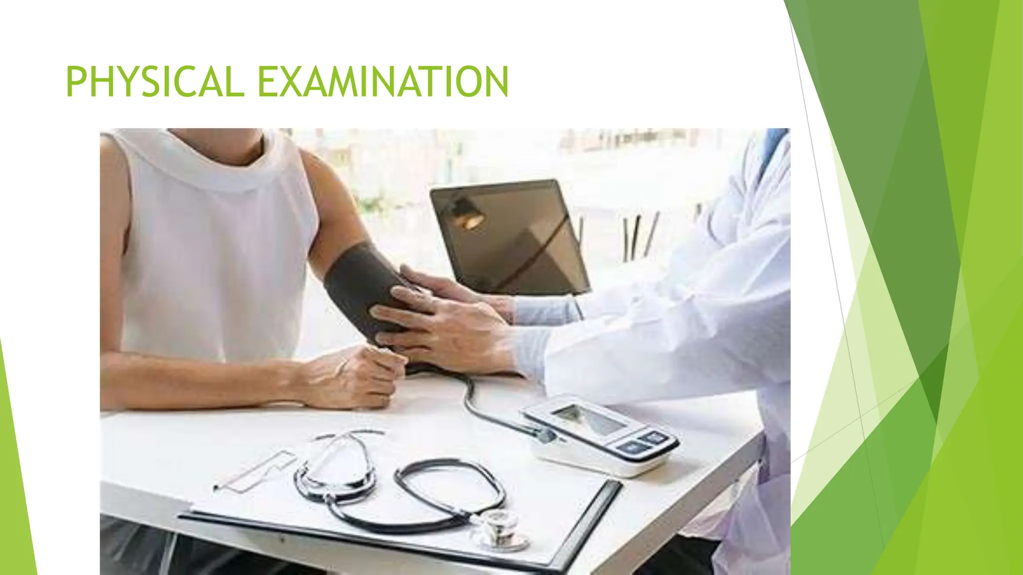
PHYSICAL EXAMINATION
• Introduce yourself to the patient
• Get patient consent
• Provide privacy
• Wash your hands
• Briefly explain to the patient what the examination
involves
• Position the patient supine
 Always start with the upper limb and proceed as below
Articles
 Inch tape
 Sphygmomanometer
 Stethoscope

 Inspection
 Palpation
 Auscultation
INSPECTION
UPPER EXTREMITIES:
The Arms
• Inspect for signs of
• Peripheral cyanosis
• Tar staining
As peripheral cyanosis
present in arterial
insufficiency or in
Raynaud syndrome
The Legs
• Expose the legs and observe from the end of the bed
• Scars
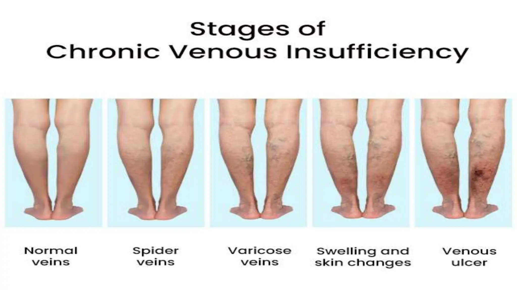
• Spider vein
• Varicose vein
• Dry flaky skin or hair loss seen in peripheral vascular
disease due to arterial insufficiency
• Skin changes
• Ulcers
• Dry Gangrene (seen in arterial insufficiency)
• Wet gangrene (seen in venous insufficiency)
Arterial ulcers are deep, painful wounds that occur when
there is inadequate blood supply, resulting in tissue damage
and slow healing.
Venous ulcers are larger, shallower sores that develop as a
consequence of impaired circulation in the veins.
ARTERIAL ULCER
VENOUS ULCER
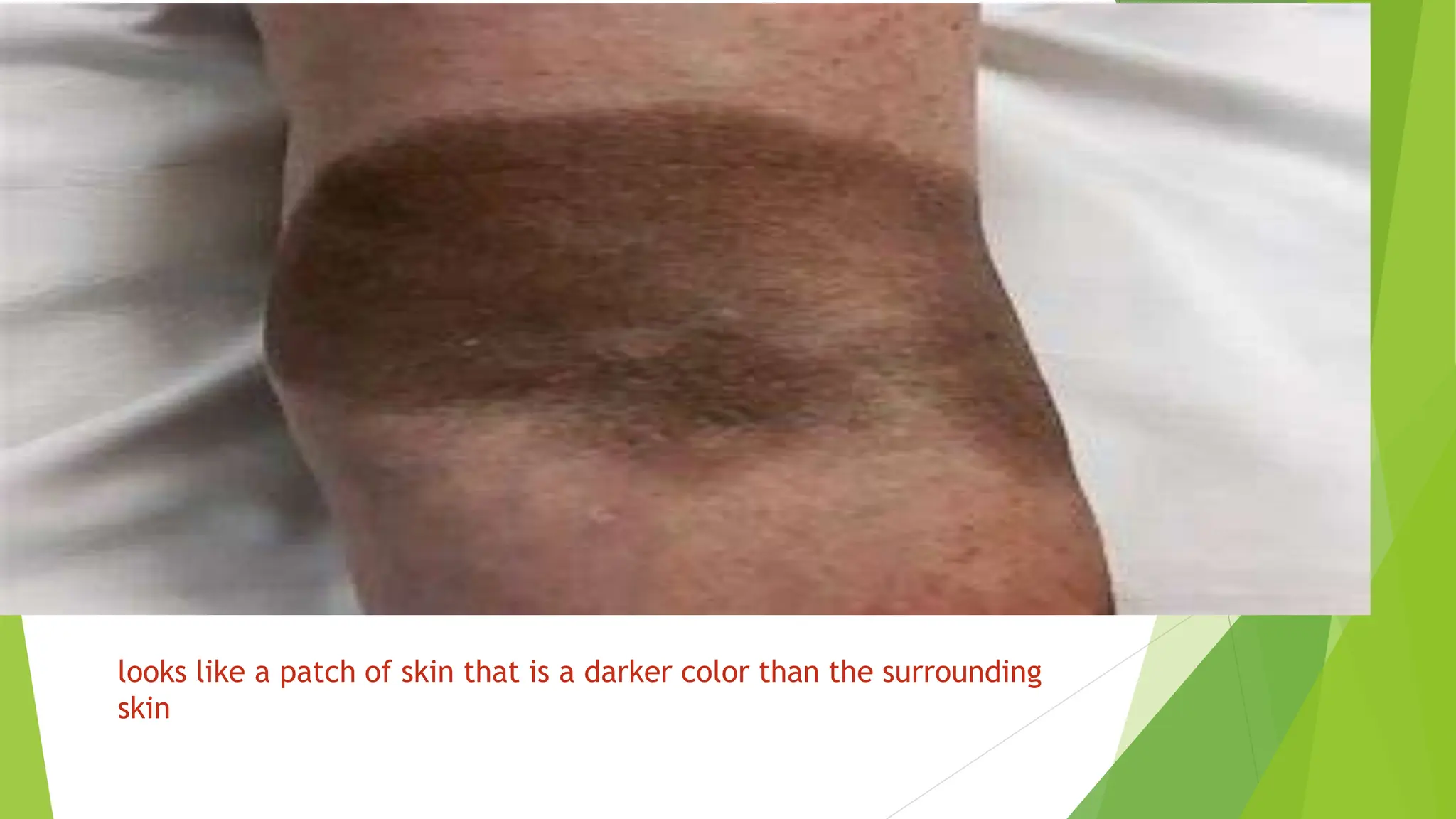
Haemosiderin Staining
Hemosiderin is an iron storage complex. Hemosiderin, a
protein that stores iron in your body, can accumulate under your
skin and in major organs. It is due to leakage, of venous blood as in
chronic venous stasis, which also occurs when the smallest blood
vessels, known as capillaries, begin to leak.
looks like a patch of skin that is a darker color than the surrounding
skin
ATROPHIE BLANCHE :
Atrophie blanche describes the result of healed ulcers.
These characteristically present as a white, atrophic stellate
scar with peripheral telangiectasias. Atrophie blanche is also
known as livedoid vasculopathy
 VARICOSE ECZEMA
Skin inflammation in the lower legs is caused
by fluid build-up. Stasis dermatitis is caused by fluid build-
up due to varicose veins, circulation issues or heart disease.
It is also called as Stasis dermatitis
THE ABDOMEN
 Look at the abdomen for obvious scars or pulsations
Assess for muscle atrophy ( sarcopenia )progressive loss of
muscle mass due to arterial insufficiency
Patients with PAD have lower exercise capacity due to
exercise‐induced pain/discomfort in the lower extremities,
leading to muscle wasting by reduction in daily physical
activity
CAPILLARY REFILL TIME
Squeeze the nail bed until it blanches, release, and observe the time
for the color to return. It is used to assess arterial blood flow to extremities.
Normal refill is less than 1-2 seconds
PALPATION
Palpate the upper and lower extremities for temperature,
moisture, pulses and edema bilaterally to assess for
symmetry.
TEMPERATURE
Palpate for temperature normally it is cold in peripheral
vascular disease, arterial insufficiency
Warmth in venous stasis, DVT, varicose vein
PULSE
The distal finger is kept hard pressed to stop the rebound pulse
coming from the palm[due to radial and ulnar arch, commonly
called as palmar arch]
ASSESS THE PULSE FOR
 Rate
 Rhythm
 Amplitude
 Force
 Symmetry
Rate:
It is the number of heart beat per minute, normal is 70-
80 beats/minute
RHYTHM
Regular at regular interval
Irregular at regular interval
irregular at irregular interval
AMPLITUDE
When a pulse wave passes below the middle finger,
expansion of the artery pushes the middle finger upward
which is called lifting of the middle finger
where stroke volume is more amplitude is high (or) vice
versa
FORCE:
 It is corresponding to systolic pressure
 Continue palpating the use by middle finger now you
have to give pressure to the proximal finger on the artery
and increase it gradually
 At one point pressure will be sufficient to stop
pressure coming to the middle finger
 The degree of pressure exerted by the proximal
finger is your observation
 If systolic blood pressure is more heavy over the proximal
finger
 Normotensive situation will require moderate
pressure over proximal pressure
 Light pressure is required in hypotensive patients
SYMMETRY
Normally pulse should be symmetrical on both side of the
extremities
Radio Radial delay
It is usually seen in subclavian artery stenosis,
Radio femoral delay
It is usually seen in aortic coarctation,
 Apex-pulse deficit has been described as a clinical sign in
patients with AF. This is the difference between the heart
rate counted from heart sounds (in terms of apex beat)
and the peripheral pulse palpated at the radial artery.
More than two beats per minutes is abnormal
ALLENS TEST
EDEMA
LYMPHEDEMA
Lymphedema is edema from impaired lymph node drainage. Surgical
removal or damage from surgery or radiation to lymph nodes and vessels
impedes lymph drainage. Lymph builds up causing lymphedema. It is
common after breast cancer treatment. Lymphedema is chronic and
progressive, so needs to be recognized and treated, such as with
compression bandages. Obesity causes lymphedema because the sheer
additional weight puts too much pressure on the lymph nodes in the groin
area, compromising the system.
This causes a fluid backup like a clogged drain. Skin can thicken, harden,
and become red, dry, and warm to the touch. It is nonpitting and feels hard
to touch unilaterally, compare the size to the other extremity:
Mild lymphedema causes an asymmetry of 1 to 3 Moderate lymphedema
causes an asymmetry of 3 to 5 Severe lymphedema is an asymmetry of
more than 5 cm
AUSCULTATION
Auscultate for abdominal bruit in case of abdominal
aneurysm
The abdominal aorta can be palpated at or slightly above the
umbilicus in the epigastrium in the supine position with the
knees bent.
BLOOD PRESSURE
 Blood pressure (BP) is the pressure of circulating blood
against the walls of blood vessels.
 It depends upon cardiac output and systemic vascular
resistance.
PULSE PRESSURE
 Pulse pressure is the difference between systolic and
diastolic blood pressure. It is measured in millimeters of
mercury (mmHg). It represents the force that the heart
generates each time it contracts. Healthy pulse pressure
is around 40 mmHg
 It is higher(wider) in hypertension, arrhythmia, aortic
regurgitation, anemia, arteriosclerosis
 It is lower(Narrower) in congestive heart failure, blood
loss
ANKLE BRACHIAL INDEX
 Measurement of blood flow in your leg arteries. Compares systolic blood
pressure in the arm with the ankle.
 Measured by Ankle systolic pressure divided by highest
brachial systolic pressure
ABI – interpretation
 >1.2 – suggests calcification of vessels (age)
 1.2-1.0 – Normal Range
 0.9 – 1.0 – Acceptable (borderline abnormal) No
referral needed
 0.8-0.9 – mild disease (manage risk factors)
 0.5 – 0.8 - moderate disease (routine referral) (mixed
ulcers )
 <0.5 – severe disease (URGENT referral) (no
compression bandaging!!!)
PULSE PARODOXUS
 Pulsus paradoxus, also paradoxic pulse or paradoxical
pulse, is an abnormally large decrease in stroke volume,
systolic blood pressure (a drop of more than 10 mmHg),
and pulse wave amplitude during inspiration
 Seen in cardiac tamponade and decreased blood to the
left heart due to lung hyperinflation (e.g. asthma, COPD)
and anaphylactic shock.
DISEASE SPECIFIC EXAMINATION
PERIPHERAL VASCULAR DISEASES
Check for 5 p’s
BUERGER TEST
 Buerger's test is performed in an assessment of arterial
sufficiency
 The vascular angle, which is also called Buerger's angle, is the
angle to which the leg has to be raised before it becomes pale,
whilst lying down
 In a limb with normal circulation the toes and sole of the foot,
stay pink, even when the limb is raised by 90 degrees. In an
ischaemic leg, elevation to 15 degrees or 30 degrees for 30 to 60
seconds may cause pallor. (This part of the test checks for
elevation pallor.) A vascular angle of less than 20 degrees
indicates severe ischemia
 From a sitting position, in normal circulation, the foot will
quickly return to pink color. Where there is peripheral
artery disease the leg will revert to the pink color more
slowly than normal and also pass through the normal
pinkness to a red-range coloring (rubor-redness) often
known as sunset foot. This is due to the dilatation of the
arterioles (post hypoxic vasodilation)
Deep Vein Thrombophlebitis (DVT)
Evaluation
Homan’s Sign
 Discomfort in the calf muscles on forced dorsiflexion
of the foot with the knee straight has been a time-
honored sign of DVT.
 Calf Circumference Measure at the widest point of the
calf and compare to other calf. Acute, unilateral,
painful swelling with asymmetry of the calves of 1 cm
or more is abnormal, refer for DVT.
 Consider this with other findings like edema,
redness, the warmth of leg (blood flow can’t return to
the heart)Calf circumference
VARICOSE VEIN
 The Trendelenburg Test or Brodie–Trendelenburg test is a
test that can be carried out as part of a physical
examination to determine the competency of the valves in
the superficial and deep veins of the legs in patients with
varicose veins
 With the patient in the supine position, the leg is flexed
at the hip and raised above heart level. The veins will
empty due to gravity or with the assistance of the
examiner's hand squeezing blood towards the heart
 A tourniquet is then applied around the upper thigh to
compress the superficial veins but not too tight as to
occlude the deeper veins. The leg is then lowered by
asking the patient to stand.
 Normally the superficial saphenous vein will fill from
below within 30–35 seconds as blood from the capillary
beds reaches the veins; if the superficial veins fill more
rapidly with the tourniquet in place there is valvular
incompetence below the level of the tourniquet in the
"deep" or "communicating" veins.
 After 20 seconds, if there has been no rapid
filling, the tourniquet is released. If there is
sudden filling at this point, it indicates that the
deep and communicating veins are competent but
the superficial veins are incompetent
 The test is reported in two parts, the initial
standing up of the patient (positive or negative
based on rapid filling) and the second phase once
the tourniquet is removed (positive or negative
based upon rapid filling).
DIAGNOSIS IN PERIPHERAL VASCULAR DISEASE
Nailfold capillaroscopy
LYMPHOSCINTIGRAPHY
LYMPH NODE ASSESSMENT
Pre auricular lymph node – palpated anterior to the tragus of
ear
Post auricular lymph node- behind the ear to mastoid
process
Submental lymph node-
Inguinal lymph node
JOURNAL REFERENCE
Sarcopenia in peripheral arterial disease: Establishing and
validating a predictive nomogram based on clinical and
computed tomography angiography indicators
Nie et al. Heliyon. 2024.
Methods: Clinical data and CTA imaging features from 281
PAD patients treated between January 1, 2019, and May 1,
2023, at two hospitals were retrospectively analyzed using
binary logistic regression to identify the independent risk
factors for sarcopenia
Conclusions:
A simple nomogram based on five independent factors,
namely age, BMI, history of CHD, WBC count, and the
severity of luminal stenosis, was developed to assist
clinicians in estimating sarcopenia risk among PAD patients.
APPLICATION OF ALLIED HEALTH
SCIENCES
 Nutrition therapist for dietary modification
 Physiotherapy
BIBLIOGRAPHY
1. Suddharth &Brunner. Textbook of Medical Surgical Nursing, 13th edition: Wolter
Kluwer publication; 2014.pgno 1450-1460.
2.Black JM, Hawks JH. Medical Surgical Nursing, 1st edition : Elseiver
publication;2019. pgno 1458-1489.
3. Woods LS, FroelicherE , Cardiac Nursing.6th edition :Wolters Kluwer
publication;2009.pgno 936-939
4.Kaur L, Kaur p. Adult Medical Surgical Nursing ,3rd edition :Lotus
publication;2008 .pgno [1080-98]
5. Workman, Ignatavicus. Medical Surgical Nursing,7th edition :Evolve
publication;2009 .pgno [1080-98]
JOURNAL REFERENCE
 Nie L, Yang Q, Song Q, Zhou Y, Zheng W, Xu Q. Sarcopenia in
peripheral arterial disease: Establishing and validating a predictive
nomogram based on clinical and computed tomography angiography
indicators. Heliyon. 2024 Mar 26;10(7):e28732. doi:
10.1016/j.heliyon.2024.e28732.
Peripheral vascular examination (2).pptx

More Related Content

Similar to Peripheral vascular examination (2).pptx

Peripheral Vascular Diseases
Peripheral Vascular DiseasesPeripheral Vascular Diseases
Peripheral Vascular Diseases
Jessie Madz
 
vasculardisorders-160920130338_8_PdfToPowerPoint.pptx
vasculardisorders-160920130338_8_PdfToPowerPoint.pptxvasculardisorders-160920130338_8_PdfToPowerPoint.pptx
vasculardisorders-160920130338_8_PdfToPowerPoint.pptx
mousaderhem1
 
coronaryheartdiseases-hanaalharbi-131024020826-phpapp02 (1).pdf
coronaryheartdiseases-hanaalharbi-131024020826-phpapp02 (1).pdfcoronaryheartdiseases-hanaalharbi-131024020826-phpapp02 (1).pdf
coronaryheartdiseases-hanaalharbi-131024020826-phpapp02 (1).pdf
Praveen Kumar
 
cardio vascular diseases.pdf
cardio vascular diseases.pdfcardio vascular diseases.pdf
cardio vascular diseases.pdf
dsptrafficknl
 
History & physical examination of cvs
History & physical examination of cvsHistory & physical examination of cvs
History & physical examination of cvs
Mandeep Duarah
 

Similar to Peripheral vascular examination (2).pptx (20)

Peripheral vascular diseases
Peripheral vascular diseasesPeripheral vascular diseases
Peripheral vascular diseases
 
Peripheral Vascular Diseases
Peripheral Vascular DiseasesPeripheral Vascular Diseases
Peripheral Vascular Diseases
 
vasculardisorders-160920130338_8_PdfToPowerPoint.pptx
vasculardisorders-160920130338_8_PdfToPowerPoint.pptxvasculardisorders-160920130338_8_PdfToPowerPoint.pptx
vasculardisorders-160920130338_8_PdfToPowerPoint.pptx
 
Vascular disorders
Vascular disordersVascular disorders
Vascular disorders
 
peripheralvasculardiseases PVHD (1).pptx
peripheralvasculardiseases PVHD (1).pptxperipheralvasculardiseases PVHD (1).pptx
peripheralvasculardiseases PVHD (1).pptx
 
Anatomy & Physiology Lecture Notes - Blood vessels & circulation
Anatomy & Physiology Lecture Notes - Blood vessels & circulationAnatomy & Physiology Lecture Notes - Blood vessels & circulation
Anatomy & Physiology Lecture Notes - Blood vessels & circulation
 
Pulmonaryedema 160624071007
Pulmonaryedema 160624071007Pulmonaryedema 160624071007
Pulmonaryedema 160624071007
 
Atherosclerosis-All about Atherosclerosis,intoduction, causes, pathophysiolog...
Atherosclerosis-All about Atherosclerosis,intoduction, causes, pathophysiolog...Atherosclerosis-All about Atherosclerosis,intoduction, causes, pathophysiolog...
Atherosclerosis-All about Atherosclerosis,intoduction, causes, pathophysiolog...
 
Peripheral vascular disease
Peripheral vascular diseasePeripheral vascular disease
Peripheral vascular disease
 
Cardiology with pharmacology for nurses
Cardiology with pharmacology for nursesCardiology with pharmacology for nurses
Cardiology with pharmacology for nurses
 
The Renal Nurse Unidad Iv
The Renal Nurse  Unidad IvThe Renal Nurse  Unidad Iv
The Renal Nurse Unidad Iv
 
pvd ppt for mpt.pptx
pvd ppt for mpt.pptxpvd ppt for mpt.pptx
pvd ppt for mpt.pptx
 
Coronary heart disease
Coronary heart diseaseCoronary heart disease
Coronary heart disease
 
coronaryheartdiseases-hanaalharbi-131024020826-phpapp02 (1).pdf
coronaryheartdiseases-hanaalharbi-131024020826-phpapp02 (1).pdfcoronaryheartdiseases-hanaalharbi-131024020826-phpapp02 (1).pdf
coronaryheartdiseases-hanaalharbi-131024020826-phpapp02 (1).pdf
 
cardio vascular diseases.pdf
cardio vascular diseases.pdfcardio vascular diseases.pdf
cardio vascular diseases.pdf
 
cardio vascular diseases.pdf
cardio vascular diseases.pdfcardio vascular diseases.pdf
cardio vascular diseases.pdf
 
History & physical examination of cvs
History & physical examination of cvsHistory & physical examination of cvs
History & physical examination of cvs
 
Test for peripheral arterial and venous circulation
Test for peripheral arterial and venous circulationTest for peripheral arterial and venous circulation
Test for peripheral arterial and venous circulation
 
medppt.pptx
medppt.pptxmedppt.pptx
medppt.pptx
 
cardio vascular diseases.pptx
cardio vascular diseases.pptxcardio vascular diseases.pptx
cardio vascular diseases.pptx
 

Recently uploaded

Failure to thrive in neonates and infants + pediatric case.pptx
Failure to thrive in neonates and infants  + pediatric case.pptxFailure to thrive in neonates and infants  + pediatric case.pptx
Failure to thrive in neonates and infants + pediatric case.pptx
claviclebrown44
 
Obat Aborsi Ampuh Usia 1,2,3,4,5,6,7 Bulan 081901222272 Obat Penggugur Kandu...
Obat Aborsi Ampuh Usia 1,2,3,4,5,6,7 Bulan  081901222272 Obat Penggugur Kandu...Obat Aborsi Ampuh Usia 1,2,3,4,5,6,7 Bulan  081901222272 Obat Penggugur Kandu...
Obat Aborsi Ampuh Usia 1,2,3,4,5,6,7 Bulan 081901222272 Obat Penggugur Kandu...
Halo Docter
 

Recently uploaded (20)

CONGENITAL HYPERTROPHIC PYLORIC STENOSIS by Dr M.KARTHIK EMMANUEL
CONGENITAL HYPERTROPHIC PYLORIC STENOSIS  by Dr M.KARTHIK EMMANUELCONGENITAL HYPERTROPHIC PYLORIC STENOSIS  by Dr M.KARTHIK EMMANUEL
CONGENITAL HYPERTROPHIC PYLORIC STENOSIS by Dr M.KARTHIK EMMANUEL
 
MOTION MANAGEMANT IN LUNG SBRT BY DR KANHU CHARAN PATRO
MOTION MANAGEMANT IN LUNG SBRT BY DR KANHU CHARAN PATROMOTION MANAGEMANT IN LUNG SBRT BY DR KANHU CHARAN PATRO
MOTION MANAGEMANT IN LUNG SBRT BY DR KANHU CHARAN PATRO
 
Lachesis Mutus- a Homoeopathic medicinel.pptx
Lachesis Mutus- a Homoeopathic medicinel.pptxLachesis Mutus- a Homoeopathic medicinel.pptx
Lachesis Mutus- a Homoeopathic medicinel.pptx
 
Top 10 Most Beautiful Russian Pornstars List 2024
Top 10 Most Beautiful Russian Pornstars List 2024Top 10 Most Beautiful Russian Pornstars List 2024
Top 10 Most Beautiful Russian Pornstars List 2024
 
Part I - Anticipatory Grief: Experiencing grief before the loss has happened
Part I - Anticipatory Grief: Experiencing grief before the loss has happenedPart I - Anticipatory Grief: Experiencing grief before the loss has happened
Part I - Anticipatory Grief: Experiencing grief before the loss has happened
 
Vesu * HiFi ℂall Girls Surat Phone No 8849756361 Elite ℂall Serviℂe Available...
Vesu * HiFi ℂall Girls Surat Phone No 8849756361 Elite ℂall Serviℂe Available...Vesu * HiFi ℂall Girls Surat Phone No 8849756361 Elite ℂall Serviℂe Available...
Vesu * HiFi ℂall Girls Surat Phone No 8849756361 Elite ℂall Serviℂe Available...
 
VIP ℂall Girls Arekere Bangalore 6378878445 WhatsApp: Me All Time Serviℂe Ava...
VIP ℂall Girls Arekere Bangalore 6378878445 WhatsApp: Me All Time Serviℂe Ava...VIP ℂall Girls Arekere Bangalore 6378878445 WhatsApp: Me All Time Serviℂe Ava...
VIP ℂall Girls Arekere Bangalore 6378878445 WhatsApp: Me All Time Serviℂe Ava...
 
Drug development life cycle indepth overview.pptx
Drug development life cycle indepth overview.pptxDrug development life cycle indepth overview.pptx
Drug development life cycle indepth overview.pptx
 
Gross Anatomy and Histology of Tongue by Dr. Rabia Inam Gandapore.pptx
Gross Anatomy and Histology of Tongue by Dr. Rabia Inam Gandapore.pptxGross Anatomy and Histology of Tongue by Dr. Rabia Inam Gandapore.pptx
Gross Anatomy and Histology of Tongue by Dr. Rabia Inam Gandapore.pptx
 
Face and Muscles of facial expression.pptx
Face and Muscles of facial expression.pptxFace and Muscles of facial expression.pptx
Face and Muscles of facial expression.pptx
 
Failure to thrive in neonates and infants + pediatric case.pptx
Failure to thrive in neonates and infants  + pediatric case.pptxFailure to thrive in neonates and infants  + pediatric case.pptx
Failure to thrive in neonates and infants + pediatric case.pptx
 
Call Girls Omr Road 7427069034 VIP Escorts Service Chennai Available 24*7 Book
Call Girls Omr Road 7427069034 VIP Escorts Service Chennai Available 24*7 BookCall Girls Omr Road 7427069034 VIP Escorts Service Chennai Available 24*7 Book
Call Girls Omr Road 7427069034 VIP Escorts Service Chennai Available 24*7 Book
 
VIP ℂall Girls Budhwar Peth Pune 8005736733 WhatsApp: Me All Time Serviℂe Ava...
VIP ℂall Girls Budhwar Peth Pune 8005736733 WhatsApp: Me All Time Serviℂe Ava...VIP ℂall Girls Budhwar Peth Pune 8005736733 WhatsApp: Me All Time Serviℂe Ava...
VIP ℂall Girls Budhwar Peth Pune 8005736733 WhatsApp: Me All Time Serviℂe Ava...
 
TEST BANK For Porth's Essentials of Pathophysiology, 5th Edition by Tommie L ...
TEST BANK For Porth's Essentials of Pathophysiology, 5th Edition by Tommie L ...TEST BANK For Porth's Essentials of Pathophysiology, 5th Edition by Tommie L ...
TEST BANK For Porth's Essentials of Pathophysiology, 5th Edition by Tommie L ...
 
Obat Aborsi Ampuh Usia 1,2,3,4,5,6,7 Bulan 081901222272 Obat Penggugur Kandu...
Obat Aborsi Ampuh Usia 1,2,3,4,5,6,7 Bulan  081901222272 Obat Penggugur Kandu...Obat Aborsi Ampuh Usia 1,2,3,4,5,6,7 Bulan  081901222272 Obat Penggugur Kandu...
Obat Aborsi Ampuh Usia 1,2,3,4,5,6,7 Bulan 081901222272 Obat Penggugur Kandu...
 
High Purity 99% PMK Ethyl Glycidate Powder CAS 28578-16-7
High Purity 99% PMK Ethyl Glycidate Powder CAS 28578-16-7High Purity 99% PMK Ethyl Glycidate Powder CAS 28578-16-7
High Purity 99% PMK Ethyl Glycidate Powder CAS 28578-16-7
 
ABO Blood grouping in-compatibility in pregnancy
ABO Blood grouping in-compatibility in pregnancyABO Blood grouping in-compatibility in pregnancy
ABO Blood grouping in-compatibility in pregnancy
 
Top 10 Most Beautiful Chinese Pornstars List 2024
Top 10 Most Beautiful Chinese Pornstars List 2024Top 10 Most Beautiful Chinese Pornstars List 2024
Top 10 Most Beautiful Chinese Pornstars List 2024
 
Creeping Stroke - Venous thrombosis presenting with pc-stroke.pptx
Creeping Stroke - Venous thrombosis presenting with pc-stroke.pptxCreeping Stroke - Venous thrombosis presenting with pc-stroke.pptx
Creeping Stroke - Venous thrombosis presenting with pc-stroke.pptx
 
ℂall Girls Chandigarh 💚8868886958 💚ℂall Girls Serviℂe Available Day and Night
ℂall Girls Chandigarh  💚8868886958  💚ℂall Girls Serviℂe Available Day and Nightℂall Girls Chandigarh  💚8868886958  💚ℂall Girls Serviℂe Available Day and Night
ℂall Girls Chandigarh 💚8868886958 💚ℂall Girls Serviℂe Available Day and Night
 

Peripheral vascular examination (2).pptx

  • 1. SEMINAR ON PERIPHERAL VASCULAR ASSESSMENT By Krishna Priya C V M.Sc Nursing 1st year
  • 2. ANATOMY The peripheral vascular system (PVS) includes all the blood vessels that exist outside the heart. The peripheral vascular system is classified as follows:  The aorta and its branches:  The arterioles  The capillaries  The venules and  veins returning blood to the heart  Lymphatic system
  • 3. LAYERS OF BLOOD VESSELS
  • 4.
  • 5. ARTERIES  Arteries are thick-walled structures that carry blood from the heart to the tissues  The intact endothelium synthesis regular of thrombosis such as prostacyclin, plasminogen, and heparin molecules.
  • 6. VEINS The walls of veins are thinner than arterioles, less muscular structure of the vein wall allows these vessels to distend more than arteries. Greater distensibility permits a large volume of blood to remain in the vein under lower pressure, for this reason, veins are referred to as capacitance vessels Because of their weaker wall structure, they are susceptible to irregular dilation
  • 7.
  • 8.
  • 9.
  • 10.
  • 11.
  • 12. LYMPHATICS SYSTEM The lymphatic system is a series of vessels and nodes that collect and filter excess tissue fluid (lymph), before returning it to the venous circulation
  • 13. The drainage of lymph begins in lymph channels, which start as blind-ended capillaries Gradually develop into vessels. These vessels travel proximally, draining through several lymph nodes. Eventually the vessels empty into lymphatic trunks (also known as collecting vessels) These eventually converge to form the right lymphatic duct and the thoracic duct. The right lymphatic duct is responsible for draining the lymph from the upper right quadrant of the body
  • 14. The thoracic duct is much larger and drains lymph from the rest of the body These two ducts then empty into the venous circulation at the subclavian veins, via the right and left venous angles.
  • 15.  Lymph is a transudative fluid that is transparent and yellow. It is formed when fluid leaves the capillary bed in tissues due to hydrostatic pressure. Roughly 10% of blood volume becomes lymph.
  • 16.
  • 18.  Demographical data: including age as Arteriosclerosis (hardening of arteries) is more prevalent among aged persons, greater than 55 years.  Occupation – clarify whether the occupation increases the risk for vascular disease including hours spent, position in occupation standing,sitting.  Present medical history : Pain in arterial insufficiency of the lower extremities, the client may complain of cramping leg pain in the calf muscles that occurs during ambulation but disappears with 1 to 2 minutes of rest. This pain is called intermittent claudication.
  • 19. As an artery becomes more stenosed, the pain may become more severe, even at rest, or pain that awakens them during the night (Rest pain).It is exacerbated by decreased cardiac output during sleep. Pain in chronic venous disease has a slow onset and is not associated with exercise or rest. The client who have venous disease may report chronic aching pain in the legs when they are in a dependent position usually a standing position. It is a throbbing and burning type of pain.
  • 20. Ask for pain over the abdomen which can radiate to the back due to the rupture of the abdominal aortic aneurysm Ask the patient whether they feel any visible pulsation in their abdomen.
  • 21.
  • 22. Edema is a more severe form of chronic venous disorder Are edema worsen at the end of day and is diminished at night by leg elevation? Any lower extremity ulcer Past medical history History of hypertension, and diabetes as they lead to increased risk of atherosclerosis that eventually leads to peripheral vascular disease. If diabetes and hypertension are present ask about their medication compliance.
  • 23.  Past surgical history Detail of any previous operations that the client has endured, particularly vascular bypass surgery. Note if there has been any previous angiography, endovascular graft or stent placement, or harvesting of arteries or veins from the lower extremities. Any history of fracture or any condition that leads to prolonged bed rest (due to increased risk of deep vein thrombosis )
  • 24. Dietary habits:  Ask about sodium, cholesterol and fat intake  Ask about the usual intake of protein and calories  Any allergic to seafoods
  • 25. FAMILY HISTORY Family history of diabetes, hypertension, and peripheral vascular disease. Personal history: History of cigarette smoking Sleep pattern whether it is disturbed due to rest pain Assess client activity level and whether it is reduced due to claudication pain. Any history of dyspnea due to pulmonarymbolism
  • 26.  Ask for any history of pain and paraesthesia in fingers and toes when exposed to cold environment.  It indicate Raynaud syndrome
  • 27. Female patients Oral contraceptive pills leads to an increased risk of atherosclerosis due to changes in lipid metabolism, and DVT risk due to increased fibrinogen levels
  • 28. Multiple pregnancies as it compresses arteries and veins to the leg leads to an increased risk of peripheral vascular disease
  • 30. • Introduce yourself to the patient • Get patient consent • Provide privacy • Wash your hands • Briefly explain to the patient what the examination involves • Position the patient supine  Always start with the upper limb and proceed as below
  • 31. Articles  Inch tape  Sphygmomanometer  Stethoscope 
  • 33. INSPECTION UPPER EXTREMITIES: The Arms • Inspect for signs of • Peripheral cyanosis • Tar staining As peripheral cyanosis present in arterial insufficiency or in Raynaud syndrome
  • 34.
  • 35. The Legs • Expose the legs and observe from the end of the bed • Scars • Spider vein • Varicose vein • Dry flaky skin or hair loss seen in peripheral vascular disease due to arterial insufficiency • Skin changes • Ulcers • Dry Gangrene (seen in arterial insufficiency) • Wet gangrene (seen in venous insufficiency)
  • 36. Arterial ulcers are deep, painful wounds that occur when there is inadequate blood supply, resulting in tissue damage and slow healing. Venous ulcers are larger, shallower sores that develop as a consequence of impaired circulation in the veins.
  • 39.
  • 40.
  • 41.
  • 42. Haemosiderin Staining Hemosiderin is an iron storage complex. Hemosiderin, a protein that stores iron in your body, can accumulate under your skin and in major organs. It is due to leakage, of venous blood as in chronic venous stasis, which also occurs when the smallest blood vessels, known as capillaries, begin to leak.
  • 43. looks like a patch of skin that is a darker color than the surrounding skin
  • 44. ATROPHIE BLANCHE : Atrophie blanche describes the result of healed ulcers. These characteristically present as a white, atrophic stellate scar with peripheral telangiectasias. Atrophie blanche is also known as livedoid vasculopathy
  • 45.
  • 46.  VARICOSE ECZEMA Skin inflammation in the lower legs is caused by fluid build-up. Stasis dermatitis is caused by fluid build- up due to varicose veins, circulation issues or heart disease. It is also called as Stasis dermatitis
  • 47.
  • 48.
  • 49. THE ABDOMEN  Look at the abdomen for obvious scars or pulsations
  • 50.
  • 51. Assess for muscle atrophy ( sarcopenia )progressive loss of muscle mass due to arterial insufficiency Patients with PAD have lower exercise capacity due to exercise‐induced pain/discomfort in the lower extremities, leading to muscle wasting by reduction in daily physical activity
  • 52.
  • 53.
  • 54. CAPILLARY REFILL TIME Squeeze the nail bed until it blanches, release, and observe the time for the color to return. It is used to assess arterial blood flow to extremities. Normal refill is less than 1-2 seconds
  • 55. PALPATION Palpate the upper and lower extremities for temperature, moisture, pulses and edema bilaterally to assess for symmetry.
  • 56. TEMPERATURE Palpate for temperature normally it is cold in peripheral vascular disease, arterial insufficiency Warmth in venous stasis, DVT, varicose vein
  • 57. PULSE
  • 58.
  • 59. The distal finger is kept hard pressed to stop the rebound pulse coming from the palm[due to radial and ulnar arch, commonly called as palmar arch]
  • 60. ASSESS THE PULSE FOR  Rate  Rhythm  Amplitude  Force  Symmetry
  • 61. Rate: It is the number of heart beat per minute, normal is 70- 80 beats/minute RHYTHM Regular at regular interval Irregular at regular interval irregular at irregular interval
  • 62. AMPLITUDE When a pulse wave passes below the middle finger, expansion of the artery pushes the middle finger upward which is called lifting of the middle finger where stroke volume is more amplitude is high (or) vice versa
  • 63.
  • 64. FORCE:  It is corresponding to systolic pressure  Continue palpating the use by middle finger now you have to give pressure to the proximal finger on the artery and increase it gradually  At one point pressure will be sufficient to stop pressure coming to the middle finger  The degree of pressure exerted by the proximal finger is your observation
  • 65.  If systolic blood pressure is more heavy over the proximal finger  Normotensive situation will require moderate pressure over proximal pressure  Light pressure is required in hypotensive patients
  • 66.
  • 67. SYMMETRY Normally pulse should be symmetrical on both side of the extremities Radio Radial delay It is usually seen in subclavian artery stenosis, Radio femoral delay It is usually seen in aortic coarctation,
  • 68.
  • 69.
  • 70.  Apex-pulse deficit has been described as a clinical sign in patients with AF. This is the difference between the heart rate counted from heart sounds (in terms of apex beat) and the peripheral pulse palpated at the radial artery. More than two beats per minutes is abnormal
  • 71.
  • 73. EDEMA
  • 74. LYMPHEDEMA Lymphedema is edema from impaired lymph node drainage. Surgical removal or damage from surgery or radiation to lymph nodes and vessels impedes lymph drainage. Lymph builds up causing lymphedema. It is common after breast cancer treatment. Lymphedema is chronic and progressive, so needs to be recognized and treated, such as with compression bandages. Obesity causes lymphedema because the sheer additional weight puts too much pressure on the lymph nodes in the groin area, compromising the system.
  • 75. This causes a fluid backup like a clogged drain. Skin can thicken, harden, and become red, dry, and warm to the touch. It is nonpitting and feels hard to touch unilaterally, compare the size to the other extremity: Mild lymphedema causes an asymmetry of 1 to 3 Moderate lymphedema causes an asymmetry of 3 to 5 Severe lymphedema is an asymmetry of more than 5 cm
  • 76.
  • 77.
  • 78. AUSCULTATION Auscultate for abdominal bruit in case of abdominal aneurysm The abdominal aorta can be palpated at or slightly above the umbilicus in the epigastrium in the supine position with the knees bent.
  • 79.
  • 80.
  • 81. BLOOD PRESSURE  Blood pressure (BP) is the pressure of circulating blood against the walls of blood vessels.  It depends upon cardiac output and systemic vascular resistance.
  • 82.
  • 83. PULSE PRESSURE  Pulse pressure is the difference between systolic and diastolic blood pressure. It is measured in millimeters of mercury (mmHg). It represents the force that the heart generates each time it contracts. Healthy pulse pressure is around 40 mmHg  It is higher(wider) in hypertension, arrhythmia, aortic regurgitation, anemia, arteriosclerosis  It is lower(Narrower) in congestive heart failure, blood loss
  • 84.
  • 85. ANKLE BRACHIAL INDEX  Measurement of blood flow in your leg arteries. Compares systolic blood pressure in the arm with the ankle.  Measured by Ankle systolic pressure divided by highest brachial systolic pressure
  • 86. ABI – interpretation  >1.2 – suggests calcification of vessels (age)  1.2-1.0 – Normal Range  0.9 – 1.0 – Acceptable (borderline abnormal) No referral needed  0.8-0.9 – mild disease (manage risk factors)  0.5 – 0.8 - moderate disease (routine referral) (mixed ulcers )  <0.5 – severe disease (URGENT referral) (no compression bandaging!!!)
  • 87. PULSE PARODOXUS  Pulsus paradoxus, also paradoxic pulse or paradoxical pulse, is an abnormally large decrease in stroke volume, systolic blood pressure (a drop of more than 10 mmHg), and pulse wave amplitude during inspiration  Seen in cardiac tamponade and decreased blood to the left heart due to lung hyperinflation (e.g. asthma, COPD) and anaphylactic shock.
  • 88.
  • 89.
  • 90.
  • 91.
  • 92.
  • 95. BUERGER TEST  Buerger's test is performed in an assessment of arterial sufficiency  The vascular angle, which is also called Buerger's angle, is the angle to which the leg has to be raised before it becomes pale, whilst lying down  In a limb with normal circulation the toes and sole of the foot, stay pink, even when the limb is raised by 90 degrees. In an ischaemic leg, elevation to 15 degrees or 30 degrees for 30 to 60 seconds may cause pallor. (This part of the test checks for elevation pallor.) A vascular angle of less than 20 degrees indicates severe ischemia
  • 96.  From a sitting position, in normal circulation, the foot will quickly return to pink color. Where there is peripheral artery disease the leg will revert to the pink color more slowly than normal and also pass through the normal pinkness to a red-range coloring (rubor-redness) often known as sunset foot. This is due to the dilatation of the arterioles (post hypoxic vasodilation)
  • 97.
  • 98. Deep Vein Thrombophlebitis (DVT) Evaluation Homan’s Sign  Discomfort in the calf muscles on forced dorsiflexion of the foot with the knee straight has been a time- honored sign of DVT.  Calf Circumference Measure at the widest point of the calf and compare to other calf. Acute, unilateral, painful swelling with asymmetry of the calves of 1 cm or more is abnormal, refer for DVT.  Consider this with other findings like edema, redness, the warmth of leg (blood flow can’t return to the heart)Calf circumference
  • 99.
  • 100.
  • 101. VARICOSE VEIN  The Trendelenburg Test or Brodie–Trendelenburg test is a test that can be carried out as part of a physical examination to determine the competency of the valves in the superficial and deep veins of the legs in patients with varicose veins  With the patient in the supine position, the leg is flexed at the hip and raised above heart level. The veins will empty due to gravity or with the assistance of the examiner's hand squeezing blood towards the heart
  • 102.  A tourniquet is then applied around the upper thigh to compress the superficial veins but not too tight as to occlude the deeper veins. The leg is then lowered by asking the patient to stand.  Normally the superficial saphenous vein will fill from below within 30–35 seconds as blood from the capillary beds reaches the veins; if the superficial veins fill more rapidly with the tourniquet in place there is valvular incompetence below the level of the tourniquet in the "deep" or "communicating" veins.
  • 103.  After 20 seconds, if there has been no rapid filling, the tourniquet is released. If there is sudden filling at this point, it indicates that the deep and communicating veins are competent but the superficial veins are incompetent  The test is reported in two parts, the initial standing up of the patient (positive or negative based on rapid filling) and the second phase once the tourniquet is removed (positive or negative based upon rapid filling).
  • 104.
  • 105. DIAGNOSIS IN PERIPHERAL VASCULAR DISEASE
  • 106.
  • 107.
  • 110. LYMPH NODE ASSESSMENT Pre auricular lymph node – palpated anterior to the tragus of ear Post auricular lymph node- behind the ear to mastoid process Submental lymph node- Inguinal lymph node
  • 111. JOURNAL REFERENCE Sarcopenia in peripheral arterial disease: Establishing and validating a predictive nomogram based on clinical and computed tomography angiography indicators Nie et al. Heliyon. 2024. Methods: Clinical data and CTA imaging features from 281 PAD patients treated between January 1, 2019, and May 1, 2023, at two hospitals were retrospectively analyzed using binary logistic regression to identify the independent risk factors for sarcopenia
  • 112. Conclusions: A simple nomogram based on five independent factors, namely age, BMI, history of CHD, WBC count, and the severity of luminal stenosis, was developed to assist clinicians in estimating sarcopenia risk among PAD patients.
  • 113. APPLICATION OF ALLIED HEALTH SCIENCES  Nutrition therapist for dietary modification  Physiotherapy
  • 114.
  • 115. BIBLIOGRAPHY 1. Suddharth &Brunner. Textbook of Medical Surgical Nursing, 13th edition: Wolter Kluwer publication; 2014.pgno 1450-1460. 2.Black JM, Hawks JH. Medical Surgical Nursing, 1st edition : Elseiver publication;2019. pgno 1458-1489. 3. Woods LS, FroelicherE , Cardiac Nursing.6th edition :Wolters Kluwer publication;2009.pgno 936-939 4.Kaur L, Kaur p. Adult Medical Surgical Nursing ,3rd edition :Lotus publication;2008 .pgno [1080-98] 5. Workman, Ignatavicus. Medical Surgical Nursing,7th edition :Evolve publication;2009 .pgno [1080-98]
  • 116. JOURNAL REFERENCE  Nie L, Yang Q, Song Q, Zhou Y, Zheng W, Xu Q. Sarcopenia in peripheral arterial disease: Establishing and validating a predictive nomogram based on clinical and computed tomography angiography indicators. Heliyon. 2024 Mar 26;10(7):e28732. doi: 10.1016/j.heliyon.2024.e28732.