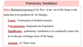
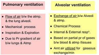
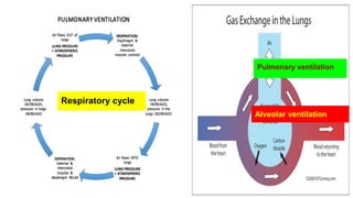
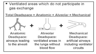
Lecture 5:Pulmonary Ventilation
Dr Shamshad
Guyton & Hall Textbook of Physiology - 13th ed. P. 465-467.
Pulmonary Ventilation
Defn: Mechanical process of air flow in or out of the lungs to the
atmp due to pr gradients & vol changes.
Cause: Contraction of skeletal muscles.
Two processes: Inspiration & Expiration.
Significance : pulmonary ventilation is to continually renew the
air in the gas exchange areas of the lungs.
Amount :6-7 litres /min
Pulmonary ventilation
● Flow of air b/w the atmp.
& the lung alveoli.
● Mechanical process
● Inspiration & Expiration
● Due to Pr gradient of air
b/w lungs & Atmp
Alveolar ventilation
● Exchange of air b/w Alveoli
& atmp.
● Chemical Process
● Internal & External respn.
● Based on partial pr of gases
b/w blood & atmp /tissues
● Amt air utilized for gaseous
exchange/min
Pulmonary ventilation
Alveolar ventilation
Respiratory cycle
In healthy adult
Pulmonary Ventilation
=Tidal Volume*Respiratory Rate
=500ml*12/minute
=6000ml/minute
AlveolarVentilation
=(Tidal Volume -Dead Space)*Resp Rate
=(500-150)ml*12/minute
=4200ml/minute
Respiratory Cycle & Respiratory Rate
Respiratory cycle : Sequence of inspiration & expiration
description of the changes in pressure, lung volumes
& airflow in one cycle.
Inspiration Rest Expiration Rest Inspiration
Respiratory Rate(RR): Number of breaths taken within a specific
amount of time (one minute).
In healthy adult:12-15 breaths/minutes
Respiratory cycle
➢Eupnea: Normal quiet respiration
➢Bradypnea: Slow rate of respiration
➢Tachypnea: Increased rate of respiration
➢Apnea: Cessation of respiration
➢Hypoventilation: Decrease in Rate & Depth of ventilation.
➢Hyperventilation: Increase in Rate & Depth of ventilation
➢Hypoapnea: Shallow breathing
➢Dyspnea: Difficulty in breathing.
Mechanics of normal & forced respiration.
Lung mechanics: It is study of forces & factors responsible for
ventilation.
Mechanics of ventilation depends on
1. Muscles & bones of thoracic cage.
2. Pleura & pleural cavity.
3. Dead space & value & types.
4. Different pressure & derived pressures.
5. Elastic property of lungs and thorax
6. Surface tension of the alveoli.
Muscles of Respiration
Quiet Inspiration: Diaphragm,External intercostal muscles
Quiet Expiration: Passive relaxation of inspiratory muscles
Forced Inspiration:Ala nostrils, scalene of neck
Sternocleidomastoids, pectoris minor,lavator scapulae,
Forced Expiration: Internal intercostal, Rectus abdominis, External
oblique muscles.
Muscles of inspiration:Elevate the chest cage
Muscles of expiration: Depress the chest cage
Thoracic cage bones: Ribs, Sternum, Vertebral column.
Diaphragm(75%)
Dome shaped- Attached to lower ribs, sternum & vertebral column.
Nerve supply: Phrenic nerve- C3,4,5.
Action :>75% of quiet inspiration . vertical dimension of thorax.
Contraction dome/pulls the dome of muscle down.
Causes flow of air into lungs.
Relaxation: dome thus decrease vertical diameter.
Causes expulsion of gases.
Moves down 1.5 cm normal (7.5cms - forced ) inspiration
Abdominal contents forced downward & forward causing ↑ in vertically
Rib margins are lifted & moved outward causing ↑ transverse diameter
Applied :Reduced movement:Obesity(moderate to severe),Pregnancy,
Tight Clothing.
Paradoxical movement of diaphragm when paralyzed
Upward movement with inspiratory drop of intrathoracic pr.
Inspiration Expiration
External intercostals (25%)
Location: Obliquely b/w ribs in forward & downward direction.
Nerve supply: Intercostal nerves (T1-11).
Action and effects:T.S+ A.P ↑by 2 mechanisms:
1: 6–10 ribs rotates upwards and outwards by a “bucket-handle
movement” → ↑ T.S.
2: upper 4 ribs rotate the sternum in upward & outward (pump-
handle movement) → ↑ in vertically .
Applied: Paralysis does not seriously alter inspiration diaphragm is
so effective but sensation of inhalation is decreases.
Inspiration Expiration
Muscles ➢ Contraction:Diaphragm
& External internal
intercostal muscles
➢ Daiphragm dome flattens
➢ Relaxation:Diaphragm &
External internal
intercostal muscles
➢ Daiphragm dome flattens
Ribs
Move
➢ up & out wards ➢ down & in wards
Thorax
Vol &Pr
➢ Vol increases &
➢ Air pr decreases
➢ Vol decreases &
➢ Air pr increases
Air flows ➔ Into lungs from atmp ➔ From lungs to atmp
Normal quiet respiration
Pleura & pleural cavity
Two layers :
1. Visceral :Attached to the lung parenchyma ,get
the same nerve supply not painful
(no pain receptors).
2. Parietal :Attached to inner aspect of thoracic
cavity, get the nerve supply from the
same,painful(contain pain receptors)
1. Intrapleural space:Space is closed space with
negative pressure , filled with pleural
fluid.
Intrapleural
space
Intrapleural Space: Has thin layer of mucoid transudate
(interstitial fluid + protein)
10-20 ml Secreted by Parietal layer.
Drained by lymphatics constant suction on pleura (-5cmH2O) of
(Mediastinum, superior - diaphragm, lateral)of parietal pleura
★ Helps create Negative pressure.
Functions:
➢ Lubricant for lungs to slide against chest wall facilitates change
in size & shape of lungs. Protects lungs from external damage.
➢ Prevents frictional irritation so membranes slide against each
other & are difficult to separate apart .
Pleural pressure
Defn:The pr of the fluid in the thin space b/w Visceral &Parital pleurae
Negative pr as compared to the atmp pr. < 760mmHg.
Beginning of the respiration is -5 cms of H2O
Function: This amount hold the lungs open to their resting position.
Negativity leads to sucking of air in lungs from the atmosphere.
During Normal quiet :the lungs and chest wall are stretched in
equal & opposite direction.
Intrapleural space increases but it being closed cavity ,negativity in it
increases more (-5 to 7.5cms of H2o)
Significance: If positive /equal to atmp pr
Leads to lungs collapse(recoil inwards)
Pneumothorax, Hemothorax, Chylothorax .
Pressures changes :During breathing
Atmospheric pressure :760mmHg/ Same=Zero
Alveolar pressure(Palv): More/same/less compared to atmp pr
Pleural pressure:(Ppl) always Negative/less than atmp pr .
Transpulmonary pr (Ptp) - Diff b/w alveolar pr & pleural pr.
(Ptp) = (Palv) - (Ppl)
Transthoracic pr (Ptt) - Diff b/w alveolar pr & body surface pr.
(Ptt) = (Palv) - (Patml)
Transchest pr (Pta)- Diff b/w alveoli pr & atmp pr.
(Pcw).= Ppl- Patm
26
Pressure relationship
Alveolar pressure
● Defn: The pr of air inside the lung alveoli
● When glottis is open:
● No air moves into the & out of the lungs
● Pr in alveoli = atmp pr =760mmHg /Zero cms of H2O
● Quiet inspiration: Alveoli opens pr = -ve /-1cm of H2o
This pulls air into alveoli in 2 seconds
● Quiet expiration: Alveolar pr =+ve/ + 1cm of H2o
This forces the inspired air out of lungs during 2-3 seconds.
TRANSMURAL PRESSURE
Thoracic cavity larger than lungs
Transmural pr:Pr across Lung Wall
Pr gradient holds thoracic wall & lungs
in close apposition
Pr gradient is balanced by the elastic forces
in the alveoli producing equilibrium
Under static conditions,
Transmural pr = the elastic recoil pr
of the compartment.
Transpulmonary pressure
● Defn:Alveolar pr- Pr on
outer space of lungs
● Measure of the elastic
forces in the lungs
● Tends to collapse the lungs
at each instant of
respiration
32
Physiological factors that affect:
Valsalva manoeuvre
Pathological factors:
Emphysema
Injury to thoracic wall will produce collapse of lungs
Dead spaces
Defn: A fraction of tidal volume of air that does not take part in
exchange of gases.
Types:
Anatomical :Vol. of air that does not reach the alveoli
Physiological: 1) Inspired air in alveoli that has no/less
blood supply.
2) Inspired air in alveoli that has normal blood flow
but less air reaches.
Normal Value:150 ml
Significance: regulation of temperature and humidification of air.
Increasing the depth of breathing is more effective in increasing
alveolar ventilation.
VT
Pulmonary ventilation 2020

Pulmonary ventilation 2020

  • 1.
  • 2.
    Guyton & HallTextbook of Physiology - 13th ed. P. 465-467.
  • 3.
    Pulmonary Ventilation Defn: Mechanicalprocess of air flow in or out of the lungs to the atmp due to pr gradients & vol changes. Cause: Contraction of skeletal muscles. Two processes: Inspiration & Expiration. Significance : pulmonary ventilation is to continually renew the air in the gas exchange areas of the lungs. Amount :6-7 litres /min
  • 4.
    Pulmonary ventilation ● Flowof air b/w the atmp. & the lung alveoli. ● Mechanical process ● Inspiration & Expiration ● Due to Pr gradient of air b/w lungs & Atmp Alveolar ventilation ● Exchange of air b/w Alveoli & atmp. ● Chemical Process ● Internal & External respn. ● Based on partial pr of gases b/w blood & atmp /tissues ● Amt air utilized for gaseous exchange/min
  • 5.
  • 6.
    In healthy adult PulmonaryVentilation =Tidal Volume*Respiratory Rate =500ml*12/minute =6000ml/minute AlveolarVentilation =(Tidal Volume -Dead Space)*Resp Rate =(500-150)ml*12/minute =4200ml/minute
  • 7.
    Respiratory Cycle &Respiratory Rate Respiratory cycle : Sequence of inspiration & expiration description of the changes in pressure, lung volumes & airflow in one cycle. Inspiration Rest Expiration Rest Inspiration Respiratory Rate(RR): Number of breaths taken within a specific amount of time (one minute). In healthy adult:12-15 breaths/minutes
  • 8.
  • 9.
    ➢Eupnea: Normal quietrespiration ➢Bradypnea: Slow rate of respiration ➢Tachypnea: Increased rate of respiration ➢Apnea: Cessation of respiration ➢Hypoventilation: Decrease in Rate & Depth of ventilation. ➢Hyperventilation: Increase in Rate & Depth of ventilation ➢Hypoapnea: Shallow breathing ➢Dyspnea: Difficulty in breathing.
  • 10.
    Mechanics of normal& forced respiration. Lung mechanics: It is study of forces & factors responsible for ventilation. Mechanics of ventilation depends on 1. Muscles & bones of thoracic cage. 2. Pleura & pleural cavity. 3. Dead space & value & types. 4. Different pressure & derived pressures. 5. Elastic property of lungs and thorax 6. Surface tension of the alveoli.
  • 12.
    Muscles of Respiration QuietInspiration: Diaphragm,External intercostal muscles Quiet Expiration: Passive relaxation of inspiratory muscles Forced Inspiration:Ala nostrils, scalene of neck Sternocleidomastoids, pectoris minor,lavator scapulae, Forced Expiration: Internal intercostal, Rectus abdominis, External oblique muscles. Muscles of inspiration:Elevate the chest cage Muscles of expiration: Depress the chest cage Thoracic cage bones: Ribs, Sternum, Vertebral column.
  • 13.
    Diaphragm(75%) Dome shaped- Attachedto lower ribs, sternum & vertebral column. Nerve supply: Phrenic nerve- C3,4,5. Action :>75% of quiet inspiration . vertical dimension of thorax. Contraction dome/pulls the dome of muscle down. Causes flow of air into lungs. Relaxation: dome thus decrease vertical diameter. Causes expulsion of gases. Moves down 1.5 cm normal (7.5cms - forced ) inspiration Abdominal contents forced downward & forward causing ↑ in vertically Rib margins are lifted & moved outward causing ↑ transverse diameter
  • 14.
    Applied :Reduced movement:Obesity(moderateto severe),Pregnancy, Tight Clothing. Paradoxical movement of diaphragm when paralyzed Upward movement with inspiratory drop of intrathoracic pr. Inspiration Expiration
  • 15.
    External intercostals (25%) Location:Obliquely b/w ribs in forward & downward direction. Nerve supply: Intercostal nerves (T1-11). Action and effects:T.S+ A.P ↑by 2 mechanisms: 1: 6–10 ribs rotates upwards and outwards by a “bucket-handle movement” → ↑ T.S. 2: upper 4 ribs rotate the sternum in upward & outward (pump- handle movement) → ↑ in vertically . Applied: Paralysis does not seriously alter inspiration diaphragm is so effective but sensation of inhalation is decreases.
  • 17.
    Inspiration Expiration Muscles ➢Contraction:Diaphragm & External internal intercostal muscles ➢ Daiphragm dome flattens ➢ Relaxation:Diaphragm & External internal intercostal muscles ➢ Daiphragm dome flattens Ribs Move ➢ up & out wards ➢ down & in wards Thorax Vol &Pr ➢ Vol increases & ➢ Air pr decreases ➢ Vol decreases & ➢ Air pr increases Air flows ➔ Into lungs from atmp ➔ From lungs to atmp Normal quiet respiration
  • 21.
    Pleura & pleuralcavity Two layers : 1. Visceral :Attached to the lung parenchyma ,get the same nerve supply not painful (no pain receptors). 2. Parietal :Attached to inner aspect of thoracic cavity, get the nerve supply from the same,painful(contain pain receptors) 1. Intrapleural space:Space is closed space with negative pressure , filled with pleural fluid. Intrapleural space
  • 22.
    Intrapleural Space: Hasthin layer of mucoid transudate (interstitial fluid + protein) 10-20 ml Secreted by Parietal layer. Drained by lymphatics constant suction on pleura (-5cmH2O) of (Mediastinum, superior - diaphragm, lateral)of parietal pleura ★ Helps create Negative pressure. Functions: ➢ Lubricant for lungs to slide against chest wall facilitates change in size & shape of lungs. Protects lungs from external damage. ➢ Prevents frictional irritation so membranes slide against each other & are difficult to separate apart .
  • 23.
    Pleural pressure Defn:The prof the fluid in the thin space b/w Visceral &Parital pleurae Negative pr as compared to the atmp pr. < 760mmHg. Beginning of the respiration is -5 cms of H2O Function: This amount hold the lungs open to their resting position. Negativity leads to sucking of air in lungs from the atmosphere. During Normal quiet :the lungs and chest wall are stretched in equal & opposite direction. Intrapleural space increases but it being closed cavity ,negativity in it increases more (-5 to 7.5cms of H2o)
  • 24.
    Significance: If positive/equal to atmp pr Leads to lungs collapse(recoil inwards) Pneumothorax, Hemothorax, Chylothorax .
  • 25.
    Pressures changes :Duringbreathing Atmospheric pressure :760mmHg/ Same=Zero Alveolar pressure(Palv): More/same/less compared to atmp pr Pleural pressure:(Ppl) always Negative/less than atmp pr . Transpulmonary pr (Ptp) - Diff b/w alveolar pr & pleural pr. (Ptp) = (Palv) - (Ppl) Transthoracic pr (Ptt) - Diff b/w alveolar pr & body surface pr. (Ptt) = (Palv) - (Patml) Transchest pr (Pta)- Diff b/w alveoli pr & atmp pr. (Pcw).= Ppl- Patm
  • 26.
  • 28.
    Alveolar pressure ● Defn:The pr of air inside the lung alveoli ● When glottis is open: ● No air moves into the & out of the lungs ● Pr in alveoli = atmp pr =760mmHg /Zero cms of H2O ● Quiet inspiration: Alveoli opens pr = -ve /-1cm of H2o This pulls air into alveoli in 2 seconds ● Quiet expiration: Alveolar pr =+ve/ + 1cm of H2o This forces the inspired air out of lungs during 2-3 seconds.
  • 30.
    TRANSMURAL PRESSURE Thoracic cavitylarger than lungs Transmural pr:Pr across Lung Wall Pr gradient holds thoracic wall & lungs in close apposition Pr gradient is balanced by the elastic forces in the alveoli producing equilibrium Under static conditions, Transmural pr = the elastic recoil pr of the compartment.
  • 31.
    Transpulmonary pressure ● Defn:Alveolarpr- Pr on outer space of lungs ● Measure of the elastic forces in the lungs ● Tends to collapse the lungs at each instant of respiration
  • 32.
    32 Physiological factors thataffect: Valsalva manoeuvre Pathological factors: Emphysema Injury to thoracic wall will produce collapse of lungs
  • 33.
    Dead spaces Defn: Afraction of tidal volume of air that does not take part in exchange of gases. Types: Anatomical :Vol. of air that does not reach the alveoli Physiological: 1) Inspired air in alveoli that has no/less blood supply. 2) Inspired air in alveoli that has normal blood flow but less air reaches. Normal Value:150 ml Significance: regulation of temperature and humidification of air. Increasing the depth of breathing is more effective in increasing alveolar ventilation. VT