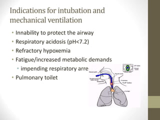
This document provides an overview of respiratory physiology and acute respiratory failure. It discusses:
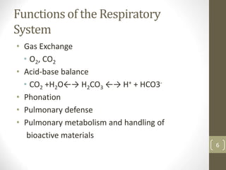
1. The functions of the respiratory system including gas exchange, acid-base balance, phonation, pulmonary defense, and metabolism.
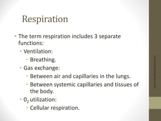
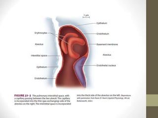
2. The three components of respiration - ventilation, gas exchange, and oxygen utilization. It describes the mechanics of ventilation and gas exchange via diffusion.
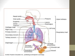
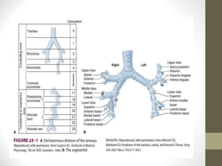
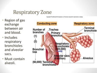
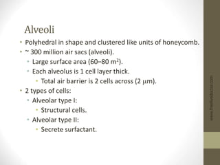
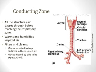
3. The conducting and respiratory zones of the lungs and structures involved in gas exchange like alveoli and surfactant.
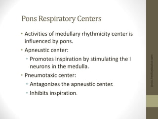
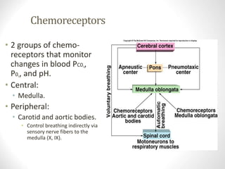
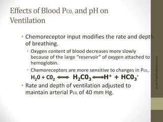
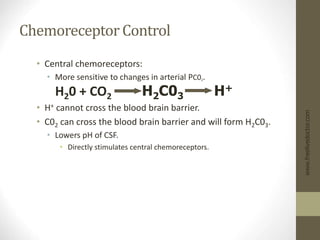
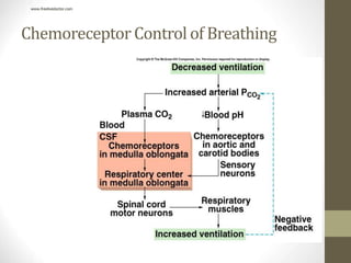
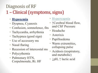
4. Control of respiration via brainstem centers that regulate rhythmic breathing and chemoreceptors that sense blood gases and pH to modulate breathing rate and depth.

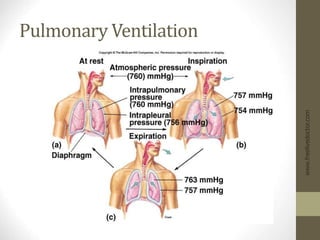
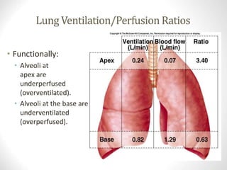
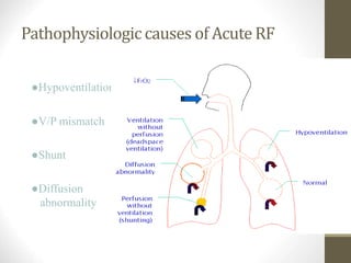
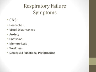
![Ventilation
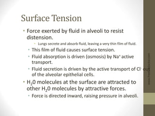
• Mechanical process that moves air
in and out of the lungs.
• [O2]of air is higher in the lungs
than in the blood, O2 diffuses from
air to the blood.
• C02 moves from the blood to the
air by diffusing down its
concentration gradient.
• Gas exchange occurs entirely by
diffusion:
• Diffusion is rapid because of the
large surface area and the small
diffusion distance.
www.freelivedoctor.com
Insert 16.1](https://image.slidesharecdn.com/1096respiratoryphysiologyposted-i-1-220921181119-d1dc8d5d/85/1096_RespiratoryPhysiologyPosted-I-1-ppt-8-320.jpg)
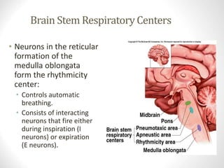
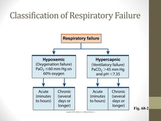
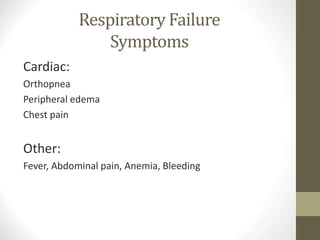
![Anatomical Dead Space
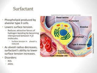
• Not all of the inspired air reached the alveoli.
• As fresh air is inhaled it is mixed with air in anatomical
dead space.
• Conducting zone and alveoli where [02] is lower than
normal and [C02] is higher than normal.
• Alveolar ventilation = F x (TV- DS).
• F = frequency (breaths/min.).
• TV = tidal volume.
• DS = dead space.
www.freelivedoctor.com](https://image.slidesharecdn.com/1096respiratoryphysiologyposted-i-1-220921181119-d1dc8d5d/85/1096_RespiratoryPhysiologyPosted-I-1-ppt-33-320.jpg)

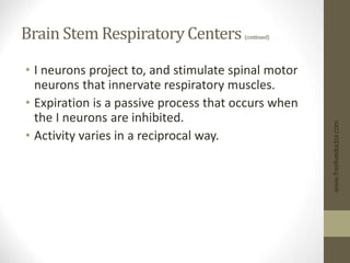
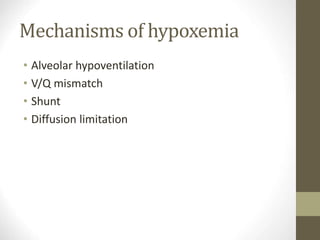
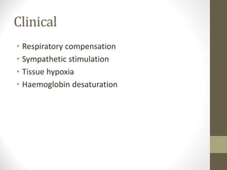
![Partial Pressures of Gases in Blood
• When a liquid or gas (blood and alveolar air) are
at equilibrium:
• The amount of gas dissolved in fluid reaches a
maximum value (Henry’s Law).
• Depends upon:
• Solubility of gas in the fluid.
• Temperature of the fluid.
• Partial pressure of the gas.
• [Gas] dissolved in a fluid depends directly on its
partial pressure in the gas mixture.
www.freelivedoctor.com](https://image.slidesharecdn.com/1096respiratoryphysiologyposted-i-1-220921181119-d1dc8d5d/85/1096_RespiratoryPhysiologyPosted-I-1-ppt-37-320.jpg)
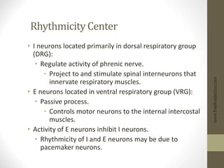
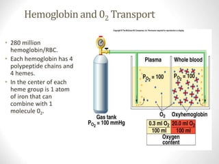
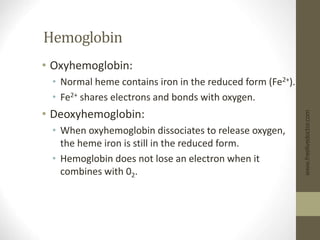
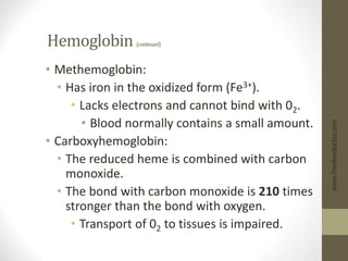
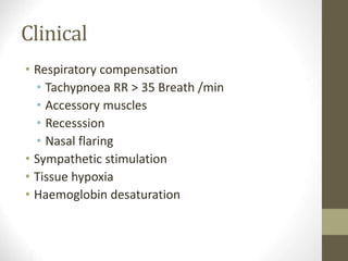
![Hemoglobin (continued)
• Oxygen-carrying capacity of blood determined by its
[hemoglobin].
• Anemia:
• [Hemoglobin] below normal.
• Polycythemia:
• [Hemoglobin] above normal.
• Hemoglobin production controlled by erythropoietin.
• Production stimulated by PC02 delivery to kidneys.
• Loading/unloading depends:
• P02 of environment.
• Affinity between hemoglobin and 02.
www.freelivedoctor.com](https://image.slidesharecdn.com/1096respiratoryphysiologyposted-i-1-220921181119-d1dc8d5d/85/1096_RespiratoryPhysiologyPosted-I-1-ppt-55-320.jpg)
![Effect of 2,3 DPG on 02 Transport
• Anemia:
• RBCs total blood [hemoglobin] falls, each RBC
produces greater amount of 2,3 DPG.
• Since RBCs lack both nuclei and
mitochondria, produce ATP through
anaerobic metabolism.
• Fetal hemoglobin (hemoglobin f):
• Has 2 g-chains in place of the b-chains.
• Hemoglobin f cannot bind to 2,3 DPG.
• Has a higher affinity for 02.
www.freelivedoctor.com](https://image.slidesharecdn.com/1096respiratoryphysiologyposted-i-1-220921181119-d1dc8d5d/85/1096_RespiratoryPhysiologyPosted-I-1-ppt-58-320.jpg)
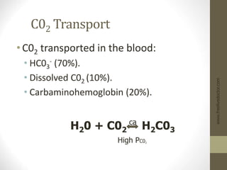
![Chloride Shift at Systemic Capillaries
• H20 + C02 H2C03 H+ + HC03
-
• At the tissues, C02 diffuses into the RBC; shifts the
reaction to the right.
• Increased [HC03
-] produced in RBC:
• HC03
- diffuses into the blood.
• RBC becomes more +.
• Cl- attracted in (Cl- shift).
• H+ released buffered by combining with
deoxyhemoglobin.
• HbC02 formed.
• Unloading of 02.
www.freelivedoctor.com](https://image.slidesharecdn.com/1096respiratoryphysiologyposted-i-1-220921181119-d1dc8d5d/85/1096_RespiratoryPhysiologyPosted-I-1-ppt-60-320.jpg)
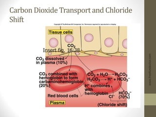
![At Pulmonary Capillaries
• H20 + C02 H2C03 H+ + HC03
-
• At the alveoli, C02 diffuses into the alveoli;
reaction shifts to the left.
• Decreased [HC03
-] in RBC, HC03
- diffuses into the
RBC.
• RBC becomes more -.
• Cl- diffuses out (reverse Cl- shift).
• Deoxyhemoglobin converted to oxyhemoglobin.
• Has weak affinity for H+.
• Gives off HbC02.
www.freelivedoctor.com](https://image.slidesharecdn.com/1096respiratoryphysiologyposted-i-1-220921181119-d1dc8d5d/85/1096_RespiratoryPhysiologyPosted-I-1-ppt-62-320.jpg)


![PERI-PROSTHETIC FRACTURE NAIL-PLATE CONSTRUCT [NPC].pptx](https://cdn.slidesharecdn.com/ss_thumbnails/drarunkumardrmohamedashrafperiprostheticfrasturenail-plateconstructnpc-260209164459-7e9d15a1-thumbnail.jpg?width=640&height=640&fit=bounds)


