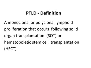
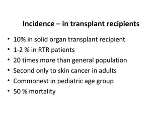
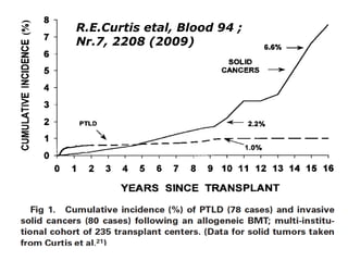
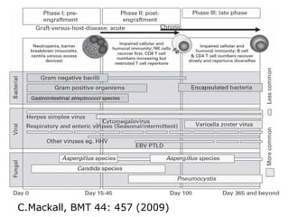
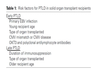
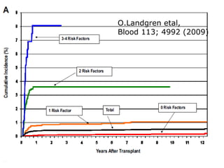
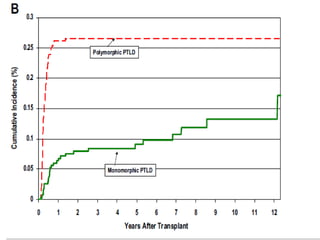
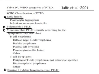
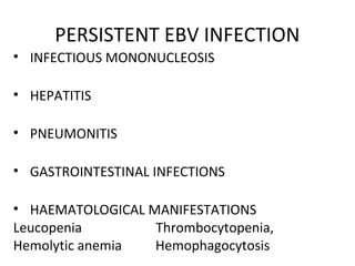
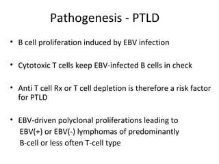
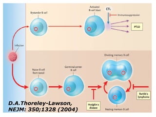
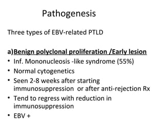
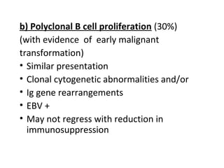
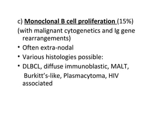
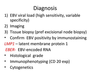
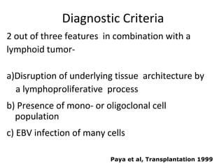
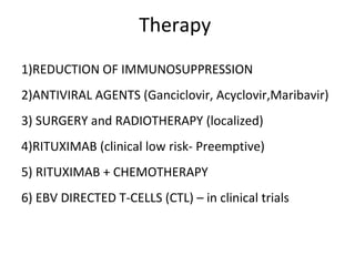
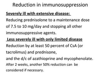
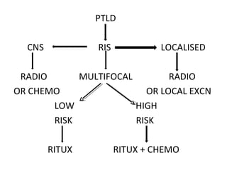
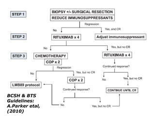
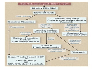
Post-transplant
lymphoproliferative
disorder
NDT
PTLD - Definition
A monoclonal or polyclonal lymphoid
proliferation that occurs following solid
organ transplantation (SOT) or
hematopoietic stem cell transplantation
(HSCT).
Incidence – in transplant recipients
• 10% in solid organ transplant recipient
• 1-2 % in RTR patients
• 20 times more than general population
• Second only to skin cancer in adults
• Commonest in pediatric age group
• 50 % mortality
Frequency A.Parker etal, BJH 149;
675 (2010)
R.E.Curtis etal, Blood 94 ;
Nr.7, 2208 (2009)
C.Mackall, BMT 44: 457 (2009)
O.Landgren etal,
Blood 113; 4992 (2009)
Jaffe et al -2001
A
B
C
D
PERSISTENT EBV INFECTION
• INFECTIOUS MONONUCLEOSIS
• HEPATITIS
• PNEUMONITIS
• GASTROINTESTINAL INFECTIONS
• HAEMATOLOGICAL MANIFESTATIONS
Leucopenia Thrombocytopenia,
Hemolytic anemia Hemophagocytosis
Pathogenesis - PTLD
• B cell proliferation induced by EBV infection
• Cytotoxic T cells keep EBV-infected B cells in check
• Anti T cell Rx or T cell depletion is therefore a risk factor
for PTLD
• EBV-driven polyclonal proliferations leading to
EBV(+) or EBV(-) lymphomas of predominantly
B-cell or less often T-cell type
D.A.Thoreley-Lawson,
NEJM: 350;1328 (2004)
Pathogenesis
Three types of EBV-related PTLD
a)Benign polyclonal proliferation /Early lesion
• Inf. Mononucleosis -like syndrome (55%)
• Normal cytogenetics
• Seen 2-8 weeks after starting
immunosuppression or after anti-rejection Rx
• Tend to regress with reduction in
immunosuppression
• EBV +
b) Polyclonal B cell proliferation (30%)
(with evidence of early malignant
transformation)
• Similar presentation
• Clonal cytogenetic abnormalities and/or
• Ig gene rearrangements
• EBV +
• May not regress with reduction in
immunosuppression
c) Monoclonal B cell proliferation (15%)
(with malignant cytogenetics and Ig gene
rearrangements)
• Often extra-nodal
• Various histologies possible:
• DLBCL, diffuse immunoblastic, MALT,
Burkitt’s-like, Plasmacytoma, HIV
associated
EBV-negative PTLD
Present much later
(median 50-60mo vs 6-10 mo)
Monomorphic
Poor outcomes , poor response to therapy
( mean survival of 1 month vs 37 months)
Increasing in frequency
Clinical featutes
• >50% present with extranodal masses
• Involved organs include solid organs and the allograft
itself
• MC (20-25% )with CNS disease
(higher than in general population)
• 15% have allograft involvement leading to allograft
dysfunction
Diagnosis
1) EBV viral load (high sensitivity, variable
specificity)
2) Imaging
3) Tissue biopsy (pref excisional node biopsy)
• Confirm EBV positivity by immunostaining
LMP1 – latent membrane protein 1
EBER- EBV-encoded RNA
• Histological grade
• Immunophenotyping (CD 20 exp)
• Cytogenetics
Diagnostic Criteria
2 out of three features in combination with a
lymphoid tumor-
a)Disruption of underlying tissue architecture by
a lymphoproliferative process
b) Presence of mono- or oligoclonal cell
population
c) EBV infection of many cells
Paya et al, Transplantation 1999
Therapy
1)REDUCTION OF IMMUNOSUPPRESSION
2)ANTIVIRAL AGENTS (Ganciclovir, Acyclovir,Maribavir)
3) SURGERY and RADIOTHERAPY (localized)
4)RITUXIMAB (clinical low risk- Preemptive)
5) RITUXIMAB + CHEMOTHERAPY
6) EBV DIRECTED T-CELLS (CTL) – in clinical trials
Reduction in immunosuppression
Severely ill with extensive disease:
Reducing prednisolone to a maintenance dose
of 7.5 to 10 mg/day and stopping all other
immunosuppressive agents.
Less severely ill with only limited disease
Reduction by at least 50 percent of CsA (or
tacrolimus) and prednisone,
and the d/c of azathioprine and mycophenolate.
After 2 weeks, another 50% reduction can be
considered if necessary.
PTLD
CNS RIS LOCALISED
RADIO MULTIFOCAL RADIO
OR CHEMO OR LOCAL EXCN
LOW HIGH
RISK RISK
RITUX RITUX + CHEMO
BCSH & BTS
Guidelines:
A.Parker etal,
(2010)
THERAPY OF PTLD with T Cells
A.Moosmann etal,
Blood 115 (14); 2960
(2010)
Outcomes
EBV – MONITORING
AT LEAST WEEKLY FOR 3 MONTHS
• FOR ALLO-SCT
• AFTER HIGH RISK SCT
• LONGER MONITORING FOR
- GVHD
- PREVIOUS EBV REACTIVATION
J.Styczynski etal, BMT 43; 757 (2009)
Prevention
1) IV ganciclovir to high-risk pts for a min of 100 days
2) Oral acyclovir in low risk patients
2) Lower target tacrolimus levels (2-5 ng/mL )
McDiarmid et al, Transplantation
PTLD

PTLD

  • 1.
  • 2.
    PTLD - Definition Amonoclonal or polyclonal lymphoid proliferation that occurs following solid organ transplantation (SOT) or hematopoietic stem cell transplantation (HSCT).
  • 3.
    Incidence – intransplant recipients • 10% in solid organ transplant recipient • 1-2 % in RTR patients • 20 times more than general population • Second only to skin cancer in adults • Commonest in pediatric age group • 50 % mortality
  • 4.
    Frequency A.Parker etal,BJH 149; 675 (2010)
  • 5.
    R.E.Curtis etal, Blood94 ; Nr.7, 2208 (2009)
  • 6.
  • 8.
  • 10.
    Jaffe et al-2001 A B C D
  • 11.
    PERSISTENT EBV INFECTION •INFECTIOUS MONONUCLEOSIS • HEPATITIS • PNEUMONITIS • GASTROINTESTINAL INFECTIONS • HAEMATOLOGICAL MANIFESTATIONS Leucopenia Thrombocytopenia, Hemolytic anemia Hemophagocytosis
  • 12.
    Pathogenesis - PTLD •B cell proliferation induced by EBV infection • Cytotoxic T cells keep EBV-infected B cells in check • Anti T cell Rx or T cell depletion is therefore a risk factor for PTLD • EBV-driven polyclonal proliferations leading to EBV(+) or EBV(-) lymphomas of predominantly B-cell or less often T-cell type
  • 13.
  • 14.
    Pathogenesis Three types ofEBV-related PTLD a)Benign polyclonal proliferation /Early lesion • Inf. Mononucleosis -like syndrome (55%) • Normal cytogenetics • Seen 2-8 weeks after starting immunosuppression or after anti-rejection Rx • Tend to regress with reduction in immunosuppression • EBV +
  • 15.
    b) Polyclonal Bcell proliferation (30%) (with evidence of early malignant transformation) • Similar presentation • Clonal cytogenetic abnormalities and/or • Ig gene rearrangements • EBV + • May not regress with reduction in immunosuppression
  • 16.
    c) Monoclonal Bcell proliferation (15%) (with malignant cytogenetics and Ig gene rearrangements) • Often extra-nodal • Various histologies possible: • DLBCL, diffuse immunoblastic, MALT, Burkitt’s-like, Plasmacytoma, HIV associated
  • 17.
    EBV-negative PTLD Present muchlater (median 50-60mo vs 6-10 mo) Monomorphic Poor outcomes , poor response to therapy ( mean survival of 1 month vs 37 months) Increasing in frequency
  • 18.
  • 19.
    • >50% presentwith extranodal masses • Involved organs include solid organs and the allograft itself • MC (20-25% )with CNS disease (higher than in general population) • 15% have allograft involvement leading to allograft dysfunction
  • 20.
    Diagnosis 1) EBV viralload (high sensitivity, variable specificity) 2) Imaging 3) Tissue biopsy (pref excisional node biopsy) • Confirm EBV positivity by immunostaining LMP1 – latent membrane protein 1 EBER- EBV-encoded RNA • Histological grade • Immunophenotyping (CD 20 exp) • Cytogenetics
  • 21.
    Diagnostic Criteria 2 outof three features in combination with a lymphoid tumor- a)Disruption of underlying tissue architecture by a lymphoproliferative process b) Presence of mono- or oligoclonal cell population c) EBV infection of many cells Paya et al, Transplantation 1999
  • 22.
    Therapy 1)REDUCTION OF IMMUNOSUPPRESSION 2)ANTIVIRALAGENTS (Ganciclovir, Acyclovir,Maribavir) 3) SURGERY and RADIOTHERAPY (localized) 4)RITUXIMAB (clinical low risk- Preemptive) 5) RITUXIMAB + CHEMOTHERAPY 6) EBV DIRECTED T-CELLS (CTL) – in clinical trials
  • 23.
    Reduction in immunosuppression Severelyill with extensive disease: Reducing prednisolone to a maintenance dose of 7.5 to 10 mg/day and stopping all other immunosuppressive agents. Less severely ill with only limited disease Reduction by at least 50 percent of CsA (or tacrolimus) and prednisone, and the d/c of azathioprine and mycophenolate. After 2 weeks, another 50% reduction can be considered if necessary.
  • 24.
    PTLD CNS RIS LOCALISED RADIOMULTIFOCAL RADIO OR CHEMO OR LOCAL EXCN LOW HIGH RISK RISK RITUX RITUX + CHEMO
  • 25.
  • 27.
    THERAPY OF PTLDwith T Cells A.Moosmann etal, Blood 115 (14); 2960 (2010)
  • 28.
  • 30.
    EBV – MONITORING ATLEAST WEEKLY FOR 3 MONTHS • FOR ALLO-SCT • AFTER HIGH RISK SCT • LONGER MONITORING FOR - GVHD - PREVIOUS EBV REACTIVATION J.Styczynski etal, BMT 43; 757 (2009)
  • 31.
    Prevention 1) IV ganciclovirto high-risk pts for a min of 100 days 2) Oral acyclovir in low risk patients 2) Lower target tacrolimus levels (2-5 ng/mL ) McDiarmid et al, Transplantation