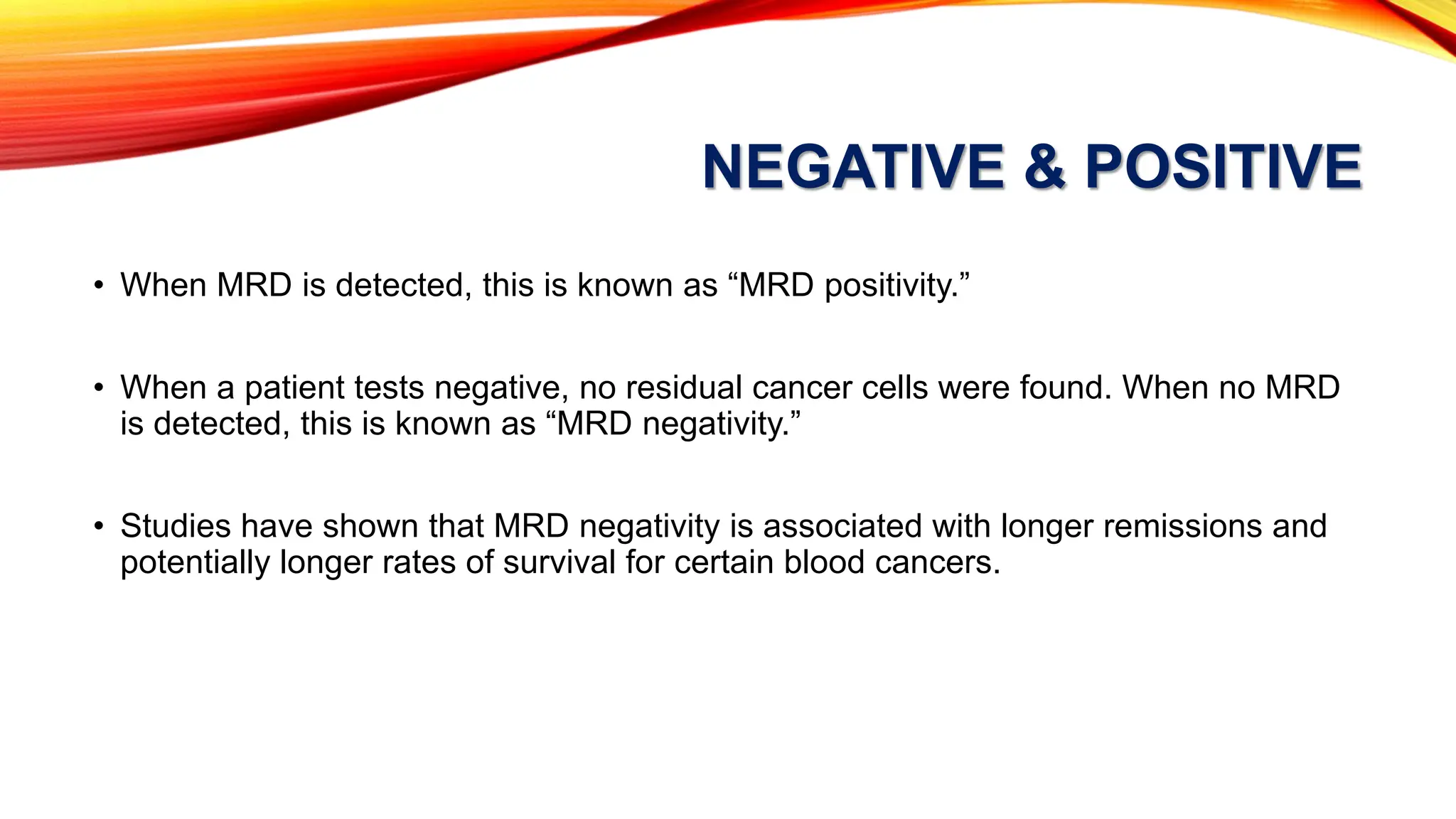
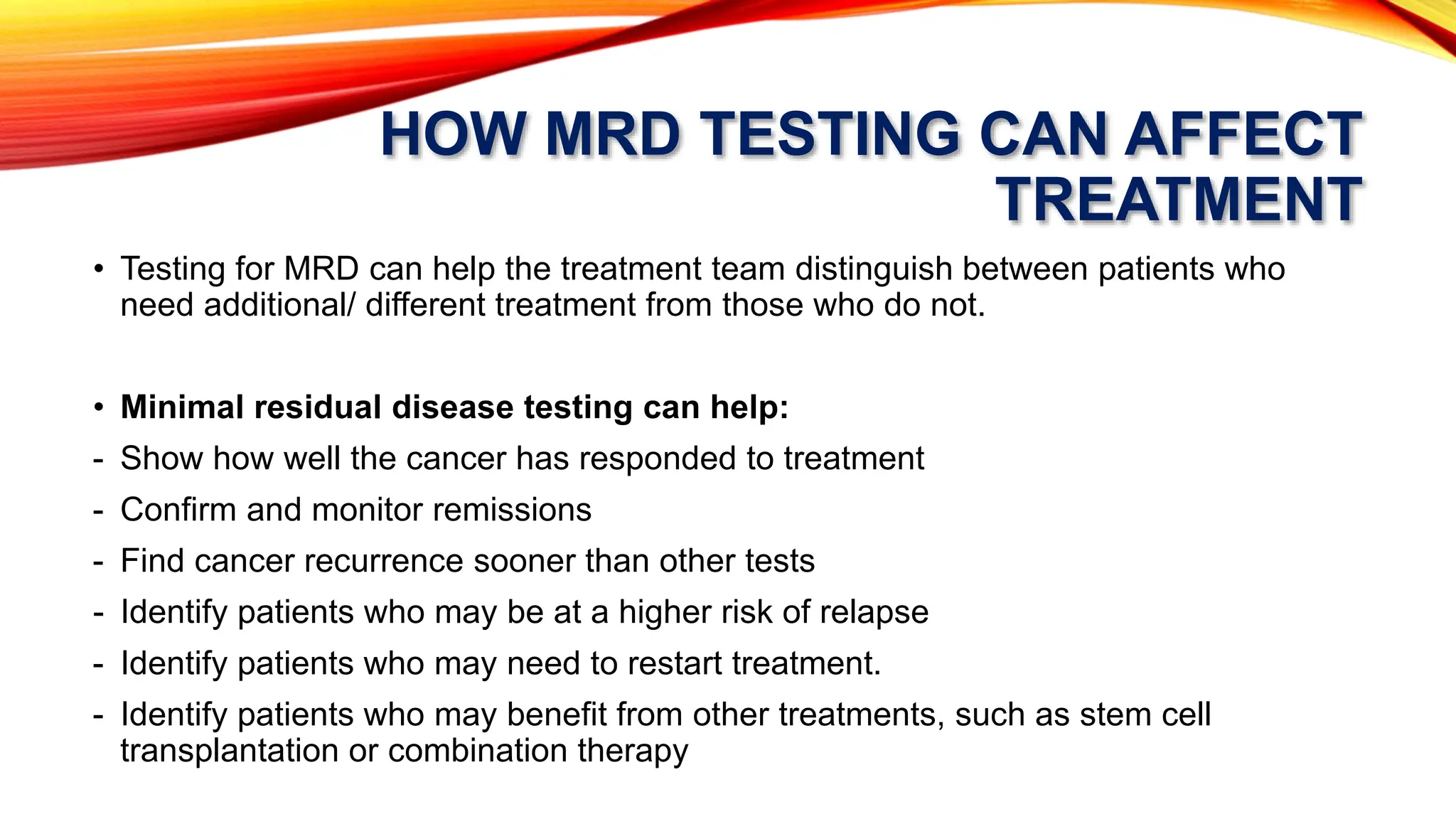
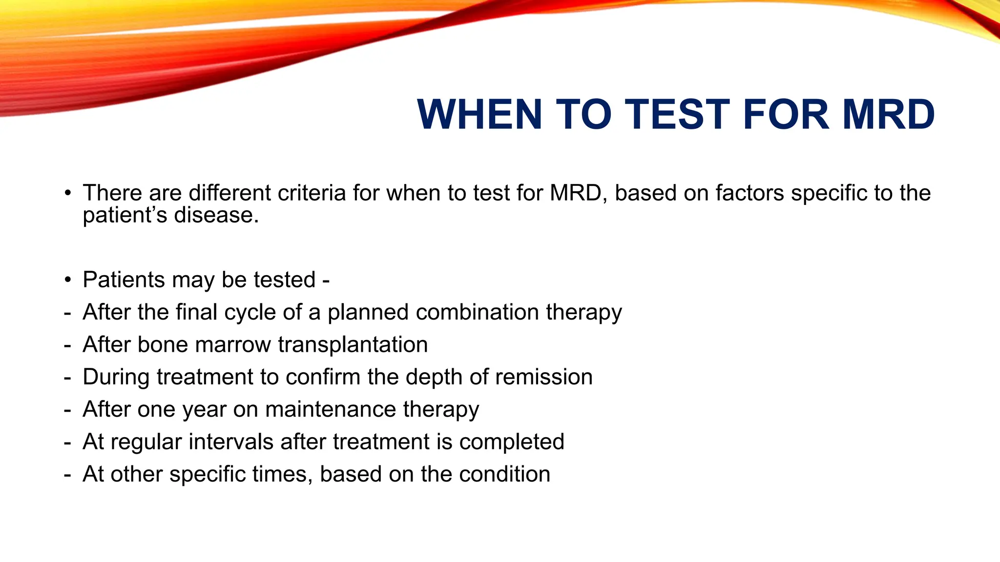
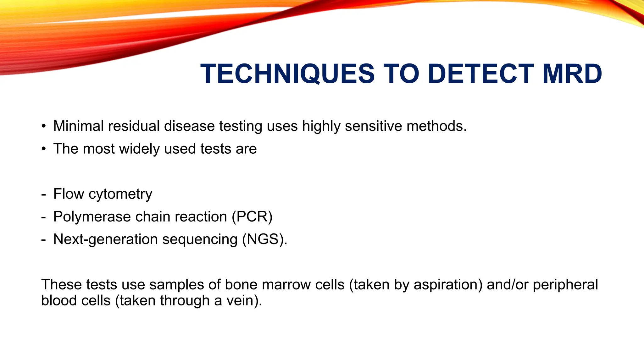
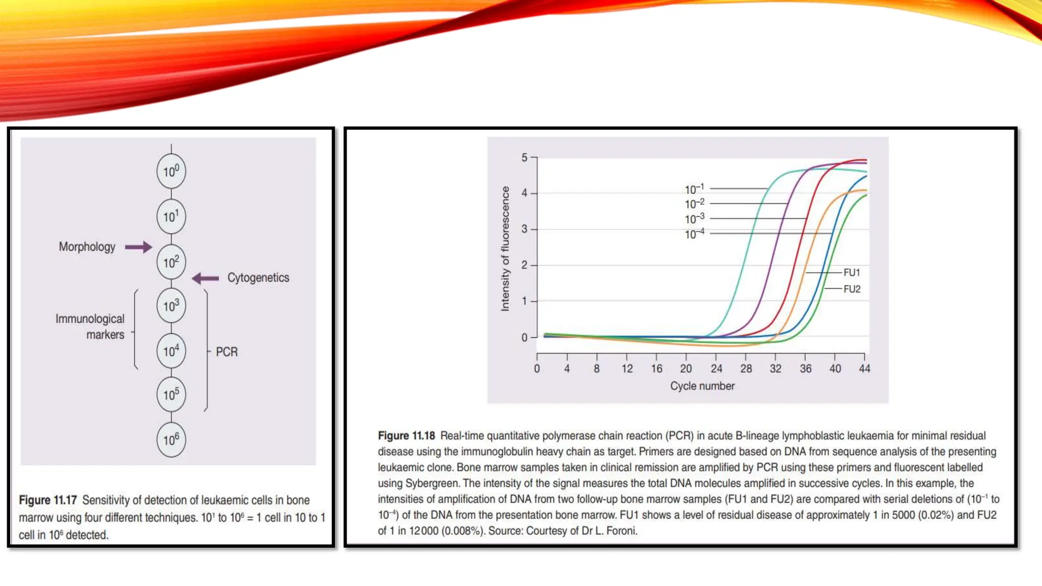
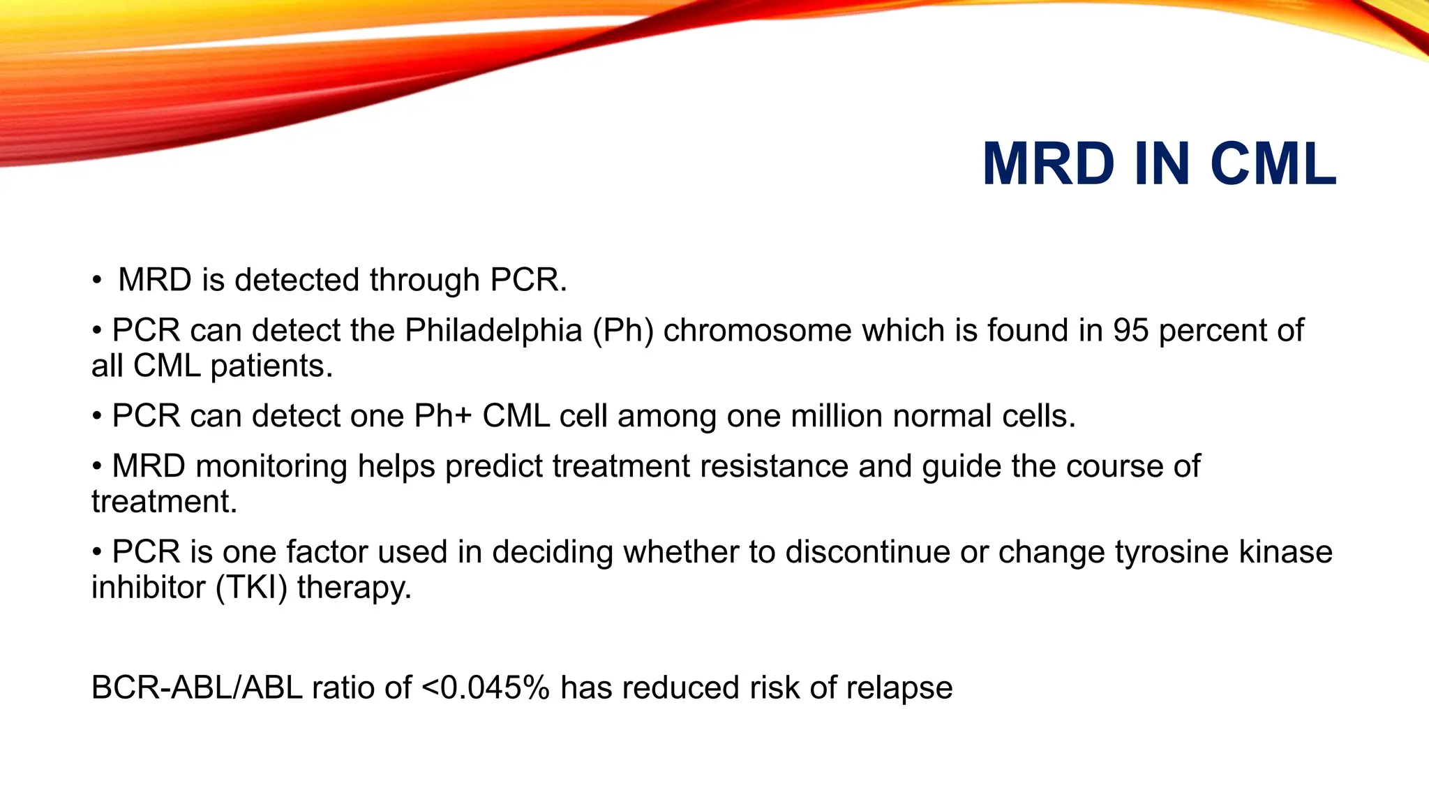
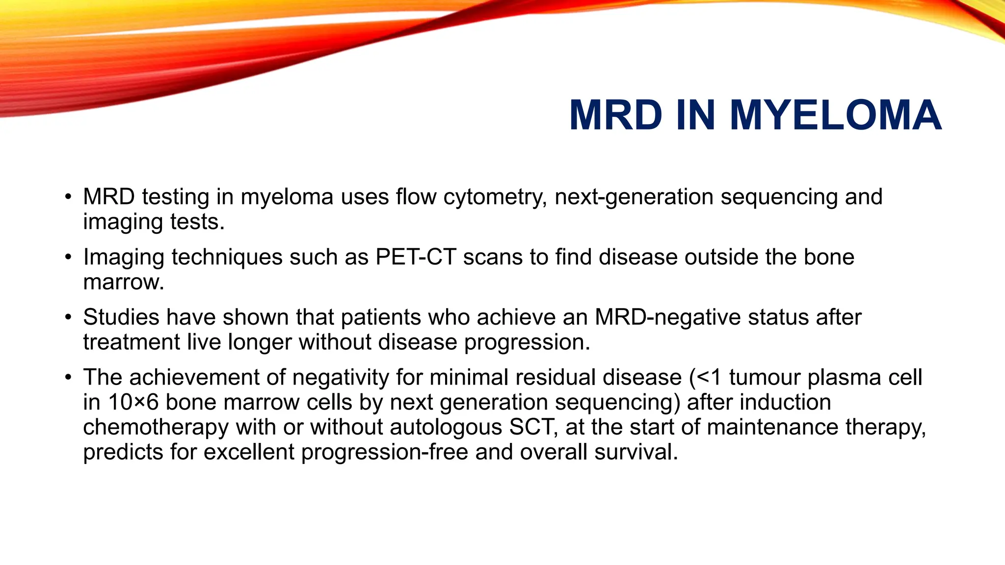
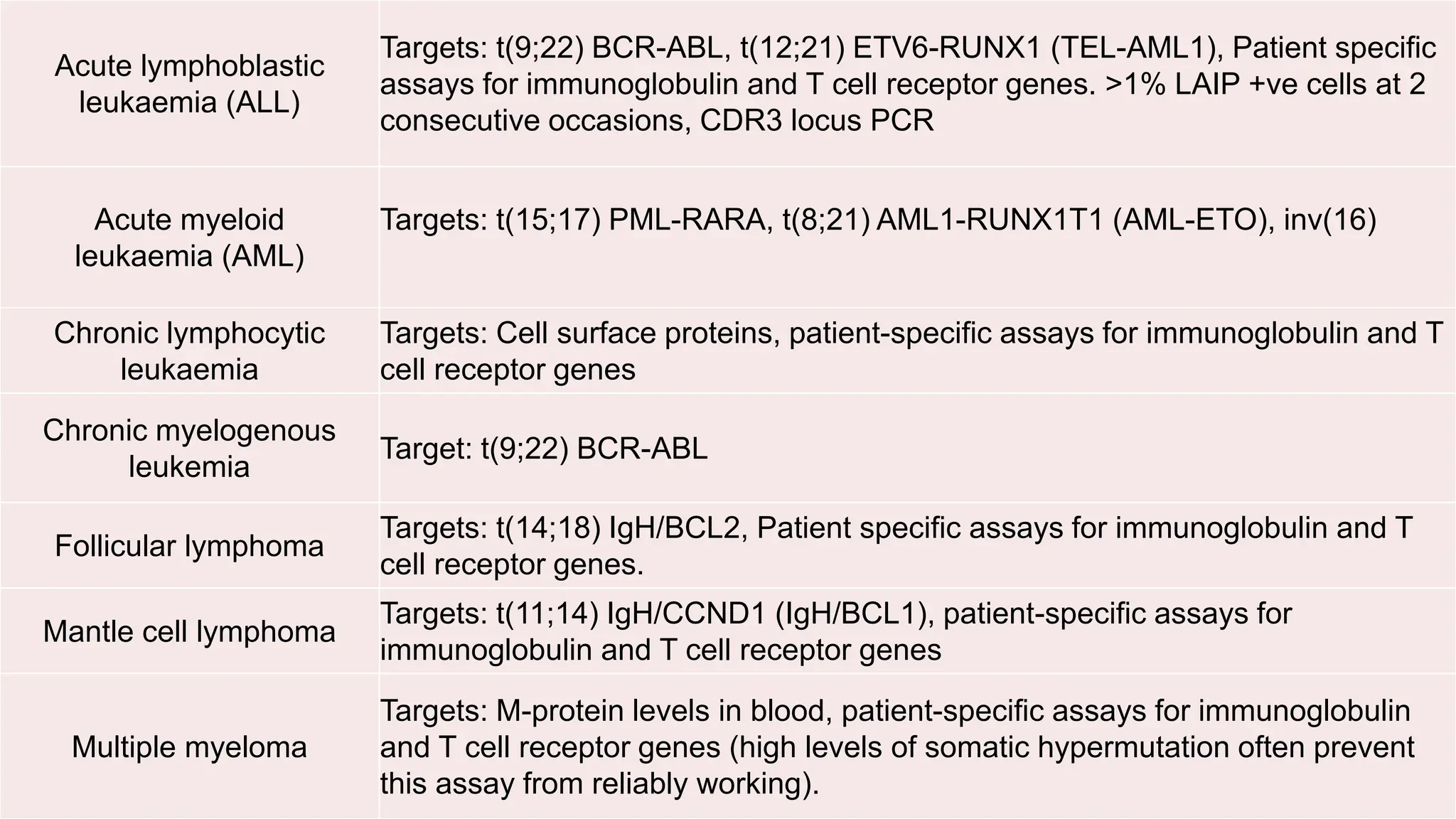
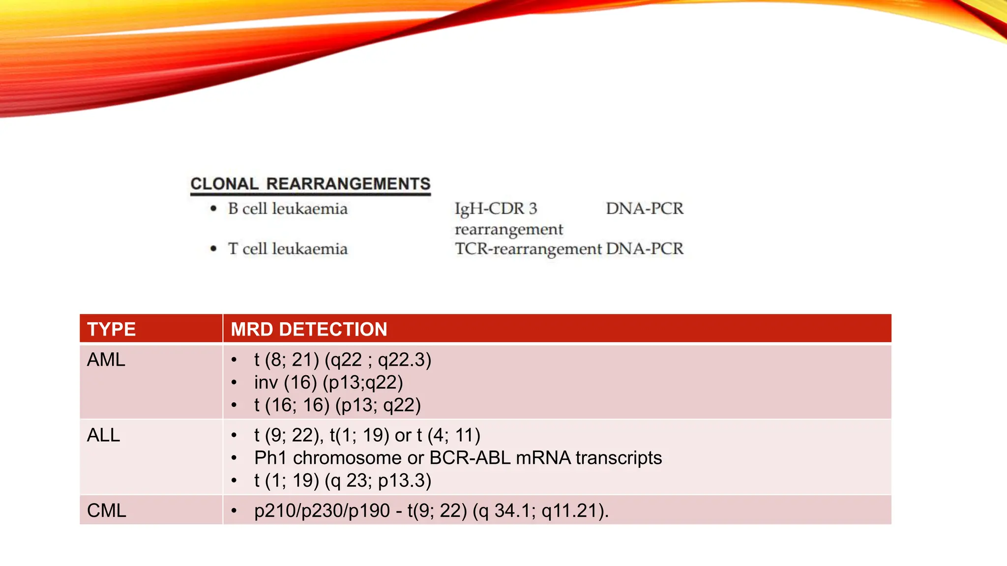
MINIMAL RESIDUAL
DISEASE
Dr. Reenaz Shaik
MD Pathology
WHAT IS MINIMAL RESIDUAL
DISEASE (MRD)?
• Minimal residual disease (MRD) refers to the small number of cancer cells that
remain in the body after treatment.
• The number of remaining cells may be so small that they do not cause any physical
signs or symptoms.
• These cells cannot even be detected through traditional methods, such as viewing
cells under a microscope and/or by tracking abnormal serum proteins in the blood.
ROLE IN PATIENT CARE
• After treating cancer, any remaining cancer cells in the body can become active and
start to multiply, causing a relapse of the disease.
• Detecting MRD may indicate that the treatment was not completely effective or that
the treatment was incomplete.
• Minimal residual disease may be present after treatment because not all of the
cancer cells responded to the therapy or because the cancer cells became resistant
to the medications used.
NEGATIVE & POSITIVE
• When MRD is detected, this is known as “MRD positivity.”
• When a patient tests negative, no residual cancer cells were found. When no MRD
is detected, this is known as “MRD negativity.”
• Studies have shown that MRD negativity is associated with longer remissions and
potentially longer rates of survival for certain blood cancers.
HOW MRD TESTING CAN AFFECT
TREATMENT
• Testing for MRD can help the treatment team distinguish between patients who
need additional/ different treatment from those who do not.
• Minimal residual disease testing can help:
- Show how well the cancer has responded to treatment
- Confirm and monitor remissions
- Find cancer recurrence sooner than other tests
- Identify patients who may be at a higher risk of relapse
- Identify patients who may need to restart treatment.
- Identify patients who may benefit from other treatments, such as stem cell
transplantation or combination therapy
WHEN TO TEST FOR MRD
• There are different criteria for when to test for MRD, based on factors specific to the
patient’s disease.
• Patients may be tested -
- After the final cycle of a planned combination therapy
- After bone marrow transplantation
- During treatment to confirm the depth of remission
- After one year on maintenance therapy
- At regular intervals after treatment is completed
- At other specific times, based on the condition
TECHNIQUES TO DETECT MRD
• Minimal residual disease testing uses highly sensitive methods.
• The most widely used tests are
- Flow cytometry
- Polymerase chain reaction (PCR)
- Next-generation sequencing (NGS).
These tests use samples of bone marrow cells (taken by aspiration) and/or peripheral
blood cells (taken through a vein).
FLOW CYTOMETRY
- Flow cytometry is a technique that evaluates individual cells by checking for the
presence or the absence of certain protein markers on the cell surface.
- A fresh bone marrow aspirate sample is required for reliable results.
- The bone marrow aspirate sample is treated with special antibodies that stick only
to the cells that have a specific protein on them.
- Based on how the flow cytometry is set up, this approach can find one cancer cell
among 100,000 bone marrow cells.
- Results can be available in less than one day
SEROLOGICAL MARKERS
• The presence of tumour-derived proteins in the serum has been used for many years as a
surrogate for minimal residual disease detection as well as for cancer screening.
• Examples of serological markers used for the evaluation of tumour burden include
- CA 15-3 and CA 27.29 (MUC1) in breast cancer
- α-fetoprotein in liver cancer
- Carcinoembryonic antigen in colon cancer
- Prostate-specific antigen in prostate cancer
- CA125 in ovarian cancer.
If a patient’s tumor overexpresses one of these proteins, the level can be monitored in the
blood. A decreasing level would indicate response to therapy; a rising level would indicate
recurrent or persistent disease.
IMMUNOHISTOCHEMISTRY
• Tumor cells also contain proteins that are absent in blood cells and thus can be used to
detect them using antibodies specific for these proteins.
• The most common technique used to detect solid tumor cells in blood, lymph nodes, or
bone marrow is immunohistochemistry.
• Ab, second Ab tagged with Biotin to detect first Ab, Detection solution: HRP +
Streptavidin which reacts with Biotin to give color.
• This technique can be used to detect very small numbers of tumor cells in bone
marrow or lymph node.
• To detect cancer cells in the blood, the cells must be first embedded in a wax block
that can be cut and stained like a tissue biopsy.
• Since the background cells will not express tumor proteins, very few tumor cells can
be visualized even in a background of many nontumor cells.
• The sensitivity of this type of assay can be increased if tumor cells are first enriched
by a selection technique such as magnetic bead selection, where magnetic particles
are coated with antibodies that bind tumor cells and then stick to tumor cells that
can be pulled out with a magnet.
CIRCULATING TUMOUR CELL ASSAYS
• The technique is based on :
- Enrichment of tumor cells using magnetic particles
- Staining of captured cells with fluorescent antibodies for cytokeratin
- Visualization of the cells on a computer microscope system that collects images for
evaluation by laboratory personnel.
This system can detect fewer than 10 tumor cells in a tube of blood, and clinical
studies have shown that the presence of circulating tumor cells predicts worse clinical
course and may be better than serum tumor markers or imaging studies for following
patients with cancer.
POLYMERASE CHAIN REACTION (PCR)
• This technique expands trace amounts of DNA so that a specific segment of DNA
can be studied.
• Polymerase chain reaction can identify malignant cells based on their characteristic
genetic abnormalities, such as mutations or chromosomal changes.
• Polymerase chain reaction essentially increases or “amplifies” small amounts of
specific pieces of either DNA or RNA to make them easier to detect and count.
• As a result, genetic abnormalities can be detected by PCR even when a very small
number of cancer cells remain.
• With PCR, it is possible to identify one cancer cell within 100,000 to one million
normal cells.
• The test is done with a bone marrow or blood sample. It may take several weeks for
results to be available.
NEXT-GENERATION SEQUENCING
(NGS)
• NGS tests can rapidly examine stretches of DNA or RNA.
• NGS can detect mutations and other genetic abnormalities in DNA extracted from a
bone marrow aspirate sample.
• This approach offers the potential for increased sensitivity - it can detect one cancer
cell in one million bone marrow cells checked.
• Test results are usually available within one week.
• Both fresh and frozen/stored samples can be used for NGS-based MRD testing.
• A test called clonoSEQ,® an NGS test designed to detect MRD in B-cell acute
lymphoblastic leukemia (ALL) and myeloma.
CELL CULTURE METHODS
• Modification of growth conditions to preferentially allow leukaemic cell growth and
inhibit normal progenitor cells; have been used to develop colony assays and detect
MRD by employing PCR on `plucked’ colonies.
• This technique is limited by the difficulty in maintaining culture conditions and also
in standardization of the cell culture methods to determine cut off levels of residual
disease.
RFLP WITH SOUTHERN BLOT
• DNA Restriction Fragment Length Polymorphism (RFLP) Analysis Combined with
Southern Blot Hybridization.
• It uses DNA labelled probes to look for chromosomal aberrations and clonal
markers.
• However, at least 1 per cent of cells under investigation should be clonal cells.
• It is a laborious, time consuming technique hampered by a low sensitivity of 1 per
cent and restricted by the number of tests that can be carried out simultaneously.
MRD IN AML
• LAIF (Leukemia associated Immunophenotype)is very useful in detection of minimal
residual disease (MED) after therapy.
• Complete remission (CR) in AML was morphology based and the criteria were
- < 5% blasts in marrow
- Absolute neutrophil count ANC > 1 x 10‘VL
- Platelet count > 100 x 109/L.
MRD IN ALL
• MRD is detected through flow cytometry, PCR and next-generation sequencing
(NGS).
• MRD is part of routine testing in the treatment of pediatric and adult ALL.
• Studies show that MRD can predict the effectiveness of a given treatment after the
induction phase of ALL treatment.
• MRD can help identify which patients are at higher risk for relapse, allowing for
earlier or additional treatments.
Poor prognosis if positive (>0.01%) at 3–6 months
MRD may also determine which patients may benefit from a bone marrow
transplantation.
• Flow cytometry can detect 1 in 10^4 -10^6 cells of the marrow.
• However, PCR and flow cytometric analysis are applied to assess the presence of
residual leukemia cells in blood/bone marrow and other tissues of the body.
• Immunophenotype with aberrant expression of CD13/ CD33 is useful in detecting
MRD.
• Detection of >1% LAIP +ve cells at 2 consecutive occasions indicates relapse in
childhood. E-ALL.
• Detection of ceils in blood/bone marrow co-expressing T-lineage antigens and TdT
or CD34 is also considered MRD as these markers are not present, on cells outside
the Thymus.
MRD IN CLL
• MRD is detected by flow cytometry and PCR.
• Patients who remain MRD negative after the end of therapy for CLL may have better
treatment outcomes.
• Patients who are MRD positive after the end of treatment may be candidates for
treatment intensification, consolidation and maintenance strategies.
MRD IN CML
• MRD is detected through PCR.
• PCR can detect the Philadelphia (Ph) chromosome which is found in 95 percent of
all CML patients.
• PCR can detect one Ph+ CML cell among one million normal cells.
• MRD monitoring helps predict treatment resistance and guide the course of
treatment.
• PCR is one factor used in deciding whether to discontinue or change tyrosine kinase
inhibitor (TKI) therapy.
BCR-ABL/ABL ratio of <0.045% has reduced risk of relapse
MRD IN LYMPHOMA
• MRD is detected through flow cytometry and PCR.
• MRD testing is used in follicular, mantle cell and diffuse large B-cell lymphoma
(DLBCL).
• MRD testing helps detect patients who are at risk of relapsing. These patients can
then receive additional treatment
• Patients who are treated for mantle cell lymphoma, DLBCL and achieve an MRD-
negative status have been shown to have longer remissions before their disease
progresses.
PCR for IgVH/TCR gene rearrangements in lymphoma cells used to detect MRD
MRD IN MYELOMA
• MRD testing in myeloma uses flow cytometry, next-generation sequencing and
imaging tests.
• Imaging techniques such as PET-CT scans to find disease outside the bone
marrow.
• Studies have shown that patients who achieve an MRD-negative status after
treatment live longer without disease progression.
• The achievement of negativity for minimal residual disease (<1 tumour plasma cell
in 10×6 bone marrow cells by next generation sequencing) after induction
chemotherapy with or without autologous SCT, at the start of maintenance therapy,
predicts for excellent progression-free and overall survival.
MOLECULAR TARGETS
1. Abnormal genes and/or transcripts (Molecular equivalents of chromosomal
alterations):
A few leukaemias are associated with constant non-random chromosomal
translocations and the consequent new tumour specific fusion genes and m-RNA
transcripts.
2. Clonality Markers: (Immunoglobulin/TCR gene rearrangements): Ig and TCR
genes are rearranged.
Acute lymphoblastic
leukaemia (ALL)
Targets: t(9;22) BCR-ABL, t(12;21) ETV6-RUNX1 (TEL-AML1), Patient specific
assays for immunoglobulin and T cell receptor genes. >1% LAIP +ve cells at 2
consecutive occasions, CDR3 locus PCR
Acute myeloid
leukaemia (AML)
Targets: t(15;17) PML-RARA, t(8;21) AML1-RUNX1T1 (AML-ETO), inv(16)
Chronic lymphocytic
leukaemia
Targets: Cell surface proteins, patient-specific assays for immunoglobulin and T
cell receptor genes
Chronic myelogenous
leukemia
Target: t(9;22) BCR-ABL
Follicular lymphoma
Targets: t(14;18) IgH/BCL2, Patient specific assays for immunoglobulin and T
cell receptor genes.
Mantle cell lymphoma
Targets: t(11;14) IgH/CCND1 (IgH/BCL1), patient-specific assays for
immunoglobulin and T cell receptor genes
Multiple myeloma
Targets: M-protein levels in blood, patient-specific assays for immunoglobulin
and T cell receptor genes (high levels of somatic hypermutation often prevent
this assay from reliably working).
TYPE MRD DETECTION
AML • t (8; 21) (q22 ; q22.3)
• inv (16) (p13;q22)
• t (16; 16) (p13; q22)
ALL • t (9; 22), t(1; 19) or t (4; 11)
• Ph1 chromosome or BCR-ABL mRNA transcripts
• t (1; 19) (q 23; p13.3)
CML • p210/p230/p190 - t(9; 22) (q 34.1; q11.21).
KEY POINTS
1. MRD is the lowest level of detectable disease by a given technique.
2. Conventional BM morphologic assessment has the low sensitivity (10–1 to 10–2).
3. Cytogenetic abnormality detected if any; is both diagnostic and prognostic: t(9;22), t(15;17),
t(8;21), t(8;14) etc.
4. FISH cytogenetics is superior to conventional cytogenetics.
5. Multi-parametric flow cytometry is useful for initial characterization and subsequent follow-
up.
6. Immunoglobulin(Ig)/TCR gene rearrangement studies are excellent molecular techniques for
clonality.
7. Detection of fusion transcripts by RT-PCR (BCR-ABL, PML-RARA,AML1-ETO) can be
routinely practiced.
8. If facilities are available, employ multiple techniques to monitor the residual disease.
9. Real time PCR technology can quantify residual disease at the molecular level.
10. Optimal approach to MRD analysis and therapeutic measures in leukaemia management
should be closely inter-linked
REFERENCES
1. Recent Advances in Hematology
2. Hoffbrand, A.V. and Moss, P.A.H. (2011) Essential Hematology. 6th Edition,
Blackwell Publishing Ltd., Oxford, UK
3. Williams Hematology, 10e Eds. Kenneth Kaushansky, et al. McGraw Hill, 2021
Minimal Residual Disease           (MRD)

Minimal Residual Disease (MRD)

  • 1.
  • 2.
    WHAT IS MINIMALRESIDUAL DISEASE (MRD)? • Minimal residual disease (MRD) refers to the small number of cancer cells that remain in the body after treatment. • The number of remaining cells may be so small that they do not cause any physical signs or symptoms. • These cells cannot even be detected through traditional methods, such as viewing cells under a microscope and/or by tracking abnormal serum proteins in the blood.
  • 3.
    ROLE IN PATIENTCARE • After treating cancer, any remaining cancer cells in the body can become active and start to multiply, causing a relapse of the disease. • Detecting MRD may indicate that the treatment was not completely effective or that the treatment was incomplete. • Minimal residual disease may be present after treatment because not all of the cancer cells responded to the therapy or because the cancer cells became resistant to the medications used.
  • 4.
    NEGATIVE & POSITIVE •When MRD is detected, this is known as “MRD positivity.” • When a patient tests negative, no residual cancer cells were found. When no MRD is detected, this is known as “MRD negativity.” • Studies have shown that MRD negativity is associated with longer remissions and potentially longer rates of survival for certain blood cancers.
  • 5.
    HOW MRD TESTINGCAN AFFECT TREATMENT • Testing for MRD can help the treatment team distinguish between patients who need additional/ different treatment from those who do not. • Minimal residual disease testing can help: - Show how well the cancer has responded to treatment - Confirm and monitor remissions - Find cancer recurrence sooner than other tests - Identify patients who may be at a higher risk of relapse - Identify patients who may need to restart treatment. - Identify patients who may benefit from other treatments, such as stem cell transplantation or combination therapy
  • 6.
    WHEN TO TESTFOR MRD • There are different criteria for when to test for MRD, based on factors specific to the patient’s disease. • Patients may be tested - - After the final cycle of a planned combination therapy - After bone marrow transplantation - During treatment to confirm the depth of remission - After one year on maintenance therapy - At regular intervals after treatment is completed - At other specific times, based on the condition
  • 7.
    TECHNIQUES TO DETECTMRD • Minimal residual disease testing uses highly sensitive methods. • The most widely used tests are - Flow cytometry - Polymerase chain reaction (PCR) - Next-generation sequencing (NGS). These tests use samples of bone marrow cells (taken by aspiration) and/or peripheral blood cells (taken through a vein).
  • 10.
    FLOW CYTOMETRY - Flowcytometry is a technique that evaluates individual cells by checking for the presence or the absence of certain protein markers on the cell surface. - A fresh bone marrow aspirate sample is required for reliable results. - The bone marrow aspirate sample is treated with special antibodies that stick only to the cells that have a specific protein on them. - Based on how the flow cytometry is set up, this approach can find one cancer cell among 100,000 bone marrow cells. - Results can be available in less than one day
  • 11.
    SEROLOGICAL MARKERS • Thepresence of tumour-derived proteins in the serum has been used for many years as a surrogate for minimal residual disease detection as well as for cancer screening. • Examples of serological markers used for the evaluation of tumour burden include - CA 15-3 and CA 27.29 (MUC1) in breast cancer - α-fetoprotein in liver cancer - Carcinoembryonic antigen in colon cancer - Prostate-specific antigen in prostate cancer - CA125 in ovarian cancer. If a patient’s tumor overexpresses one of these proteins, the level can be monitored in the blood. A decreasing level would indicate response to therapy; a rising level would indicate recurrent or persistent disease.
  • 12.
    IMMUNOHISTOCHEMISTRY • Tumor cellsalso contain proteins that are absent in blood cells and thus can be used to detect them using antibodies specific for these proteins. • The most common technique used to detect solid tumor cells in blood, lymph nodes, or bone marrow is immunohistochemistry. • Ab, second Ab tagged with Biotin to detect first Ab, Detection solution: HRP + Streptavidin which reacts with Biotin to give color. • This technique can be used to detect very small numbers of tumor cells in bone marrow or lymph node.
  • 13.
    • To detectcancer cells in the blood, the cells must be first embedded in a wax block that can be cut and stained like a tissue biopsy. • Since the background cells will not express tumor proteins, very few tumor cells can be visualized even in a background of many nontumor cells. • The sensitivity of this type of assay can be increased if tumor cells are first enriched by a selection technique such as magnetic bead selection, where magnetic particles are coated with antibodies that bind tumor cells and then stick to tumor cells that can be pulled out with a magnet.
  • 14.
    CIRCULATING TUMOUR CELLASSAYS • The technique is based on : - Enrichment of tumor cells using magnetic particles - Staining of captured cells with fluorescent antibodies for cytokeratin - Visualization of the cells on a computer microscope system that collects images for evaluation by laboratory personnel. This system can detect fewer than 10 tumor cells in a tube of blood, and clinical studies have shown that the presence of circulating tumor cells predicts worse clinical course and may be better than serum tumor markers or imaging studies for following patients with cancer.
  • 15.
    POLYMERASE CHAIN REACTION(PCR) • This technique expands trace amounts of DNA so that a specific segment of DNA can be studied. • Polymerase chain reaction can identify malignant cells based on their characteristic genetic abnormalities, such as mutations or chromosomal changes. • Polymerase chain reaction essentially increases or “amplifies” small amounts of specific pieces of either DNA or RNA to make them easier to detect and count. • As a result, genetic abnormalities can be detected by PCR even when a very small number of cancer cells remain. • With PCR, it is possible to identify one cancer cell within 100,000 to one million normal cells. • The test is done with a bone marrow or blood sample. It may take several weeks for results to be available.
  • 16.
    NEXT-GENERATION SEQUENCING (NGS) • NGStests can rapidly examine stretches of DNA or RNA. • NGS can detect mutations and other genetic abnormalities in DNA extracted from a bone marrow aspirate sample. • This approach offers the potential for increased sensitivity - it can detect one cancer cell in one million bone marrow cells checked. • Test results are usually available within one week. • Both fresh and frozen/stored samples can be used for NGS-based MRD testing. • A test called clonoSEQ,® an NGS test designed to detect MRD in B-cell acute lymphoblastic leukemia (ALL) and myeloma.
  • 17.
    CELL CULTURE METHODS •Modification of growth conditions to preferentially allow leukaemic cell growth and inhibit normal progenitor cells; have been used to develop colony assays and detect MRD by employing PCR on `plucked’ colonies. • This technique is limited by the difficulty in maintaining culture conditions and also in standardization of the cell culture methods to determine cut off levels of residual disease.
  • 18.
    RFLP WITH SOUTHERNBLOT • DNA Restriction Fragment Length Polymorphism (RFLP) Analysis Combined with Southern Blot Hybridization. • It uses DNA labelled probes to look for chromosomal aberrations and clonal markers. • However, at least 1 per cent of cells under investigation should be clonal cells. • It is a laborious, time consuming technique hampered by a low sensitivity of 1 per cent and restricted by the number of tests that can be carried out simultaneously.
  • 19.
    MRD IN AML •LAIF (Leukemia associated Immunophenotype)is very useful in detection of minimal residual disease (MED) after therapy. • Complete remission (CR) in AML was morphology based and the criteria were - < 5% blasts in marrow - Absolute neutrophil count ANC > 1 x 10‘VL - Platelet count > 100 x 109/L.
  • 20.
    MRD IN ALL •MRD is detected through flow cytometry, PCR and next-generation sequencing (NGS). • MRD is part of routine testing in the treatment of pediatric and adult ALL. • Studies show that MRD can predict the effectiveness of a given treatment after the induction phase of ALL treatment. • MRD can help identify which patients are at higher risk for relapse, allowing for earlier or additional treatments. Poor prognosis if positive (>0.01%) at 3–6 months MRD may also determine which patients may benefit from a bone marrow transplantation.
  • 21.
    • Flow cytometrycan detect 1 in 10^4 -10^6 cells of the marrow. • However, PCR and flow cytometric analysis are applied to assess the presence of residual leukemia cells in blood/bone marrow and other tissues of the body. • Immunophenotype with aberrant expression of CD13/ CD33 is useful in detecting MRD. • Detection of >1% LAIP +ve cells at 2 consecutive occasions indicates relapse in childhood. E-ALL. • Detection of ceils in blood/bone marrow co-expressing T-lineage antigens and TdT or CD34 is also considered MRD as these markers are not present, on cells outside the Thymus.
  • 23.
    MRD IN CLL •MRD is detected by flow cytometry and PCR. • Patients who remain MRD negative after the end of therapy for CLL may have better treatment outcomes. • Patients who are MRD positive after the end of treatment may be candidates for treatment intensification, consolidation and maintenance strategies.
  • 24.
    MRD IN CML •MRD is detected through PCR. • PCR can detect the Philadelphia (Ph) chromosome which is found in 95 percent of all CML patients. • PCR can detect one Ph+ CML cell among one million normal cells. • MRD monitoring helps predict treatment resistance and guide the course of treatment. • PCR is one factor used in deciding whether to discontinue or change tyrosine kinase inhibitor (TKI) therapy. BCR-ABL/ABL ratio of <0.045% has reduced risk of relapse
  • 25.
    MRD IN LYMPHOMA •MRD is detected through flow cytometry and PCR. • MRD testing is used in follicular, mantle cell and diffuse large B-cell lymphoma (DLBCL). • MRD testing helps detect patients who are at risk of relapsing. These patients can then receive additional treatment • Patients who are treated for mantle cell lymphoma, DLBCL and achieve an MRD- negative status have been shown to have longer remissions before their disease progresses. PCR for IgVH/TCR gene rearrangements in lymphoma cells used to detect MRD
  • 26.
    MRD IN MYELOMA •MRD testing in myeloma uses flow cytometry, next-generation sequencing and imaging tests. • Imaging techniques such as PET-CT scans to find disease outside the bone marrow. • Studies have shown that patients who achieve an MRD-negative status after treatment live longer without disease progression. • The achievement of negativity for minimal residual disease (<1 tumour plasma cell in 10×6 bone marrow cells by next generation sequencing) after induction chemotherapy with or without autologous SCT, at the start of maintenance therapy, predicts for excellent progression-free and overall survival.
  • 27.
    MOLECULAR TARGETS 1. Abnormalgenes and/or transcripts (Molecular equivalents of chromosomal alterations): A few leukaemias are associated with constant non-random chromosomal translocations and the consequent new tumour specific fusion genes and m-RNA transcripts. 2. Clonality Markers: (Immunoglobulin/TCR gene rearrangements): Ig and TCR genes are rearranged.
  • 28.
    Acute lymphoblastic leukaemia (ALL) Targets:t(9;22) BCR-ABL, t(12;21) ETV6-RUNX1 (TEL-AML1), Patient specific assays for immunoglobulin and T cell receptor genes. >1% LAIP +ve cells at 2 consecutive occasions, CDR3 locus PCR Acute myeloid leukaemia (AML) Targets: t(15;17) PML-RARA, t(8;21) AML1-RUNX1T1 (AML-ETO), inv(16) Chronic lymphocytic leukaemia Targets: Cell surface proteins, patient-specific assays for immunoglobulin and T cell receptor genes Chronic myelogenous leukemia Target: t(9;22) BCR-ABL Follicular lymphoma Targets: t(14;18) IgH/BCL2, Patient specific assays for immunoglobulin and T cell receptor genes. Mantle cell lymphoma Targets: t(11;14) IgH/CCND1 (IgH/BCL1), patient-specific assays for immunoglobulin and T cell receptor genes Multiple myeloma Targets: M-protein levels in blood, patient-specific assays for immunoglobulin and T cell receptor genes (high levels of somatic hypermutation often prevent this assay from reliably working).
  • 30.
    TYPE MRD DETECTION AML• t (8; 21) (q22 ; q22.3) • inv (16) (p13;q22) • t (16; 16) (p13; q22) ALL • t (9; 22), t(1; 19) or t (4; 11) • Ph1 chromosome or BCR-ABL mRNA transcripts • t (1; 19) (q 23; p13.3) CML • p210/p230/p190 - t(9; 22) (q 34.1; q11.21).
  • 31.
    KEY POINTS 1. MRDis the lowest level of detectable disease by a given technique. 2. Conventional BM morphologic assessment has the low sensitivity (10–1 to 10–2). 3. Cytogenetic abnormality detected if any; is both diagnostic and prognostic: t(9;22), t(15;17), t(8;21), t(8;14) etc. 4. FISH cytogenetics is superior to conventional cytogenetics. 5. Multi-parametric flow cytometry is useful for initial characterization and subsequent follow- up. 6. Immunoglobulin(Ig)/TCR gene rearrangement studies are excellent molecular techniques for clonality. 7. Detection of fusion transcripts by RT-PCR (BCR-ABL, PML-RARA,AML1-ETO) can be routinely practiced. 8. If facilities are available, employ multiple techniques to monitor the residual disease. 9. Real time PCR technology can quantify residual disease at the molecular level. 10. Optimal approach to MRD analysis and therapeutic measures in leukaemia management should be closely inter-linked
  • 32.
    REFERENCES 1. Recent Advancesin Hematology 2. Hoffbrand, A.V. and Moss, P.A.H. (2011) Essential Hematology. 6th Edition, Blackwell Publishing Ltd., Oxford, UK 3. Williams Hematology, 10e Eds. Kenneth Kaushansky, et al. McGraw Hill, 2021