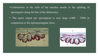
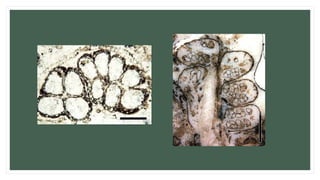
ANGIOPTERIS
BY,
G. STEPHY AROKIYA
MARY
24PBO106
TABLE OF CONTENTS
• Systematic Position
• Introduction
• Angiopteris
• External Morphology
• Internal Structure
• Spore Producing Organs
• Gametophyte
• Reproduction
• Conclusion
SYSTEMATIC POSITION
KINGDOM - PLANTAE
DIVISION - PTERIDOPHYTA
SUB – DIVISION - PTEROPSIDA
CLASS - EUSPORANGIATE
ORDER - MARATTIALES
FAMILY - ANGIOPTERIDACEAE
GENUS - ANGIOPTERIS
INTRODUCTION
• The class Eusporangiate is a small group of primitive ferns.
• This class is represented by 17 Genera and 30 Species.
The members of this class shows the following distinguishing features:
• Sporangia are borne on the fertile spikes (Ophioglossum), or on the
abaxial side of the lamina (Angiopteris, Marattia).
• The Sporangium develops from a group of initial cells.
• The Sporangial Jacket is more than one cell in thickness.
• They produce only one type of spores, i.e., they are homosporous.
• The class Eusporangiate has only two orders – Marattiales and
Ophioglossales.
• The Order Marattiales includes those eusporangiate ferns which bear
sporangia on the abaxial side of the lamina.
• They mostly occur in the tropical rain forests.
• There are seven living genera; of these Archangiopteris,
Macroglossum, Protomarattia and Christensenia are very small.
• Angiopteris, Marattia and Danaea are conspicuous because of their
massive fleshy axis and large fronds.
• The young leaves are circinate vernation (coiled) and each leaf has two
stipules at it’s base.
• The Sporangium opens by slits on either side or by a pore.
• The gametophyte is large, thalloid and green.
ANGIOPTERIS
• Angiopteris has some 100 species overall.
• They are distributed in Japan, Tropical
Australia, New Caledonia, Madagascar
and Polynesia.
• Angiopteris evecta (King Fern) occurs
throughout India up to an elevation of
2,500 meters.
EXTERNAL MORPHOLOGY
• The Plant Body is Sporophytic i.e., the plant body is differentiated into root,
stem and leaves.
• The stem is large, upright and 60cm or more in height.
• Angiopteris is shielded by fleshy stipules.
• The leaves are Spirally arranged and form a Crown at the apex of the stem.
• They are Pinnately compound, often large, 2 – 6 meters long.
• A conspicuous swelling, called Pulvinus, is present at the base of each pinna.
• The venation is open, dichotomously branched and veins are sub –
parallel.
• The Young leaves shows typical Circinate vernation.
• Numerous Adventitious roots arises from the basal part of the stem.
They are somewhat fleshy, branched and mycorrhizic.
• These adventitious roots arise endogenously and are inserted below the
leaves.
• The young stem grows by means of a single narrow apical cell, which is
replaced by group of initials at maturity.
INTERNAL STRUCTURE
The Internal structure of the
Angiopteris consists of the
following, They are:
• Stem
• Root
• Pinna
ANATOMY OF THE STEM
• The anatomy of the stem is complex and it varies with the age of the
plant.
• The Parenchymatous cortex has numerous mucilagenous ducts.
• The fleshy nature of the stem is due to the absence of thick walled cells.
• Mechanical support to the stem is provided by thick leaf bases.
• The young stem is Protostelic. The stele is surrounded by a distinct
layer of endodermis.
• As the stem grows older, the stele becomes amphiphloic siphonostele
with distinct leaf gaps.
• In a mature stem, however, the stele breaks into many meristeles due to
overlapping leaf gaps.
• It appears to be polycyclic as several curved sheets of vascular strands
are arranged in the concentric rings.
• Root traces, however, arise only from the meristeles of the inner rings.
T.S. OF YOUNG STEM
T.S. OF MATURE STEM
ANATOMY OF ROOT
• The internal structure of root is relatively simple.
• The epidermal cells of the root forms the root hairs.
• Some root hairs are multicellular.
• There is a large parenchymatous cortex with many mucilagenous canals.
• The Stele is surrounded by a single layer of endodermis and one or two layers of
pericycle.
• The Stele is Polyarch with 4 – 10 (or more ) long and thin xylem arms.
• A small parenchymatous pith is present in the centre of the stele.
T.S. OF ROOT
ANATOMY OF PINNA
• The internal structure of a pinna is similar to that of the
dicotyledonous leaf.
• Both, the lower and the upper epidermis are single layered.
• Stomata are confined to the lower epidermis.
• The mesophyll is differentiated into 1 – 2 layers of
elongated palisade like cells, followed by spongy
parenchyma.
SPORE PRODUCING ORGANS
• The sporangia forms linear, oblong or boat shaped sori on the lower
surface of the pinnae near the margin.
• Each Sorus has 8 – 14 or more sporangia which develops over a
veinlet.
• The sporangia of a sorus are free and in this feature Angiopteris
differs from other members of the Marattiales where sporangia fuse
to form synangia.
• The sori are non – indusiate, instead few multicellular hairs are present
along the margin of the sorus.
• The development of sporangium is of eusporangiate type.
• In Angiopteris, sporangia of a sorus do not initiate individually, instead the
entire sorus arises as a Surface receptacular meristem.
• All surface cells of this meristem divides periclinally and then groups of
hypodermal cells differentiate as Sporangial initials.
• Adjacent groups of sporangial initials are separated by sterile
intersporangial areas.
• Each group of Sporangial initial forms a sorus.
• The mature Sporangium is large, massive and sessile.
• The proximal part of sporangium is directly attached to the
receptacle.
• The wall of sporangium is several layered : The outermost layer has
stomata and the innermost layer has the Sporogenous tissue, which
forms the Tapetum.
• An arch of slightly thick walled cells, the annulus, is differentiated in
the sporangial wall.
• It extends vertically from one end to the other across the top of the
sporangium.
• Contractions in the cells of the annulus results in the splitting of
sporangium along the line of the dehiscence.
• The spore output per sporangium is very large (1400 – 7500) in
comparison to the leptosporangiate ferns.
GAMETOPHYTE
• Germination of spores takes place within a few days after their shedding.
• The spores forms large, perennial prothalli.
• The Prothallus is dark – green, mycorrhizic and thalloid, resembling very
much with thallose liverworts.
• It contains a many celled thick central cushion and thin lateral wing –
like extensions.
• The growth of the central mycorrhizal cushion continues for quite a long
time.
REPRODUCTION
• Angiopteris can reproduce Vegetatively or by Producing spores.
• Vegetative reproduction occurs when fleshy stipules located at the base of
each petiole produce proliferous buds that break off on suitable substrate and
produce new plants.
• The spores produced by sporophyte stage germinate and produce the
gametophyte, usually called the Prothallium, that bears both sex organs –
The Antheridium and Archegonium.
• After gamete production, the fertilization occurs in humid
environments, a new sporophyte (fern) emerges.
• Angiopteris evecta takes many years before it is capable of producing
spores.
• Adult leaves are covered in thousands of sporangia, each of which
produces 1,440 spores.
USES…..
• Angiopteris have antibacterial and antifungal properties in them.
• Angiopteris evecta is often cultivated as an ornamental plant in gardens, parks and
botanical gardens.
• The rhizome of Angiopteris evecta is used by the members of Tangsa tribe, both as
famine food and a remedy for snake bites and insect stings.
• The leaves of Angiopteris evecta are pounded to relieve coughs.
• The roots of Angiopteris evecta are used to stop bleeding after a miscarriage.
• The rhizome is used medicinally in Thailand.
CONCLUSION
• Angiopteris are invasive species.
• Angiopteris is unique among the ferns in having explosively
dispersed spores, which are thought to be caused by airspace
between the spore layers.
• These species have the unique spore dispersal mechanism in them.
THANK
YOU

ANGIOPTERIS modified.pptxgyhhhuhhghhhhhh

  • 1.
  • 2.
    TABLE OF CONTENTS •Systematic Position • Introduction • Angiopteris • External Morphology • Internal Structure • Spore Producing Organs • Gametophyte • Reproduction • Conclusion
  • 3.
    SYSTEMATIC POSITION KINGDOM -PLANTAE DIVISION - PTERIDOPHYTA SUB – DIVISION - PTEROPSIDA CLASS - EUSPORANGIATE ORDER - MARATTIALES FAMILY - ANGIOPTERIDACEAE GENUS - ANGIOPTERIS
  • 4.
    INTRODUCTION • The classEusporangiate is a small group of primitive ferns. • This class is represented by 17 Genera and 30 Species. The members of this class shows the following distinguishing features: • Sporangia are borne on the fertile spikes (Ophioglossum), or on the abaxial side of the lamina (Angiopteris, Marattia). • The Sporangium develops from a group of initial cells. • The Sporangial Jacket is more than one cell in thickness.
  • 5.
    • They produceonly one type of spores, i.e., they are homosporous. • The class Eusporangiate has only two orders – Marattiales and Ophioglossales. • The Order Marattiales includes those eusporangiate ferns which bear sporangia on the abaxial side of the lamina. • They mostly occur in the tropical rain forests. • There are seven living genera; of these Archangiopteris, Macroglossum, Protomarattia and Christensenia are very small.
  • 6.
    • Angiopteris, Marattiaand Danaea are conspicuous because of their massive fleshy axis and large fronds. • The young leaves are circinate vernation (coiled) and each leaf has two stipules at it’s base. • The Sporangium opens by slits on either side or by a pore. • The gametophyte is large, thalloid and green.
  • 7.
    ANGIOPTERIS • Angiopteris hassome 100 species overall. • They are distributed in Japan, Tropical Australia, New Caledonia, Madagascar and Polynesia. • Angiopteris evecta (King Fern) occurs throughout India up to an elevation of 2,500 meters.
  • 8.
    EXTERNAL MORPHOLOGY • ThePlant Body is Sporophytic i.e., the plant body is differentiated into root, stem and leaves. • The stem is large, upright and 60cm or more in height. • Angiopteris is shielded by fleshy stipules. • The leaves are Spirally arranged and form a Crown at the apex of the stem. • They are Pinnately compound, often large, 2 – 6 meters long. • A conspicuous swelling, called Pulvinus, is present at the base of each pinna.
  • 9.
    • The venationis open, dichotomously branched and veins are sub – parallel. • The Young leaves shows typical Circinate vernation. • Numerous Adventitious roots arises from the basal part of the stem. They are somewhat fleshy, branched and mycorrhizic. • These adventitious roots arise endogenously and are inserted below the leaves. • The young stem grows by means of a single narrow apical cell, which is replaced by group of initials at maturity.
  • 11.
    INTERNAL STRUCTURE The Internalstructure of the Angiopteris consists of the following, They are: • Stem • Root • Pinna
  • 12.
    ANATOMY OF THESTEM • The anatomy of the stem is complex and it varies with the age of the plant. • The Parenchymatous cortex has numerous mucilagenous ducts. • The fleshy nature of the stem is due to the absence of thick walled cells. • Mechanical support to the stem is provided by thick leaf bases. • The young stem is Protostelic. The stele is surrounded by a distinct layer of endodermis.
  • 13.
    • As thestem grows older, the stele becomes amphiphloic siphonostele with distinct leaf gaps. • In a mature stem, however, the stele breaks into many meristeles due to overlapping leaf gaps. • It appears to be polycyclic as several curved sheets of vascular strands are arranged in the concentric rings. • Root traces, however, arise only from the meristeles of the inner rings.
  • 14.
    T.S. OF YOUNGSTEM T.S. OF MATURE STEM
  • 15.
    ANATOMY OF ROOT •The internal structure of root is relatively simple. • The epidermal cells of the root forms the root hairs. • Some root hairs are multicellular. • There is a large parenchymatous cortex with many mucilagenous canals. • The Stele is surrounded by a single layer of endodermis and one or two layers of pericycle. • The Stele is Polyarch with 4 – 10 (or more ) long and thin xylem arms. • A small parenchymatous pith is present in the centre of the stele.
  • 16.
  • 17.
    ANATOMY OF PINNA •The internal structure of a pinna is similar to that of the dicotyledonous leaf. • Both, the lower and the upper epidermis are single layered. • Stomata are confined to the lower epidermis. • The mesophyll is differentiated into 1 – 2 layers of elongated palisade like cells, followed by spongy parenchyma.
  • 18.
    SPORE PRODUCING ORGANS •The sporangia forms linear, oblong or boat shaped sori on the lower surface of the pinnae near the margin. • Each Sorus has 8 – 14 or more sporangia which develops over a veinlet. • The sporangia of a sorus are free and in this feature Angiopteris differs from other members of the Marattiales where sporangia fuse to form synangia.
  • 19.
    • The soriare non – indusiate, instead few multicellular hairs are present along the margin of the sorus. • The development of sporangium is of eusporangiate type. • In Angiopteris, sporangia of a sorus do not initiate individually, instead the entire sorus arises as a Surface receptacular meristem. • All surface cells of this meristem divides periclinally and then groups of hypodermal cells differentiate as Sporangial initials.
  • 20.
    • Adjacent groupsof sporangial initials are separated by sterile intersporangial areas. • Each group of Sporangial initial forms a sorus. • The mature Sporangium is large, massive and sessile. • The proximal part of sporangium is directly attached to the receptacle.
  • 21.
    • The wallof sporangium is several layered : The outermost layer has stomata and the innermost layer has the Sporogenous tissue, which forms the Tapetum. • An arch of slightly thick walled cells, the annulus, is differentiated in the sporangial wall. • It extends vertically from one end to the other across the top of the sporangium.
  • 22.
    • Contractions inthe cells of the annulus results in the splitting of sporangium along the line of the dehiscence. • The spore output per sporangium is very large (1400 – 7500) in comparison to the leptosporangiate ferns.
  • 24.
    GAMETOPHYTE • Germination ofspores takes place within a few days after their shedding. • The spores forms large, perennial prothalli. • The Prothallus is dark – green, mycorrhizic and thalloid, resembling very much with thallose liverworts. • It contains a many celled thick central cushion and thin lateral wing – like extensions. • The growth of the central mycorrhizal cushion continues for quite a long time.
  • 25.
    REPRODUCTION • Angiopteris canreproduce Vegetatively or by Producing spores. • Vegetative reproduction occurs when fleshy stipules located at the base of each petiole produce proliferous buds that break off on suitable substrate and produce new plants. • The spores produced by sporophyte stage germinate and produce the gametophyte, usually called the Prothallium, that bears both sex organs – The Antheridium and Archegonium.
  • 26.
    • After gameteproduction, the fertilization occurs in humid environments, a new sporophyte (fern) emerges. • Angiopteris evecta takes many years before it is capable of producing spores. • Adult leaves are covered in thousands of sporangia, each of which produces 1,440 spores.
  • 28.
    USES….. • Angiopteris haveantibacterial and antifungal properties in them. • Angiopteris evecta is often cultivated as an ornamental plant in gardens, parks and botanical gardens. • The rhizome of Angiopteris evecta is used by the members of Tangsa tribe, both as famine food and a remedy for snake bites and insect stings. • The leaves of Angiopteris evecta are pounded to relieve coughs. • The roots of Angiopteris evecta are used to stop bleeding after a miscarriage. • The rhizome is used medicinally in Thailand.
  • 29.
    CONCLUSION • Angiopteris areinvasive species. • Angiopteris is unique among the ferns in having explosively dispersed spores, which are thought to be caused by airspace between the spore layers. • These species have the unique spore dispersal mechanism in them.
  • 30.