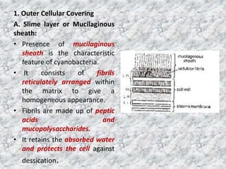
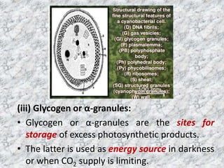
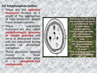
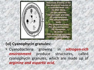
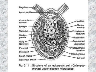
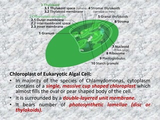
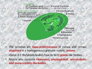
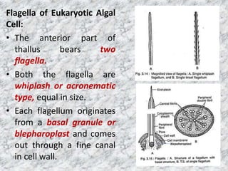
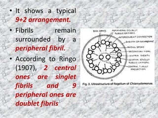
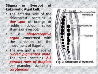
The document summarizes the cell structure of prokaryotic and eukaryotic algal cells. Prokaryotic cells like cyanobacteria have an outer cellular covering including a mucilaginous sheath and cell wall, as well as a cytoplasm differentiated into a pigmented chromoplasm and central colorless centroplasm containing DNA. Eukaryotic algal cells have a cell wall, plasma lemma, and protoplast containing organelles like a chloroplast, mitochondria, and flagella. The chloroplast contains thylakoids and pyrenoids and the cell has structures like an eyespot.
Algae are chlorophyll bearing autotrophic bodies with thalloid plant body. Thallus may be unicellular to multicellular, microscopic or macroscopic in structure.
The plant body in algae is always a thallus. It is not differentiated in root, stem and leaves. Algae range in size from minute unicellular plants (less than 1 µ in diameter in some planktons) to very large highly differentiated multicellular forms e.g., some sea-weeds.
Their forms may be colonial (loose or integrated by inter-connections of protoplasmic strands), filamentous (branched or un-branched), septate (branched or un-branched), non-septate or branched, multinucleate siphonaceous tube where the nuclear divisions occur without usual septa formation.
The algae reproduce by vegetative, asexual, and sexual methods. Vegetative reproduction is by fragmentation, where each fragment develops into a thallus. Asexual reproduction is by the production of flagellated zoospores which on germination give rise to new plants.
Heterothallic species have sexes that reside in different individuals. . The term is applied particularly to distinguish heterothallic fungi, which require two compatible partners to produce sexual spores, from homothallic ones, which are capable of sexual reproduction from a single organism.
About 20,000 species.
Eukaryotic cell and contain all the membrane bound organelles.
Thallus is green due to the presence of green pigment chlorophyll.
Chlorophyll is contained in chloroplast.
Pyrenoids embedded in chloroplast.
Cytoplasm contains vacuoles.
Motile cell of primitive forms contains eye spot or stigma.
Reserve carbohydrates are in the form of starch.
Cell wall invariably contains cellulose.
Produce motile reproductive bodies generally with two or four flagella.
Most are aquatic but some are subarial.
Several species of ulvales and siphonales are marine.
Some strains of chlorella are thermophilic.
Species of chlamydomonas and some chlorococcales occur in snow.
Coloechaete nitellarum is endophytic.
Cephaleuros is parasitic – cause ‘red rust of tea’.
Live epizoically on or endozoically within the bodies of lower animals – chlorella is found in hydra; chlorella beneath the scales of fish; characium on the antennae of mosquito.
Green algae in assosciation with the fungi constitute lichens.
• Gymnosperms (Gymnos = naked, Sperma = seed) include the small group of plants with naked seeds.
• The Gymnosperms originated in the Devonian period of the Paleozoic Era and formed the supreme vegetation in the Mesozoic Era.
This lecture is about classification of algae. In this presentation outline of Fritsch's and Smith's classifications are given. Helpful for B. Sc. students.
Biology Class 11 Chapter 8
FOR FURTHER DETAILS YOU CAN WATCH THE RELATED VIDEO AT THE GIVEN LINK
https://www.youtube.com/channel/UCxo06Nj-QWo_7SNvMyDnJCQ?view_as=subscriber
General bacteriology / /certified fixed orthodontic courses by Indian dental...Indian dental academy
The Indian Dental Academy is the Leader in continuing dental education , training dentists in all aspects of dentistry and offering a wide range of dental certified courses in different formats.
Indian dental academy provides dental crown & Bridge,rotary endodontics,fixed orthodontics,
Dental implants courses.for details pls visit www.indiandentalacademy.com ,or call
00919248678078
Bacteria are unicellular, procaryotic microorganisms which have diverse shape size and structures. Bacteria are found almost everywhere on Earth. Even the human body is full of bacteria, and in fact is estimated to contain more bacterial cells than human cells. Most bacteria in the body are harmless, and some are even helpful. A relatively small number of species cause disease.
Specially for Science students.
Understand cell completely with easy language. Easy language for students who are not good in English.
Students who are in high school just read it with conscious mind and grab the points which will help in understanding cell easily as well as it will help you to score good in tests/exams.
For students who are in universities colleges need to understand it completely by putting some efforts.
Dear Students, this is the PPT to get the idea on Parts of Garden. The parts of garden are really very nice to read and know. You can built your garden with your own interest.
Dear students, how are you all?!. This PPT will give a basic idea for planning, designing and principles of Garden. You all can use this PPT as notes for your exams.
Dear students, in this ppt you will able to understand about the Incomplete dominance. Incomplete dominance is an allelic interaction. In incomplete dominance, both alleles of a character express their character in the F1 generation.
Students able to understand that who helps to transport in plants, Mechanism of transport in plants, physical forces involved in transport, Behavior with different solutions.
Chemotaxonomy is a little bit difficult task for the students to learn and understand. This slide helps the teachers and students to take class and understood it in a liable way
Richard's entangled aventures in wonderlandRichard Gill
Since the loophole-free Bell experiments of 2020 and the Nobel prizes in physics of 2022, critics of Bell's work have retreated to the fortress of super-determinism. Now, super-determinism is a derogatory word - it just means "determinism". Palmer, Hance and Hossenfelder argue that quantum mechanics and determinism are not incompatible, using a sophisticated mathematical construction based on a subtle thinning of allowed states and measurements in quantum mechanics, such that what is left appears to make Bell's argument fail, without altering the empirical predictions of quantum mechanics. I think however that it is a smoke screen, and the slogan "lost in math" comes to my mind. I will discuss some other recent disproofs of Bell's theorem using the language of causality based on causal graphs. Causal thinking is also central to law and justice. I will mention surprising connections to my work on serial killer nurse cases, in particular the Dutch case of Lucia de Berk and the current UK case of Lucy Letby.
Cancer cell metabolism: special Reference to Lactate PathwayAADYARAJPANDEY1
Normal Cell Metabolism:
Cellular respiration describes the series of steps that cells use to break down sugar and other chemicals to get the energy we need to function.
Energy is stored in the bonds of glucose and when glucose is broken down, much of that energy is released.
Cell utilize energy in the form of ATP.
The first step of respiration is called glycolysis. In a series of steps, glycolysis breaks glucose into two smaller molecules - a chemical called pyruvate. A small amount of ATP is formed during this process.
Most healthy cells continue the breakdown in a second process, called the Kreb's cycle. The Kreb's cycle allows cells to “burn” the pyruvates made in glycolysis to get more ATP.
The last step in the breakdown of glucose is called oxidative phosphorylation (Ox-Phos).
It takes place in specialized cell structures called mitochondria. This process produces a large amount of ATP. Importantly, cells need oxygen to complete oxidative phosphorylation.
If a cell completes only glycolysis, only 2 molecules of ATP are made per glucose. However, if the cell completes the entire respiration process (glycolysis - Kreb's - oxidative phosphorylation), about 36 molecules of ATP are created, giving it much more energy to use.
IN CANCER CELL:
Unlike healthy cells that "burn" the entire molecule of sugar to capture a large amount of energy as ATP, cancer cells are wasteful.
Cancer cells only partially break down sugar molecules. They overuse the first step of respiration, glycolysis. They frequently do not complete the second step, oxidative phosphorylation.
This results in only 2 molecules of ATP per each glucose molecule instead of the 36 or so ATPs healthy cells gain. As a result, cancer cells need to use a lot more sugar molecules to get enough energy to survive.
introduction to WARBERG PHENOMENA:
WARBURG EFFECT Usually, cancer cells are highly glycolytic (glucose addiction) and take up more glucose than do normal cells from outside.
Otto Heinrich Warburg (; 8 October 1883 – 1 August 1970) In 1931 was awarded the Nobel Prize in Physiology for his "discovery of the nature and mode of action of the respiratory enzyme.
WARNBURG EFFECT : cancer cells under aerobic (well-oxygenated) conditions to metabolize glucose to lactate (aerobic glycolysis) is known as the Warburg effect. Warburg made the observation that tumor slices consume glucose and secrete lactate at a higher rate than normal tissues.
Slide 1: Title Slide
Extrachromosomal Inheritance
Slide 2: Introduction to Extrachromosomal Inheritance
Definition: Extrachromosomal inheritance refers to the transmission of genetic material that is not found within the nucleus.
Key Components: Involves genes located in mitochondria, chloroplasts, and plasmids.
Slide 3: Mitochondrial Inheritance
Mitochondria: Organelles responsible for energy production.
Mitochondrial DNA (mtDNA): Circular DNA molecule found in mitochondria.
Inheritance Pattern: Maternally inherited, meaning it is passed from mothers to all their offspring.
Diseases: Examples include Leber’s hereditary optic neuropathy (LHON) and mitochondrial myopathy.
Slide 4: Chloroplast Inheritance
Chloroplasts: Organelles responsible for photosynthesis in plants.
Chloroplast DNA (cpDNA): Circular DNA molecule found in chloroplasts.
Inheritance Pattern: Often maternally inherited in most plants, but can vary in some species.
Examples: Variegation in plants, where leaf color patterns are determined by chloroplast DNA.
Slide 5: Plasmid Inheritance
Plasmids: Small, circular DNA molecules found in bacteria and some eukaryotes.
Features: Can carry antibiotic resistance genes and can be transferred between cells through processes like conjugation.
Significance: Important in biotechnology for gene cloning and genetic engineering.
Slide 6: Mechanisms of Extrachromosomal Inheritance
Non-Mendelian Patterns: Do not follow Mendel’s laws of inheritance.
Cytoplasmic Segregation: During cell division, organelles like mitochondria and chloroplasts are randomly distributed to daughter cells.
Heteroplasmy: Presence of more than one type of organellar genome within a cell, leading to variation in expression.
Slide 7: Examples of Extrachromosomal Inheritance
Four O’clock Plant (Mirabilis jalapa): Shows variegated leaves due to different cpDNA in leaf cells.
Petite Mutants in Yeast: Result from mutations in mitochondrial DNA affecting respiration.
Slide 8: Importance of Extrachromosomal Inheritance
Evolution: Provides insight into the evolution of eukaryotic cells.
Medicine: Understanding mitochondrial inheritance helps in diagnosing and treating mitochondrial diseases.
Agriculture: Chloroplast inheritance can be used in plant breeding and genetic modification.
Slide 9: Recent Research and Advances
Gene Editing: Techniques like CRISPR-Cas9 are being used to edit mitochondrial and chloroplast DNA.
Therapies: Development of mitochondrial replacement therapy (MRT) for preventing mitochondrial diseases.
Slide 10: Conclusion
Summary: Extrachromosomal inheritance involves the transmission of genetic material outside the nucleus and plays a crucial role in genetics, medicine, and biotechnology.
Future Directions: Continued research and technological advancements hold promise for new treatments and applications.
Slide 11: Questions and Discussion
Invite Audience: Open the floor for any questions or further discussion on the topic.
(May 29th, 2024) Advancements in Intravital Microscopy- Insights for Preclini...Scintica Instrumentation
Intravital microscopy (IVM) is a powerful tool utilized to study cellular behavior over time and space in vivo. Much of our understanding of cell biology has been accomplished using various in vitro and ex vivo methods; however, these studies do not necessarily reflect the natural dynamics of biological processes. Unlike traditional cell culture or fixed tissue imaging, IVM allows for the ultra-fast high-resolution imaging of cellular processes over time and space and were studied in its natural environment. Real-time visualization of biological processes in the context of an intact organism helps maintain physiological relevance and provide insights into the progression of disease, response to treatments or developmental processes.
In this webinar we give an overview of advanced applications of the IVM system in preclinical research. IVIM technology is a provider of all-in-one intravital microscopy systems and solutions optimized for in vivo imaging of live animal models at sub-micron resolution. The system’s unique features and user-friendly software enables researchers to probe fast dynamic biological processes such as immune cell tracking, cell-cell interaction as well as vascularization and tumor metastasis with exceptional detail. This webinar will also give an overview of IVM being utilized in drug development, offering a view into the intricate interaction between drugs/nanoparticles and tissues in vivo and allows for the evaluation of therapeutic intervention in a variety of tissues and organs. This interdisciplinary collaboration continues to drive the advancements of novel therapeutic strategies.
Multi-source connectivity as the driver of solar wind variability in the heli...Sérgio Sacani
The ambient solar wind that flls the heliosphere originates from multiple
sources in the solar corona and is highly structured. It is often described
as high-speed, relatively homogeneous, plasma streams from coronal
holes and slow-speed, highly variable, streams whose source regions are
under debate. A key goal of ESA/NASA’s Solar Orbiter mission is to identify
solar wind sources and understand what drives the complexity seen in the
heliosphere. By combining magnetic feld modelling and spectroscopic
techniques with high-resolution observations and measurements, we show
that the solar wind variability detected in situ by Solar Orbiter in March
2022 is driven by spatio-temporal changes in the magnetic connectivity to
multiple sources in the solar atmosphere. The magnetic feld footpoints
connected to the spacecraft moved from the boundaries of a coronal hole
to one active region (12961) and then across to another region (12957). This
is refected in the in situ measurements, which show the transition from fast
to highly Alfvénic then to slow solar wind that is disrupted by the arrival of
a coronal mass ejection. Our results describe solar wind variability at 0.5 au
but are applicable to near-Earth observatories.
3. The two main parts of cyanobacterial
cell.
• Outer Cellular Covering
• Cytoplasm.
4. 1. Outer Cellular Covering
A. Slime layer or Mucilaginous
sheath:
• Presence of mucilaginous
sheath is the characteristic
feature of cyanobacteria.
• It consists of fibrils
reticulately arranged within
the matrix to give a
homogeneous appearance.
• Fibrils are made up of peptic
acids and
mucopolysaccharides.
• It retains the absorbed water
and protects the cell against
dessication.
5. B. Cell Wall:
• It is present between the slime layer and
plasma membrane.
• It is a rigid and complex structure and
resembles the cell wall of bacteria.
• It is made of four layers.
• Carr and Whitton (1973) named all these
four layers as L I, L II, L III and L IV.
• L I is a transparent space and occurs
between the L II and plasmembrane.
• L II and L III are mucopolymer, made up of
alanine, glucosamine, peptidoglycan,
muramic acid, glutamic acid and α-
diaminopimelic acid.
• The L IV is undulating, wavy and made of
liposaccharides and proteins.
6. C. Plasma Membrane:
• It is present below the cell wall.
• It is made up of protein-lipid-protein
layers.
• The cytoplasmic membrane and its
invaginations are the sites of biochemical
functions, normally associated with the
membrane bounded structures like
mitochondria, endoplasmic reticulum
and Golgi bodies of the eukaryotic cells.
7. 2. Cytoplasm of Cyanobacterial Cell:
• It is differentiated into two regions
– Chromoplasm
– Centroplasm
a. Chromoplasm:
• It is the outer or peripheral pigmented region.
• This region consists of flattened vesicle like
structures called thylakoids or photosynthetic
lamellae.
• These lamellae contain chlorophyll V, carotenoids
and three phycobilins - C-phycocyanin,
allophycocyanin and C-phycoerythrin.
8. • Photosynthetic lamellae are arranged in
parallel rows close to the periphery of
the cell or they are distributed
irregularly throughout the cell.
• In between the lamellae, occur certain
granules of 400 A° diameter.
• These granules contain phycobilin
pigment and are called cyanosomes y or
phycobilisomes.
9. (2) Centroplasm:
• It is the inner or central colourless region.
• It is often called nucleoid or incipient
nucleus.
• It consists of DNA fibrils. DNA is not
surrounded with protein materials
(histones).
• Like bacteria, small circular DNA segments
occur in addition to nucleoid. These are
known as plasmids or transposons.
• 70S ribosomes are also present in this region
10.
11. 4. The Cytoplasm:
• The cytoplasm of cyanobacterial cell, like that
of bacteria, is incredibly boring.
• It lacks eukaryotic organelles such as
chloroplasts, mitochondria, endoplasmic
reticulum, Golgi bodies.
• But, it possesses photosynthetic apparatus,
ribosomes, and a large number of subcellular
inclusions such as glycogen or α-granules,
polyphosphate bodies, polyhedral bodies,
cyanophycin granules, and the genetic
material.
12. (i) Photosynthetic apparatus:
• In place of the chloroplasts of photosynthetic
eukaryotes, cyanobacteria have flattened
vesicular structures called thylakoids or
lamellae, which resemble the individual
thylakoids of the true chloroplasts of
photosynthetic eukaryotes.
13. • The lamellae or thylakoids are both
physiologically or structurally complex and
possess photosynthetic pigments.
• The principal pigment of all cyanobacteria is
chlorophyll a.
• In addition, there are β-carotene and other
accessory pigments, namely, phycobiliproteins.
• The phycobiliproteins are phycocyanin (PC),
allophycocyanin (AP), allophycocyanin-B (APB),
and phycoerythrin.
• By possessing phycocyanin and phycoerythrin
accessory pigments, the cyanobacteria resemble
with red algae.
14. • However, the necessary pigments of these
organisms are generally organized into
organelles called phycobilisomes and trap
light energy of lower wavelengths, which
cannot be absorbed by chlorophyll a, and pass
it on to the chlorophyll a.
• This is the reason why cyanobacteria, like
green algae, can exploit deeper waters where
the quality and quantity of illumination is
inappropriate for the photosynthetic plants.
15.
16. (ii) Ribosomes:
• These are the sites of protein synthesis.
Cyanobacterian ribosomes occur freely in the
cytoplasm and are identical to those of
bacteria in being 70S ribosomes.
17. (iii) Glycogen or α-granules:
• Glycogen or α-granules are the sites for
storage of excess photosynthetic products.
• The latter is used as energy source in darkness
or when CO2 supply is limiting.
18. (iv) Polyphosphate bodies:
• These are the spherical
structures formed as a
result of the aggregation
of high molecular weight
linear polyphosphates.
• These subcellular
inclusions are also called
metachromatin granules
or volutin granules and
serve as phosphate stores
and are consumed during
periods of phosphate
starvation.
• These structures develop
mostly in those
cyanobacteria that grow
in a phosphate-rich
environment.
19. (v) Polyhedral bodies:
• All cyanobacteria store their ribulose 1, 5-
bisphosphate carboxylase (RUBP carboxylase)
enzyme in structures referred to as polyhedral
bodies.
20. (vi) Cyanophycin granules:
• Cyanobacteria growing in nitrogen-rich
environment produce structures, called
cyanophycin granules, which are made up of
arginine and aspartic acid.
21. (vii) Genetic material:
• The genetic material of cyanobacteria is made up
of naked DNA fibrils found dispersed in the
central region of the cytoplasm.
• Like other prokaryotes, they lack membrane-
bound organized nucleus.
• The exact number of genomes per cell is not yet
known
• it has recently been reported that Agmenellum
contains 2, 3 or more copies of its genetic
material.
• The molecular weight of the cyanobacterial
genome is considered to range from 2.7 to 7.5 x
109 daltons.
22. (viii) Plasmids:
• All the naturally occurring plasmids in
cyanobacteria are phenotypically cryptic.
• They are covalently closed circular DNAs and
their genetic compositions and complete
function is not yet known.
• However, plasmid-mediated transfer of
genetic material has been reported in certain
cyanobacteria.
25. Cell Wall of Eukaryotic Algal Cell:
• The cell is bounded by a thin, cellulose cell
wall.
• Cellulose layer is finely striated with parallel
cellulose fibrils
• In many species there is a pectose layer
external to it which dissolves in water and
forms a mucilaginous pectin layer.
• According to Roberts et. al. (1972), Hills (1973)
the cell wall in C. Reinhardt consists of seven
layers.
26. Plasma Lemma of Eukaryotic Algal Cell:
• It is present just below the cell wall and consists of
two opaque layers which remain separated by less
opaque zone.
Protoplast of Eukaryotic Algal Cell:
• It is bounded by plasma lemma.
• It is differentiated into
– cytoplasm
– nucleus
– chloroplast with one or more pyrenoids
– mitochondria
– Golgi bodies
– two contractile vacuoles
– a red eye spot and
– two flagella
27. Chloroplast of Eukaryotic Algal Cell:
• In majority of the species of Chlamydomonas, cytoplasm
contains of a single, massive cup shaped chloroplast which
almost fills the oval or pear shaped body of the cell.
• It is surrounded by a double-layered unit membrane.
• It bears number of photosynthetic lamellae (disc or
thylakoids).
28. • The lamellae are lippo-proteinaceous in nature and remain
dispersed in a homogeneous granular matrix, stroma.
• About 3-7 thylakoids bodies fuse to form grana like bodies.
• Matrix also contains ribosomes, plastoglobuli, microtubules
and many crystals like bodies.
29. Flagella of Eukaryotic Algal
Cell:
• The anterior part of
thallus bears two
flagella.
• Both the flagella are
whiplash or acronematic
type, equal in size.
• Each flagellum originates
from a basal granule or
blepharoplast and comes
out through a fine canal
in cell wall.
blepaharoplast
30. • It shows a typical
9+2 arrangement.
• Fibrils remain
surrounded by a
peripheral fibril.
• According to Ringo
(1907), 2 central
ones are singlet
fibrils and 9
peripheral ones are
doublet fibrils
31. Stigma or Eyespot of
Eukaryotic Algal Cell:
• The anterior side of the
chloroplast contains a
tiny spot of orange or
reddish colour called
stigma or eyespot.
• It is photoreceptive
organ concerned with
the direction of the
movement of flagella.
• The eye spot is made of
curved pigmented plate.
The plate contains 2-3
parallel rows of droplets
or granules containing
carotenoids