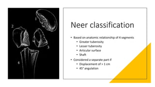
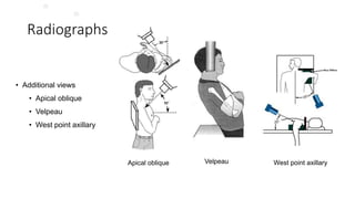
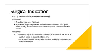
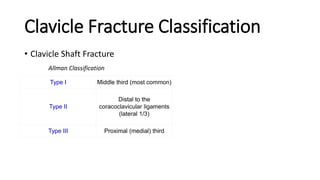
Upper Extremity
Fracture Principle
(SHOULDER)
Marsa - Orthopaedi & Traumatologi Bandung
Proximal Humerus
Fracture
Classification
A0
• Organizes fractures into 3 main
groups and additional subgroups
based on
• Fracture location
• Status of the surgical neck
• Presence/absence of
dislocation
AO
classification
Type A: extra-articular unifocal (either tuberosity +/- surgical
neck of the humerus)
•A1: extra-articular unifocal fracture
•A2: extra-articular unifocal fracture with impacted metaphyseal fracture
•A3: extra-articular unifocal fracture with non-impacted metaphyseal fracture
Type B: extra-articular bifocal (both tuberosities +/- surgical
neck of the humerus or glenohumeral dislocation)
•B1: extra-articular bifocal fractures with impacted metaphyseal fracture
•B2: extra-articular bifocal fractures with non-impacted metaphyseal fracture
•B3: extra-articular bifocal fractures with glenohumeral joint dislocation
Type C: extra-articular (anatomical neck) but with compromise
to the vascular supply of the articular segment
•C1: anatomical neck fracture, minimally displaced
•C2: anatomical neck fracture, displaced and impacted
•C3: anatomical neck fracture with glenohumeral joint dislocation
Neer classification
• Based on anatomic relationship of 4 segments
• Greater tuberosity
• Lesser tuberosity
• Articular surface
• Shaft
• Considered a separate part if
• Displacement of > 1 cm
• 45° angulation
NEER
Classification
Fracture lines involve 1-4 parts
None of the parts are displaced (i.E <1 cm and <45
degrees)
One-part
fracture
Fracture lines involve 2-4 parts
One part is displaced (i.E >1 cm or >45 degrees)
Two-part
fracture
Fracture lines involve 3-4 parts
Two parts are displaced (i.E >1 cm or >45 degrees)
Three-part
fracture
Fracture lines involve more than 4 parts
Three parts are displaced (i.E., >1 cm or >45 degrees) with
respect to the 4th
Four-part
fracture
Radiological
Parametric
Radiographs
• Complete trauma series
• True AP (grashey)
• Scapular Y
• Axillary
Scapular Y (normal)
True AP (grashey)
Axillary
Radiographs
• Additional views
• Apical oblique
• Velpeau
• West point axillary
Apical oblique Velpeau West point axillary
• Findings
• Combined cortical thickness (medial +
lateral thickness >4 mm)
• Studies suggest correlation with
increased lateral plate pullout strength
• Pseudosubluxation (inferior humeral head
subluxation) caused by blood in the
capsule and muscular atony
CT scan
• CT scans are not necessary for all proximal humerus fractures, especially if minimally displaced.
• They can be very helpful for assessing complex injuries, particularly involving the humeral head,
or with significant comminution.
• CT scans aid assessment of:
• Fracture morphology (including the number of fragments)
• Bone stock of the tuberosities and humeral head fragment
• Degree of comminution
• Size of fixable fragments
• Length of posteromedial metaphyseal extension
MRI
• Indications
• Rarely indicated
• Useful to identify associated
rotator cuff injury
Surgical Indication
• CRPP (closed reduction percutaneous pinning)
• Indications
• 2-part surgical neck fractures
• 3-part and valgus-impacted 4-part fractures in patients with good
bone quality, minimal metaphyseal comminution, and intact medial
calcar
• Outcomes
• Considerably higher complication rate compared to ORIF, HA, and RSA
• Axillary nerve at risk with lateral pins
• Musculocutaneous nerve, cephalic vein, and bicep tendon at risk
with anterior pins
ORIF
•Indications
•Greater tuberosity displaced > 5mm
•Displaced 2-part fractures
•3-, and 4-part fractures in younger patients
•Head-splitting fractures in younger patients
•Outcomes
•Medial support necessary for fractures with
posteromedial comminution
•Consider use of a fibula strut if concerned about
medial support or bone quality
•Calcar screw placement critical to decrease varus
collapse of head
Intramedullary
nailing
• Indications
• Surgical neck fractures or 3-part
greater tuberosity fractures in
younger patients
• Combined proximal humerus and
humeral shaft fractures
• Outcomes
• Biomechanically inferior with
torsional stress compared to plates
• Favorable rates of fracture healing
and ROM compared to ORIF
Arthroplasty
Indications
• Hemiarthroplasty
• In younger patients (40-65 years old) with complex fracture-
dislocations or head-splitting components that may fail fixation
• Recommended use of convertible stems to permit easier conversion
to RSA if necessary in future
• Reverse total shoulder
• Low-demand elderly individuals with non-
reconstructible tuberosities and poor bone stock
• Older patients with fracture-dislocation
Arthroplasty
Outcomes
• Improved results if
• Anatomic tuberosity reduction and healing
• Restoration of humeral height and version
• Humeral height is best judged from the superior border of
the pectoralis major insertion
• Poor results with
• Tuberosity nonunion or malunion
• Retroversion of humeral component > 40°
Clavicle Fracture Classification
• Clavicle Shaft Fracture
Allman Classification
Type I Middle third (most common)
Type II
Distal to the
coracoclavicular ligaments
(lateral 1/3)
Type III Proximal (medial) third
Radiologic Parametric
views sitting/standing upright, standard AP view of
bilateral shoulders
15° cephalic tilt (ZANCA view) determine superior/inferior
displacement
Surgical Indication
• open reduction internal fixation
• indications
• controversial: adolescent fractures with significant shortening(>2cm)
• absolute
• open fxs
• displaced fracture with soft-tissue at risk from tenting
• subclavian artery or vein injury
Clavicle Fracture Classification (distal Clavicle)
• Neer Classification (type I)
• extraarticular fracture occurring lateral to CC ligaments
• conoid and/or trapezoid ligament remain intact
• minimal displacement
• stable
Clavicle Fracture Classification (distal Clavicle)
Type IIA Type IIB Type III
Type IV Type V
Radiologic Parametric
• upright AP of bilateral shoulders
• axillary lateral
• 15° cephalic tilt (zanca view)
• helps to determine superior/inferior displacement
Surgical Indication
• open reduction internal fixation
• Absolute
• open or impending open fractures
• subclavian artery or vein injury
• floating shoulder (distal clavicle and scapula neck fractures with > 10mm of displacement)
• symptomatic nonunion
• relative
• unstable fracture patterns (Neer Type IIA, IIB, V)
• brachial plexus injury (questionable because 66% have spontaneous return)
• closed head injury
• seizure disorder
• polytrauma patient
Clavicle Fracture Classification (midshaft clavicle)
• Neer Classification
• Nondisplaced -<100% displacement
• Displaced >100% displacement
Clavicle Fracture Classification (midshaft clavicle)
Radiologic Parametric
• upright AP of bilateral shoulders
• axillary lateral
• 15° cephalic tilt (zanca view)
• helps to determine superior/inferior displacement
Surgical Indication
• Closed reduction and intramedullary fixation vs. open
reduction internal fixation
• Absolute indication
• Open fractures
• Displaced fractures with skin tenting
• Subclavian artery or vein injury
• Floating shoulder
• Symptomatic nonunion
• Symptomatic malunion
Surgical Indication
• Closed reduction and intramedullary fixation vs. open
reduction internal fixation
• Relative and controversial indications
• Displaced with >2 cm shortening
• bilateral displaced clavicle fractures
• brachial plexus injury (questionable because 66% have spontaneous
return)
• closed head injury
• seizure disorder
• polytrauma patient
Scapular Fracture Classification
Classification is based on the location of the fracture and includes
• coracoid fractures
• acromial fractures
• glenoid fractures
• scapular neck fractures
• look for associated AC joint separation or clavicle fracture
• known as "floating shoulder"
• scapular body fractures
• described based on anatomic location
• scapulothoracic dissociation
Coracoid Fracture Classification
Acromial Fracture Classification
Ideberg Classification of Glenoid Fracture
Radiographs
recommended views
true AP, scapular Y and axillary lateral view
open reduction internal fixation
•Indications
•glenohumeral instability
• > 25% glenoid involvement with subluxation of humerus
• > 5mm of glenoid articular surface step off or major gap
• excessive medialization of glenoid
•displaced scapula neck fx
• with > 40 degrees angulation or 1 cm translation
•open fracture
•loss of rotator cuff function
•coracoid fx with > 1cm of displacement
•"double disruption" of the superior shoulder suspensory complex
ACROMIOCLAVICULAR JOINT
DISRUPTION
Classification
• Rockwood classification
Radiographs
• Required views
• bilateral anteroposterior (AP) view of AC joints
• axillary lateral view
• Zanca view
Radiographs
• Additional views
• cross-body adduction view (Basmania)
• weighted stress views
Surgical Indication
• CC interval restoration (ORIF vs. Ligament Reconstruction)
• Indication
• acute type IV, V or VI injuries
• acute type III injuries in laborers, elite athletes, patients with cosmetic
concerns
• chronic type III injuries that failed non-op treatment
• Contraindication
• patient unlikely to comply with postoperative rehabilitation
• skin problems over fixation approach site
THANK YOU

Upper extremity (shoulder fracture

  • 1.
    Upper Extremity Fracture Principle (SHOULDER) Marsa- Orthopaedi & Traumatologi Bandung
  • 2.
    Proximal Humerus Fracture Classification A0 • Organizesfractures into 3 main groups and additional subgroups based on • Fracture location • Status of the surgical neck • Presence/absence of dislocation
  • 3.
    AO classification Type A: extra-articularunifocal (either tuberosity +/- surgical neck of the humerus) •A1: extra-articular unifocal fracture •A2: extra-articular unifocal fracture with impacted metaphyseal fracture •A3: extra-articular unifocal fracture with non-impacted metaphyseal fracture Type B: extra-articular bifocal (both tuberosities +/- surgical neck of the humerus or glenohumeral dislocation) •B1: extra-articular bifocal fractures with impacted metaphyseal fracture •B2: extra-articular bifocal fractures with non-impacted metaphyseal fracture •B3: extra-articular bifocal fractures with glenohumeral joint dislocation Type C: extra-articular (anatomical neck) but with compromise to the vascular supply of the articular segment •C1: anatomical neck fracture, minimally displaced •C2: anatomical neck fracture, displaced and impacted •C3: anatomical neck fracture with glenohumeral joint dislocation
  • 4.
    Neer classification • Basedon anatomic relationship of 4 segments • Greater tuberosity • Lesser tuberosity • Articular surface • Shaft • Considered a separate part if • Displacement of > 1 cm • 45° angulation
  • 6.
    NEER Classification Fracture lines involve1-4 parts None of the parts are displaced (i.E <1 cm and <45 degrees) One-part fracture Fracture lines involve 2-4 parts One part is displaced (i.E >1 cm or >45 degrees) Two-part fracture Fracture lines involve 3-4 parts Two parts are displaced (i.E >1 cm or >45 degrees) Three-part fracture Fracture lines involve more than 4 parts Three parts are displaced (i.E., >1 cm or >45 degrees) with respect to the 4th Four-part fracture
  • 7.
  • 8.
    Radiographs • Complete traumaseries • True AP (grashey) • Scapular Y • Axillary Scapular Y (normal) True AP (grashey) Axillary
  • 9.
    Radiographs • Additional views •Apical oblique • Velpeau • West point axillary Apical oblique Velpeau West point axillary
  • 10.
    • Findings • Combinedcortical thickness (medial + lateral thickness >4 mm) • Studies suggest correlation with increased lateral plate pullout strength • Pseudosubluxation (inferior humeral head subluxation) caused by blood in the capsule and muscular atony
  • 11.
    CT scan • CTscans are not necessary for all proximal humerus fractures, especially if minimally displaced. • They can be very helpful for assessing complex injuries, particularly involving the humeral head, or with significant comminution. • CT scans aid assessment of: • Fracture morphology (including the number of fragments) • Bone stock of the tuberosities and humeral head fragment • Degree of comminution • Size of fixable fragments • Length of posteromedial metaphyseal extension
  • 12.
    MRI • Indications • Rarelyindicated • Useful to identify associated rotator cuff injury
  • 13.
    Surgical Indication • CRPP(closed reduction percutaneous pinning) • Indications • 2-part surgical neck fractures • 3-part and valgus-impacted 4-part fractures in patients with good bone quality, minimal metaphyseal comminution, and intact medial calcar • Outcomes • Considerably higher complication rate compared to ORIF, HA, and RSA • Axillary nerve at risk with lateral pins • Musculocutaneous nerve, cephalic vein, and bicep tendon at risk with anterior pins
  • 14.
    ORIF •Indications •Greater tuberosity displaced> 5mm •Displaced 2-part fractures •3-, and 4-part fractures in younger patients •Head-splitting fractures in younger patients •Outcomes •Medial support necessary for fractures with posteromedial comminution •Consider use of a fibula strut if concerned about medial support or bone quality •Calcar screw placement critical to decrease varus collapse of head
  • 15.
    Intramedullary nailing • Indications • Surgicalneck fractures or 3-part greater tuberosity fractures in younger patients • Combined proximal humerus and humeral shaft fractures • Outcomes • Biomechanically inferior with torsional stress compared to plates • Favorable rates of fracture healing and ROM compared to ORIF
  • 16.
    Arthroplasty Indications • Hemiarthroplasty • Inyounger patients (40-65 years old) with complex fracture- dislocations or head-splitting components that may fail fixation • Recommended use of convertible stems to permit easier conversion to RSA if necessary in future • Reverse total shoulder • Low-demand elderly individuals with non- reconstructible tuberosities and poor bone stock • Older patients with fracture-dislocation
  • 17.
    Arthroplasty Outcomes • Improved resultsif • Anatomic tuberosity reduction and healing • Restoration of humeral height and version • Humeral height is best judged from the superior border of the pectoralis major insertion • Poor results with • Tuberosity nonunion or malunion • Retroversion of humeral component > 40°
  • 18.
    Clavicle Fracture Classification •Clavicle Shaft Fracture Allman Classification Type I Middle third (most common) Type II Distal to the coracoclavicular ligaments (lateral 1/3) Type III Proximal (medial) third
  • 19.
    Radiologic Parametric views sitting/standingupright, standard AP view of bilateral shoulders 15° cephalic tilt (ZANCA view) determine superior/inferior displacement
  • 20.
    Surgical Indication • openreduction internal fixation • indications • controversial: adolescent fractures with significant shortening(>2cm) • absolute • open fxs • displaced fracture with soft-tissue at risk from tenting • subclavian artery or vein injury
  • 21.
    Clavicle Fracture Classification(distal Clavicle) • Neer Classification (type I) • extraarticular fracture occurring lateral to CC ligaments • conoid and/or trapezoid ligament remain intact • minimal displacement • stable
  • 22.
    Clavicle Fracture Classification(distal Clavicle) Type IIA Type IIB Type III Type IV Type V
  • 23.
    Radiologic Parametric • uprightAP of bilateral shoulders • axillary lateral • 15° cephalic tilt (zanca view) • helps to determine superior/inferior displacement
  • 24.
    Surgical Indication • openreduction internal fixation • Absolute • open or impending open fractures • subclavian artery or vein injury • floating shoulder (distal clavicle and scapula neck fractures with > 10mm of displacement) • symptomatic nonunion • relative • unstable fracture patterns (Neer Type IIA, IIB, V) • brachial plexus injury (questionable because 66% have spontaneous return) • closed head injury • seizure disorder • polytrauma patient
  • 25.
    Clavicle Fracture Classification(midshaft clavicle) • Neer Classification • Nondisplaced -<100% displacement • Displaced >100% displacement
  • 26.
  • 27.
    Radiologic Parametric • uprightAP of bilateral shoulders • axillary lateral • 15° cephalic tilt (zanca view) • helps to determine superior/inferior displacement
  • 28.
    Surgical Indication • Closedreduction and intramedullary fixation vs. open reduction internal fixation • Absolute indication • Open fractures • Displaced fractures with skin tenting • Subclavian artery or vein injury • Floating shoulder • Symptomatic nonunion • Symptomatic malunion
  • 29.
    Surgical Indication • Closedreduction and intramedullary fixation vs. open reduction internal fixation • Relative and controversial indications • Displaced with >2 cm shortening • bilateral displaced clavicle fractures • brachial plexus injury (questionable because 66% have spontaneous return) • closed head injury • seizure disorder • polytrauma patient
  • 30.
    Scapular Fracture Classification Classificationis based on the location of the fracture and includes • coracoid fractures • acromial fractures • glenoid fractures • scapular neck fractures • look for associated AC joint separation or clavicle fracture • known as "floating shoulder" • scapular body fractures • described based on anatomic location • scapulothoracic dissociation
  • 31.
  • 32.
  • 33.
    Ideberg Classification ofGlenoid Fracture
  • 35.
    Radiographs recommended views true AP,scapular Y and axillary lateral view
  • 36.
    open reduction internalfixation •Indications •glenohumeral instability • > 25% glenoid involvement with subluxation of humerus • > 5mm of glenoid articular surface step off or major gap • excessive medialization of glenoid •displaced scapula neck fx • with > 40 degrees angulation or 1 cm translation •open fracture •loss of rotator cuff function •coracoid fx with > 1cm of displacement •"double disruption" of the superior shoulder suspensory complex
  • 37.
  • 38.
  • 39.
    Radiographs • Required views •bilateral anteroposterior (AP) view of AC joints • axillary lateral view • Zanca view
  • 40.
    Radiographs • Additional views •cross-body adduction view (Basmania) • weighted stress views
  • 41.
    Surgical Indication • CCinterval restoration (ORIF vs. Ligament Reconstruction) • Indication • acute type IV, V or VI injuries • acute type III injuries in laborers, elite athletes, patients with cosmetic concerns • chronic type III injuries that failed non-op treatment • Contraindication • patient unlikely to comply with postoperative rehabilitation • skin problems over fixation approach site
  • 42.