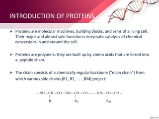
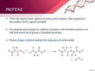
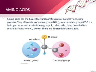
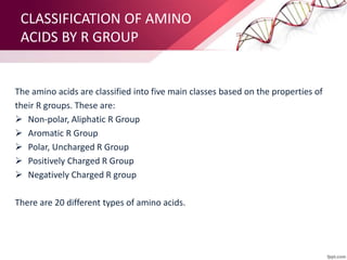
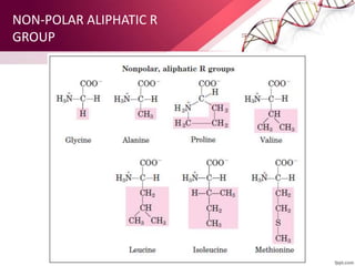
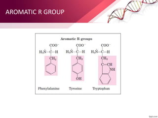
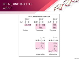
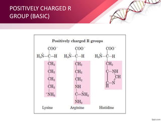
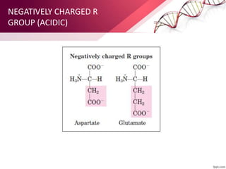
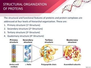
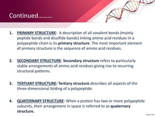
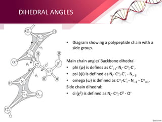
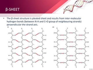
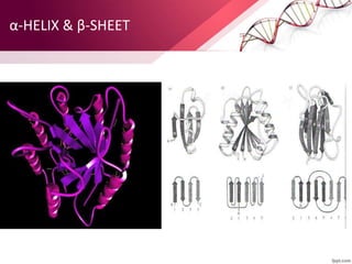
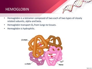
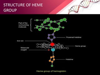
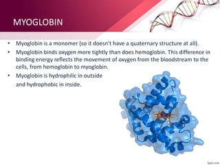
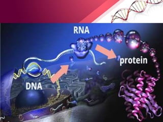
PROTEINS
PRESENTED BY:
DEEPIKA KAITHAL
M.Sc. SEMESTER IV
DEPARTMENT OF
APPLIED PHYSICS
Archana Soni archi22ss@gmail.com
CONTENTS
 INTRODUCTION
 TYPES OF PROTEINS
 AMINO ACIDS
 CLASSIFICATION OF AMINO ACIDS
 STRUCTURAL ORGANIZATION OF PROTEINS
 AMINO ACIDS AND BASES
 PEPTIDE BONDS
 DIHEDRAL ANGLES
 HEMOGLOBIN AND MYOGLOBIN
INTRODUCTION OF PROTEINS
 Proteins are molecular machines, building blocks, and arms of a living cell.
Their major and almost sole function is enzymatic catalysis of chemical
conversions in and around the cell.
 Proteins are polymers: they are built up by amino acids that are linked into
a peptide chain.
 The chain consists of a chemically regular backbone (“main chain”) from
which various side chains (R1, R2, . . . ,RM) project:
PROTEINS
 There are twenty main species of amino acid residues. Their position in
the protein chain is gene-encoded.
 The peptide bond allows for rotation of protein and therefore protein can
fold and orient the R group in favorable positions.
 Protein shape is determined by the sequence of amino acids.
TYPES OF PROTEINS
According to their “environmental conditions” and general structure, proteins can
be roughly divided into three classes:
1) FIBROUS PROTEIN: It is usually water-deficient aggregates; their structure is
usually highly hydrogen bonded; highly regular and maintained mainly by
interactions between various chains.
2) MEMBRANE PROTEINS: It reside in a water-deficient membrane environment
(although they partly project into water). The intra-membrane portions are
highly regular and highly hydrogen-bonded; but restricted in size by the
membrane thickness.
3) GLOBULAR PROTEINS: They are less regular and their structure is maintained
by interactions of chain with itself and sometimes by chain interactions with
cofactors.
AMINO ACIDS
• Amino acids are the basic structural constituents of naturally occurring
proteins. They all consists of amino group (NH+
3), a carboxylate group (COO-), a
hydrogen atom and a substituent group, R, called side chain, bounded to a
central carbon atom (C atom). There are 20 standard amino acid.
CLASSIFICATION OF AMINO
ACIDS BY R GROUP
The amino acids are classified into five main classes based on the properties of
their R groups. These are:
 Non-polar, Aliphatic R Group
 Aromatic R Group
 Polar, Uncharged R Group
 Positively Charged R Group
 Negatively Charged R group
There are 20 different types of amino acids.
NON-POLAR ALIPHATIC R
GROUP
AROMATIC R GROUP
POLAR, UNCHARGED R
GROUP
POSITIVELY CHARGED R
GROUP (BASIC)
NEGATIVELY CHARGED R
GROUP (ACIDIC)
STRUCTURAL ORGANIZATION
OF PROTEINS
The structural and functional features of proteins and protein complexes are
addressed at four levels of hierarchal organization. These are:
1. Primary structure (1o-Structure)
2. Secondary structure (2o-Structure)
3. Tertiary structure (3o-Structure)
4. Quaternary structure (4o-Structure)
Continued………
1. PRIMARY STRUCTURE: A description of all covalent bonds (mainly
peptide bonds and disulfide bonds) linking amino acid residues in a
polypeptide chain is its primary structure. The most important element
of primary structure is the sequence of amino acid residues.
2. SECONDARY STRUCTURE: Secondary structure refers to particularly
stable arrangements of amino acid residues giving rise to recurring
structural patterns.
3. TERTIARY STRUCTURE: Tertiary structure describes all aspects of the
three-dimensional folding of a polypeptide.
4. QUATERNARY STRUCTURE: When a protein has two or more polypeptide
subunits, their arrangement in space is referred to as quaternary
structure.
DIHEDRAL ANGLES
• Diagram showing a polypeptide chain with a
side group.
Main chain angle/ Backbone dihedral
• phi (φ) is defines as C’i-1- Ni- C
i-C’i.
• psi (ψ) is defined as Ni- C
i-C’i - Ni+1.
• omega (ω) is defined as C
i-C’i - Ni+1 - C
i+1.
Side chain dihedral:
• ci (χ1) is defined as Ni- C
i-C - O
α-HELIX
• The α-helix is a stable right-handed helical structure and is also called a 3.613 –
helix. It is a rod-like structure.
β-SHEET
• The β-sheet structure is pleated sheet and results from inter-molecular
hydrogen bonds (between N-H and C=O group of neighbouring strands)
perpendicular the strand axis.
α-HELIX & β-SHEET
HEMOGLOBIN
• Hemoglobin is a tetramer composed of two each of two types of closely
related subunits, alpha and beta.
• Hemoglobin transport O₂ from lungs to tissues.
• Hemoglobin is hydrophilic.
STRUCTURE OF HEME
GROUP
MYOGLOBIN
• Myoglobin is a monomer (so it doesn't have a quaternary structure at all).
• Myoglobin binds oxygen more tightly than does hemoglobin. This difference in
binding energy reflects the movement of oxygen from the bloodstream to the
cells, from hemoglobin to myoglobin.
• Myoglobin is hydrophilic in outside
and hydrophobic in inside.
Proteins ppt
Proteins ppt

Proteins ppt

  • 1.
    PROTEINS PRESENTED BY: DEEPIKA KAITHAL M.Sc.SEMESTER IV DEPARTMENT OF APPLIED PHYSICS Archana Soni archi22ss@gmail.com
  • 2.
    CONTENTS  INTRODUCTION  TYPESOF PROTEINS  AMINO ACIDS  CLASSIFICATION OF AMINO ACIDS  STRUCTURAL ORGANIZATION OF PROTEINS  AMINO ACIDS AND BASES  PEPTIDE BONDS  DIHEDRAL ANGLES  HEMOGLOBIN AND MYOGLOBIN
  • 3.
    INTRODUCTION OF PROTEINS Proteins are molecular machines, building blocks, and arms of a living cell. Their major and almost sole function is enzymatic catalysis of chemical conversions in and around the cell.  Proteins are polymers: they are built up by amino acids that are linked into a peptide chain.  The chain consists of a chemically regular backbone (“main chain”) from which various side chains (R1, R2, . . . ,RM) project:
  • 4.
    PROTEINS  There aretwenty main species of amino acid residues. Their position in the protein chain is gene-encoded.  The peptide bond allows for rotation of protein and therefore protein can fold and orient the R group in favorable positions.  Protein shape is determined by the sequence of amino acids.
  • 5.
    TYPES OF PROTEINS Accordingto their “environmental conditions” and general structure, proteins can be roughly divided into three classes: 1) FIBROUS PROTEIN: It is usually water-deficient aggregates; their structure is usually highly hydrogen bonded; highly regular and maintained mainly by interactions between various chains. 2) MEMBRANE PROTEINS: It reside in a water-deficient membrane environment (although they partly project into water). The intra-membrane portions are highly regular and highly hydrogen-bonded; but restricted in size by the membrane thickness. 3) GLOBULAR PROTEINS: They are less regular and their structure is maintained by interactions of chain with itself and sometimes by chain interactions with cofactors.
  • 6.
    AMINO ACIDS • Aminoacids are the basic structural constituents of naturally occurring proteins. They all consists of amino group (NH+ 3), a carboxylate group (COO-), a hydrogen atom and a substituent group, R, called side chain, bounded to a central carbon atom (C atom). There are 20 standard amino acid.
  • 7.
    CLASSIFICATION OF AMINO ACIDSBY R GROUP The amino acids are classified into five main classes based on the properties of their R groups. These are:  Non-polar, Aliphatic R Group  Aromatic R Group  Polar, Uncharged R Group  Positively Charged R Group  Negatively Charged R group There are 20 different types of amino acids.
  • 8.
  • 9.
  • 10.
  • 11.
  • 12.
  • 13.
    STRUCTURAL ORGANIZATION OF PROTEINS Thestructural and functional features of proteins and protein complexes are addressed at four levels of hierarchal organization. These are: 1. Primary structure (1o-Structure) 2. Secondary structure (2o-Structure) 3. Tertiary structure (3o-Structure) 4. Quaternary structure (4o-Structure)
  • 14.
    Continued……… 1. PRIMARY STRUCTURE:A description of all covalent bonds (mainly peptide bonds and disulfide bonds) linking amino acid residues in a polypeptide chain is its primary structure. The most important element of primary structure is the sequence of amino acid residues. 2. SECONDARY STRUCTURE: Secondary structure refers to particularly stable arrangements of amino acid residues giving rise to recurring structural patterns. 3. TERTIARY STRUCTURE: Tertiary structure describes all aspects of the three-dimensional folding of a polypeptide. 4. QUATERNARY STRUCTURE: When a protein has two or more polypeptide subunits, their arrangement in space is referred to as quaternary structure.
  • 15.
    DIHEDRAL ANGLES • Diagramshowing a polypeptide chain with a side group. Main chain angle/ Backbone dihedral • phi (φ) is defines as C’i-1- Ni- C i-C’i. • psi (ψ) is defined as Ni- C i-C’i - Ni+1. • omega (ω) is defined as C i-C’i - Ni+1 - C i+1. Side chain dihedral: • ci (χ1) is defined as Ni- C i-C - O
  • 16.
    α-HELIX • The α-helixis a stable right-handed helical structure and is also called a 3.613 – helix. It is a rod-like structure.
  • 17.
    β-SHEET • The β-sheetstructure is pleated sheet and results from inter-molecular hydrogen bonds (between N-H and C=O group of neighbouring strands) perpendicular the strand axis.
  • 18.
  • 19.
    HEMOGLOBIN • Hemoglobin isa tetramer composed of two each of two types of closely related subunits, alpha and beta. • Hemoglobin transport O₂ from lungs to tissues. • Hemoglobin is hydrophilic.
  • 20.
  • 21.
    MYOGLOBIN • Myoglobin isa monomer (so it doesn't have a quaternary structure at all). • Myoglobin binds oxygen more tightly than does hemoglobin. This difference in binding energy reflects the movement of oxygen from the bloodstream to the cells, from hemoglobin to myoglobin. • Myoglobin is hydrophilic in outside and hydrophobic in inside.