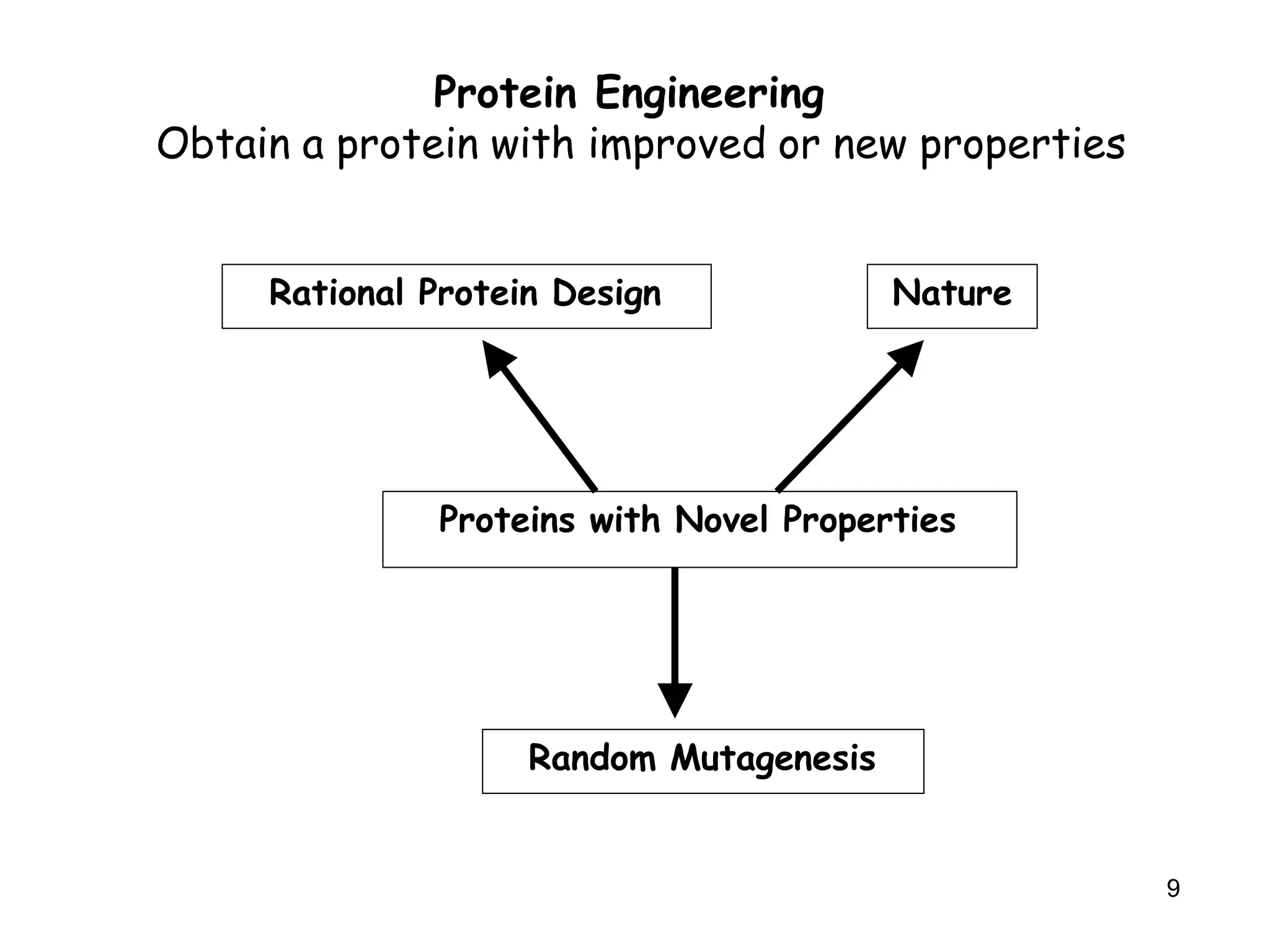
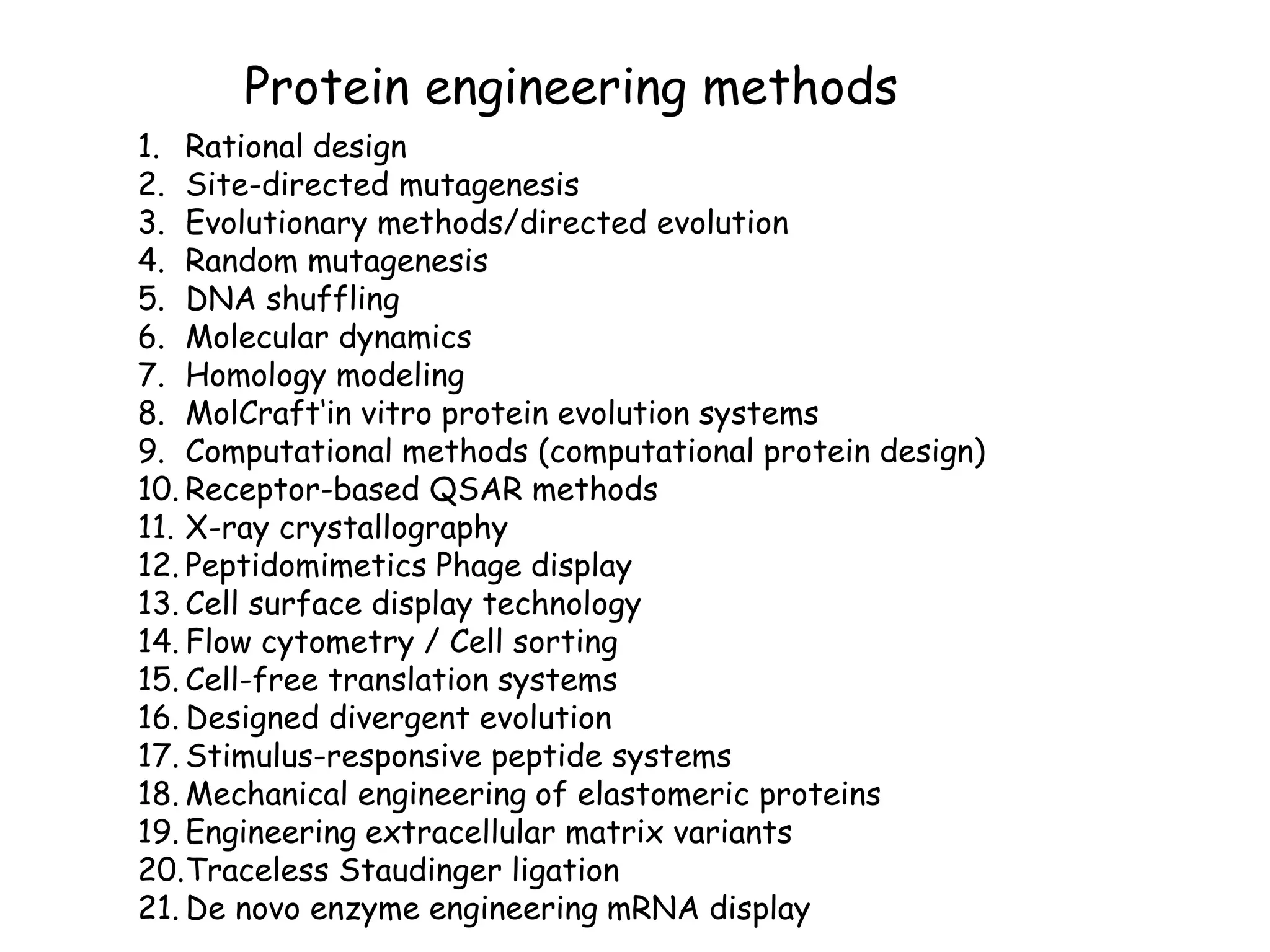
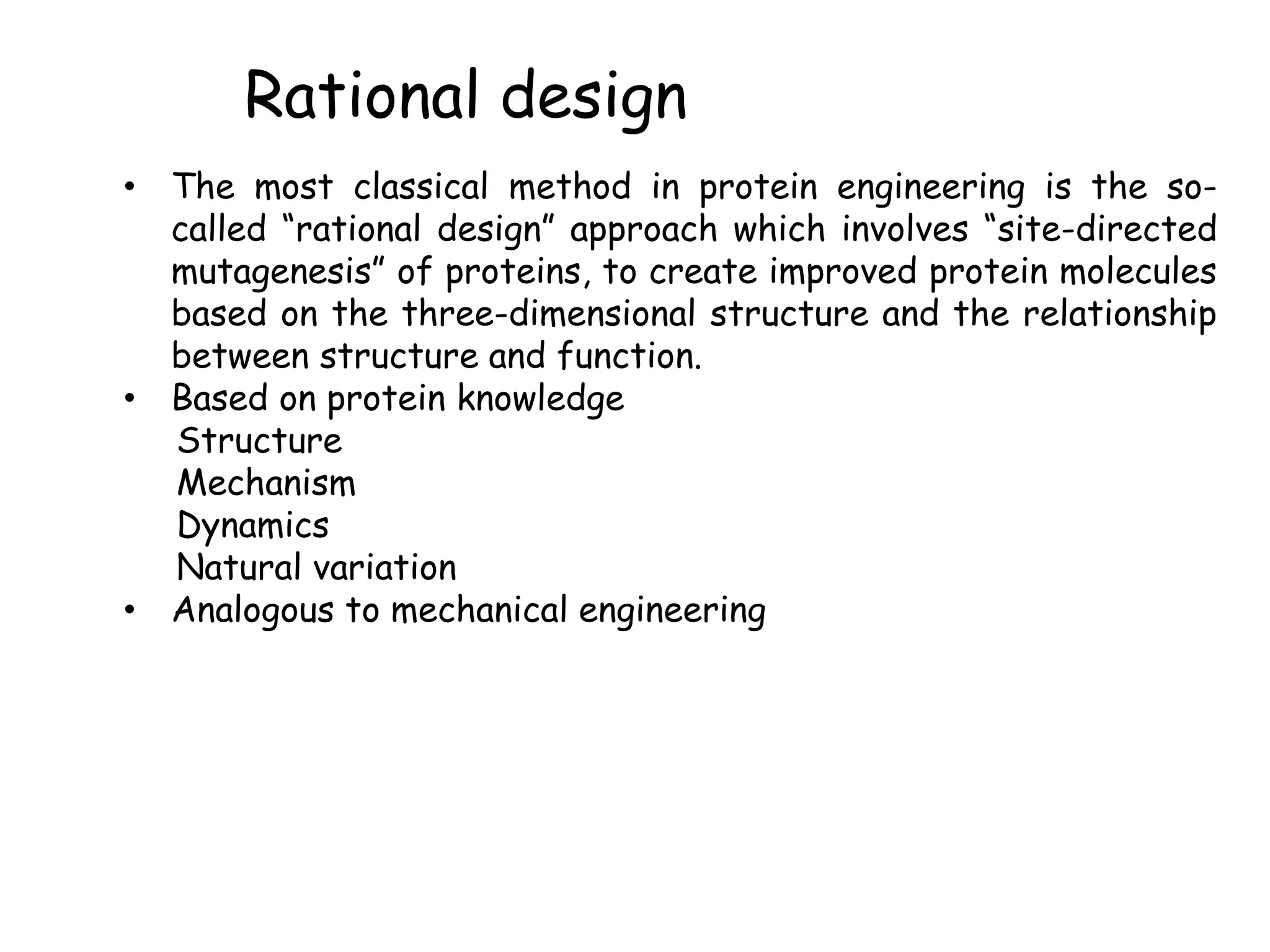
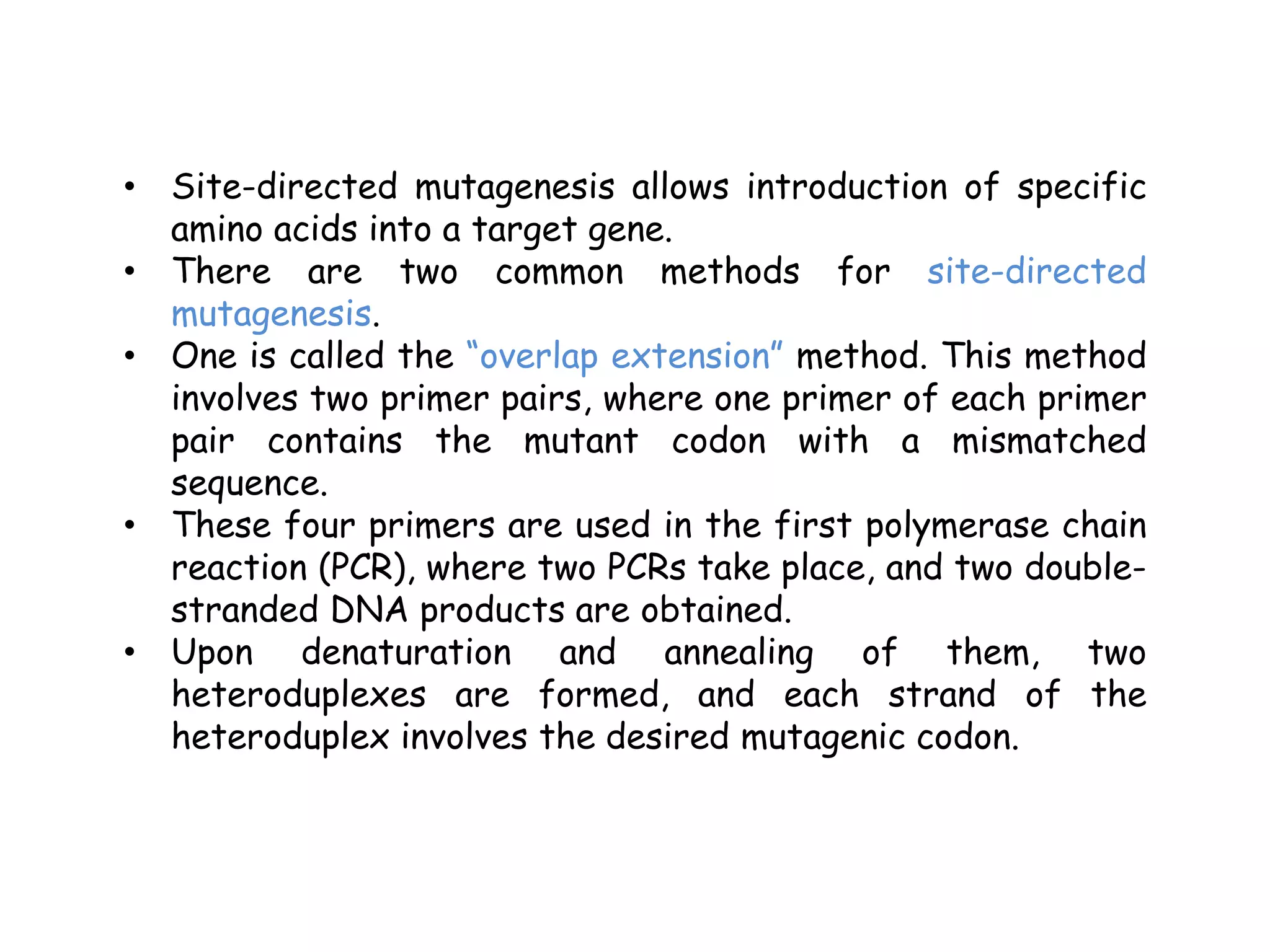
Protein Engineering
1
Himanshu Kamboj
Assistant Professor
Contents
 Protein engineering
 Objectives of protein engineering
 Rationale of protein engineering
 Protein engineering methods
 Rational design -site-directed mutagenesis methods
 Advantages and disadvantages of rational design
 Directed evolution -random mutagenesis
 Advantages and disadvantages of directed evolution
 Peptidomimetics
 Classification of peptidomimetics
 Advantages and disadvantages of peptidomimetics
 Flow cytometry
 Instrumentation
 Principle
 components
Protein Engineering
Protein engineering can be defined as
the modification of protein structure with
recombinant DNA technology or
chemical treatment to get a desirable
function for better use in medicine,
industry and agriculture.
OBJECTIVES OF PROTEIN
ENGINEERING
The objectives of protein engineering is
as follows –
(a) to create a superior enzyme to
catalyze the production of high value
specific chemicals.
(b) to produce enzyme in large quantities.
(c) to produce biological
compounds(include synthetic peptide,
storage protein, and synthetic drugs)
superior to natural one.
RATIONAL OF PROTEIN
ENGINEERING
For industrial application an enzyme,
should possess some characteristics in
addition to those of enzymes in cells.
These characteristics are :-
(1) enzyme should be robust with long
life.
(2) enzyme should be able to use the
substrate supplied in the industry even it
differs from that in the cell.
(3) enzyme should be able to work
under conditions, e.g. extreme of pH,
temperature and concentration of the
industry even if they differ from those in
the cell.
 In view of above, the enzyme should be
engineered to meet the altered needs.
Therefore efforts have been made to
alter the properties of enzymes.
These are some character that one
might have to change in a predictable
manner in protein engineering or
enzyme engineering to get the desired
function :-
Kinetic properties of enzyme-turnover
and Michaelis constant, Km.
Thermo stability and the optimum
temperature for the enzyme.
Stability and activity of enzyme in
nonaqueous solvents.
Substrate and reaction specificity.
Cofactor requirements
Optimum PH.
Molecular weight and subunit structure.
Therefore for a particular class of
enzymes, variation in nature may occur
for each of the above properties, so that
one may like to combine all the optimum
properties to the most efficient form of
the enzyme.
For an e.g. glucose isomerases, which
convert glucose into other isomers like
fructose and are used to make high
fructose corn syrup vital for soft drink
industries.
Protein Engineering
Obtain a protein with improved or new properties
Proteins with Novel Properties
Rational Protein Design Nature
Random Mutagenesis
9
Protein engineering methods
1. Rational design
2. Site-directed mutagenesis
3. Evolutionary methods/directed evolution
4. Random mutagenesis
5. DNA shuffling
6. Molecular dynamics
7. Homology modeling
8. MolCraft‘in vitro protein evolution systems
9. Computational methods (computational protein design)
10. Receptor-based QSAR methods
11. X-ray crystallography
12. Peptidomimetics Phage display
13. Cell surface display technology
14. Flow cytometry / Cell sorting
15. Cell-free translation systems
16. Designed divergent evolution
17. Stimulus-responsive peptide systems
18. Mechanical engineering of elastomeric proteins
19. Engineering extracellular matrix variants
20.Traceless Staudinger ligation
21. De novo enzyme engineering mRNA display
Rational design
• The most classical method in protein engineering is the so-
called “rational design” approach which involves “site-directed
mutagenesis” of proteins, to create improved protein molecules
based on the three-dimensional structure and the relationship
between structure and function.
• Based on protein knowledge
Structure
Mechanism
Dynamics
Natural variation
• Analogous to mechanical engineering
• Site-directed mutagenesis allows introduction of specific
amino acids into a target gene.
• There are two common methods for site-directed
mutagenesis.
• One is called the “overlap extension” method. This method
involves two primer pairs, where one primer of each primer
pair contains the mutant codon with a mismatched
sequence.
• These four primers are used in the first polymerase chain
reaction (PCR), where two PCRs take place, and two double-
stranded DNA products are obtained.
• Upon denaturation and annealing of them, two
heteroduplexes are formed, and each strand of the
heteroduplex involves the desired mutagenic codon.
• The other site-directed mutagenesis method is called
“whole plasmid single round PCR”.
• This method forms the basis of the commercial “Quik
Change Site-Directed Mutagenesis Kit” from Stratagene.
• It requires two oligonucleotide primers with the desired
mutation(s) which are complementary to the opposite
strands of a double-stranded DNA plasmid template.
• Using DNA polymerase PCR takes place, and both strands
of the template are replicated without displacing the
primers and a mutated plasmid is obtained with breaks
that do not overlap.
• DpnI methylase is then used for selective digestion to
obtain a circular, nicked vector with the mutant gene.
• Upon transformation of the nicked vector into competent
cells, the nick in the DNA is repaired, and a circular,
mutated plasmid is obtained
Site-directed mutagenesis methods
14
Site-directed mutagenesis methods – PCR based
15
Site-directed mutagenesis methods –
Oligonucleotide - directed method
16
Advantages and disadvantages
Advantages
• Intellectually satisfying
• Controlled outcome
• Range of available techniques
• Increasing computational power
Disadvantages
• Requires deep understanding
 Natural variation
 Structure
 Dynamics
 Mechanism
• High failure rate
Failures rarely reported
18
Directed Evolution
 This method mimics natural evolution and generally
produce superior results to rational design
 This also know as irrational design
 An additional technique known as DNA shuffling mixes
and matches the pieces of variants in order to produce
better results.
 In this random mutagenesis play an important role.
19
Directed Evolution – Random mutagenesis
- NO structural information required
- NO understanding of the mechanism required
General Procedure:
Generation of genetic diversity
 Random mutagenesis: when an organism exposed to physical or
chemical mutagen, mutations are induced randomly in all genes of
the organism.
Hence, this process of generating mutations is known as Random
mutagenesis.
Identification of successful variants
 Screening and seletion
20
Directed Evolution
- A simple and common technique for random mutagenesis is
“saturation mutagenesis”.
- It involves the replacement of a single amino acid within a protein
with each of the natural amino acids, and provides all possible
variations at that site. “
- Localized or region-specific random mutagenesis” is another
technique which is a combination of rational and random approaches
of protein engineering.
- It includes the simultaneous replacement of a few amino acid
residues in a specific region, to obtain proteins with new
specificities.
21
- This technique also makes use of overlap extension, and the whole-
plasmid, single round PCR mutagenesis, as in the case of site-
directed mutagenesis.
- However, the major difference here is that the codons for the
selected amino acids are randomized, such that a mixture of 64
different forward and 64 different reverse primers are used,
based on a statistical mixture of four bases and three nucleotides
in a randomized codon
22
Random Mutagenesis (PCR based)
with degenerated primers (saturation mutagenesis)
23
Random Mutagenesis (PCR based)
with degenerated primers (saturation mutagenesis)
24
Random Mutagenesis (PCR based)
Error –prone PCR
-> PCR with low fidelity !!!
Achieved by:
- Increased Mg2+ concentration
- Addition of Mn2+
- Not equal concentration of the
four dNTPs
- Use of dITP
- Increasing amount of Taq
polymerase (Polymerase with NO
proof reading function)
25
Random Mutagenesis (PCR based)
DNA Shuffling
26
DNase I treatment (Fragmentation,
10-50 bp, Mn2+)
Reassembly (PCR without primers,
Extension and Recombination)
PCR amplification
Random Mutagenesis (PCR based)
Family Shuffling
Genes coming from the same
gene family -> highly
homologous
-> Family shuffling
27
Advantages and disadvantages
Advantages
• Simple concepts
• Widely applicable
• High success rate
• Requires no knowledge of protein (or of mutations)
Disadvantages
• Starting activity range
Requires some starting activity
Can rarely improve native activity
• Requires high-throughput assay
Typically enzyme-specific
Screens can be expensive
Synergistic mutations hard to find
• Understanding results is not trivial: Especially to apply to another protein
• You get what you screen for: Surrogate substrates can yield artefacts, Side-
effects if not constrained
29
Peptidomimetics
 Peptidomimetics are compounds whose essential elements
(pharmacophore) mimic a natural peptide or protein in 3D
space and which retain the ability to interact with the
biological target and produce the same biological effect.
 It involves mimicking or blocking the activity of enzymes
or natural peptides upon design and synthesis of peptide
analogs that are metabolically stable.
 Peptidomimetics is an important approach for bioorganic
and medical chemistry.
 It includes a variety of synthesis methods such as the
use of a common intermediate, solid phase synthesis and
combinatorial approaches.
 Peptidomimetics are designed to avoid some of the
problems associated with a natural peptide for example
Stability against proteolysis (duration of activity)
Poor bioavailability.
Receptor selectivity or potency (often can be
substantially improved)
30
Peptidomimetics
 The design process begins by developing structure–
activity relationships (SAR) that can define a minimal
active sequence or major pharmacophore elements, and
identify the key residues that are responsible for the
biological effect.
 Then structural constraints are applied to probe the 3D
arrangement(s) of these features. In this process, the
peptide complexity is reduced and the basic
pharmacophore model is defined by its critical structural
features in 3D space.
 This model then supports the re-assembly of the critical
elements and non-peptide variants on a modified scaffold
that presents the optimized pharmacophore to the
receptor
31
Classification of Peptidomimetics
 Type-I peptidomimetics or pseudopeptides: These are
synthesized by structure based drug design. These
peptidomimetics are closely similar to peptide backbone
while retaining functional groups that makes important
contacts with binding sites of the receptors.
 Type-II peptidomimetics or functional mimetics: These
peptidomimetics are synthesized by molecular modeling
and high throughput screening (HTS) etc. These are
small non-peptide molecule that binds to a peptide
receptor.
32
Classification of Peptidomimetics
 Type-III peptidomimetics or topographical mimetics:
These are synthesized by structure based drug design
which represents that they possess novel templates,
which appear unrelated to the original peptides but
contain the essential groups, positioned on a novel non-
peptide scaffold to serve as topographical mimetics.
 Type-IV Peptidomimetics or non-peptide mimetics: hese
are synthesized by Group Replacement Assisted Binding
(GRAB) technique of drug design.
33
Advantages
1. Conformationally restrained structures can minimize
binding to non-target receptors and enhance the
activity at the desired receptor.
2. Addition of hydrophobic residues and/or replacement
of amide bonds results in better transport properties
through cellular membranes.
3. Isosteres, retro-inverso peptides, cyclic peptides and
non-peptidomimetics all reduce the rate of degradation
by peptidases and other enzymes.
34
Disadvantages
1. Limited stability towards proteolysis by peptidases in the
gastrointestinal tract and in serum (t1/2 on the order of
minutes)
2. Poor transport properties from the intestine to the blood
and across the blood-brain barrier due to high MW and lack
of specific transport systems
3. Rapid excretion through the liver and/or kidneys
4. Inherent flexibility enables interaction with multiple
receptors besides the target, and could result in undesired
side-effects
35
Flow cytometry
Flow= flow
Cyto= cell
Metry= measurement
“Flow cytometry”, a powerful method for single cell analysis,
is also used in protein engineering studies. A variety of
examples are available where the sorting was done according
to ligand binding in antibody and peptide surface display
studies, or enzyme engineering of intra- and extracellular
enzymes.
36
Flow cytometry
The advantages and disadvantages of random mutagenesis
methods used in protein engineering were also determined
and compared to each other in detail.
Based on the nucleotide substitution method used, these
random mutagenesis methods were divided into four major
groups:
enzyme-based methods,
synthetic chemistry-based methods,
whole cell methods and
combined methods.
Their comparison was made according to a variety of
parameters such as controllable mutation frequency,
technical robustness, cost-effectiveness, etc
37
HISTORY
• Mark Fulwyler was the inventor of the forerunner of the
modern flow cytometer.
• This was developed in 1965
• First fluorescence based flow cytometer was developed
in 1986by Wolfgang Gohde.
• The flow cytometer was originally called pulse
cytophotometry.
• In 1988, the name was officially changed to “flow
cytometry” at the Conference of the American
Engineering Foundation in Pensacola, Florida.
38
THE INSTRUMENT
• Modern flow cytometers can analyze several thousand
particles every second.
• It can also actively separate and isolate particles having
specified property.
 A flow cytometry is composed of three main systems
namely, fluidics, optics, electronics
• Each cell is subjected to a laser beam.
• The light reflected from each cell is captured.
• Information is then interpreted statistically by a
software
39
COMPONENTS
Liquid stream system: carries and aligns cells so that they
pass through a single file.
Measuring system: measure the impedance.
optical system: lamps, high power and low power lasers
Detector and analogue system
computer
40
PRINCIPLE
o Beam of light of single wavelength is directed onto
hydrodynamically focused stream of liquid
o Detectors are placed where the stream passed through
the light beam.
o The light scattered by the cells are detected.
o The amount of light scattered is measured.
o This is analysed and results are interpreted.
41
HYDRODYNAMIC FOCUSING
Cells to be analyzed are suspended in liquid and forced
through a small aperture. A tube is used through which the
sheath fluid is pumped. Cells along with the fluid are forced
through this narrow aperture. Cells move in a single file or
line. Laser hits each cell and data from each cell can be
read.
SCATTERING
Cells pass through the laser. Light gets refracted or
scattered in all angles.
2 types of scatter are analyzed:
• Forward scatter: scatter in the forward direction
• Side scatter: light scattered in very large angles.
42
FLOURESCENCE IN FLOW CYTOMETRY
Most common method of studying cellular characteristics.
Antibodies are tagged with a flourophore. The antibody
binds to cells. When laser hits the flourophore, the molecule
gets excited. Signal is emitted which can be detected.
DETECTION OF FLOURESCENCE
Fluorescent t light is travels the same path as side scatter.
The light is directed through a series of filters and mirrors.
Particular wavelength of light is detected by the
appropriate detector.
43
MEASURABLE PARAMETERS
o Volume and morphological complexity of cells
o Cell pigments such as chlorophyll
o Cell cycle analysis, cell kinetics
o Chromosomal analysis
o Protein modification
o Antigens
o Enzymatic activity
o Membrane fluidity
Thank you

Protein engineering and its techniques himanshu

  • 1.
  • 2.
    Contents  Protein engineering Objectives of protein engineering  Rationale of protein engineering  Protein engineering methods  Rational design -site-directed mutagenesis methods  Advantages and disadvantages of rational design  Directed evolution -random mutagenesis  Advantages and disadvantages of directed evolution  Peptidomimetics  Classification of peptidomimetics  Advantages and disadvantages of peptidomimetics  Flow cytometry  Instrumentation  Principle  components
  • 3.
    Protein Engineering Protein engineeringcan be defined as the modification of protein structure with recombinant DNA technology or chemical treatment to get a desirable function for better use in medicine, industry and agriculture.
  • 4.
    OBJECTIVES OF PROTEIN ENGINEERING Theobjectives of protein engineering is as follows – (a) to create a superior enzyme to catalyze the production of high value specific chemicals. (b) to produce enzyme in large quantities. (c) to produce biological compounds(include synthetic peptide, storage protein, and synthetic drugs) superior to natural one.
  • 5.
    RATIONAL OF PROTEIN ENGINEERING Forindustrial application an enzyme, should possess some characteristics in addition to those of enzymes in cells. These characteristics are :- (1) enzyme should be robust with long life. (2) enzyme should be able to use the substrate supplied in the industry even it differs from that in the cell. (3) enzyme should be able to work under conditions, e.g. extreme of pH, temperature and concentration of the industry even if they differ from those in the cell.
  • 6.
     In viewof above, the enzyme should be engineered to meet the altered needs. Therefore efforts have been made to alter the properties of enzymes. These are some character that one might have to change in a predictable manner in protein engineering or enzyme engineering to get the desired function :-
  • 7.
    Kinetic properties ofenzyme-turnover and Michaelis constant, Km. Thermo stability and the optimum temperature for the enzyme. Stability and activity of enzyme in nonaqueous solvents. Substrate and reaction specificity. Cofactor requirements Optimum PH. Molecular weight and subunit structure.
  • 8.
    Therefore for aparticular class of enzymes, variation in nature may occur for each of the above properties, so that one may like to combine all the optimum properties to the most efficient form of the enzyme. For an e.g. glucose isomerases, which convert glucose into other isomers like fructose and are used to make high fructose corn syrup vital for soft drink industries.
  • 9.
    Protein Engineering Obtain aprotein with improved or new properties Proteins with Novel Properties Rational Protein Design Nature Random Mutagenesis 9
  • 10.
    Protein engineering methods 1.Rational design 2. Site-directed mutagenesis 3. Evolutionary methods/directed evolution 4. Random mutagenesis 5. DNA shuffling 6. Molecular dynamics 7. Homology modeling 8. MolCraft‘in vitro protein evolution systems 9. Computational methods (computational protein design) 10. Receptor-based QSAR methods 11. X-ray crystallography 12. Peptidomimetics Phage display 13. Cell surface display technology 14. Flow cytometry / Cell sorting 15. Cell-free translation systems 16. Designed divergent evolution 17. Stimulus-responsive peptide systems 18. Mechanical engineering of elastomeric proteins 19. Engineering extracellular matrix variants 20.Traceless Staudinger ligation 21. De novo enzyme engineering mRNA display
  • 11.
    Rational design • Themost classical method in protein engineering is the so- called “rational design” approach which involves “site-directed mutagenesis” of proteins, to create improved protein molecules based on the three-dimensional structure and the relationship between structure and function. • Based on protein knowledge Structure Mechanism Dynamics Natural variation • Analogous to mechanical engineering
  • 12.
    • Site-directed mutagenesisallows introduction of specific amino acids into a target gene. • There are two common methods for site-directed mutagenesis. • One is called the “overlap extension” method. This method involves two primer pairs, where one primer of each primer pair contains the mutant codon with a mismatched sequence. • These four primers are used in the first polymerase chain reaction (PCR), where two PCRs take place, and two double- stranded DNA products are obtained. • Upon denaturation and annealing of them, two heteroduplexes are formed, and each strand of the heteroduplex involves the desired mutagenic codon.
  • 13.
    • The othersite-directed mutagenesis method is called “whole plasmid single round PCR”. • This method forms the basis of the commercial “Quik Change Site-Directed Mutagenesis Kit” from Stratagene. • It requires two oligonucleotide primers with the desired mutation(s) which are complementary to the opposite strands of a double-stranded DNA plasmid template. • Using DNA polymerase PCR takes place, and both strands of the template are replicated without displacing the primers and a mutated plasmid is obtained with breaks that do not overlap. • DpnI methylase is then used for selective digestion to obtain a circular, nicked vector with the mutant gene. • Upon transformation of the nicked vector into competent cells, the nick in the DNA is repaired, and a circular, mutated plasmid is obtained
  • 14.
  • 15.
  • 16.
    Site-directed mutagenesis methods– Oligonucleotide - directed method 16
  • 17.
    Advantages and disadvantages Advantages •Intellectually satisfying • Controlled outcome • Range of available techniques • Increasing computational power Disadvantages • Requires deep understanding  Natural variation  Structure  Dynamics  Mechanism • High failure rate Failures rarely reported
  • 18.
    18 Directed Evolution  Thismethod mimics natural evolution and generally produce superior results to rational design  This also know as irrational design  An additional technique known as DNA shuffling mixes and matches the pieces of variants in order to produce better results.  In this random mutagenesis play an important role.
  • 19.
    19 Directed Evolution –Random mutagenesis - NO structural information required - NO understanding of the mechanism required General Procedure: Generation of genetic diversity  Random mutagenesis: when an organism exposed to physical or chemical mutagen, mutations are induced randomly in all genes of the organism. Hence, this process of generating mutations is known as Random mutagenesis. Identification of successful variants  Screening and seletion
  • 20.
    20 Directed Evolution - Asimple and common technique for random mutagenesis is “saturation mutagenesis”. - It involves the replacement of a single amino acid within a protein with each of the natural amino acids, and provides all possible variations at that site. “ - Localized or region-specific random mutagenesis” is another technique which is a combination of rational and random approaches of protein engineering. - It includes the simultaneous replacement of a few amino acid residues in a specific region, to obtain proteins with new specificities.
  • 21.
    21 - This techniquealso makes use of overlap extension, and the whole- plasmid, single round PCR mutagenesis, as in the case of site- directed mutagenesis. - However, the major difference here is that the codons for the selected amino acids are randomized, such that a mixture of 64 different forward and 64 different reverse primers are used, based on a statistical mixture of four bases and three nucleotides in a randomized codon
  • 22.
  • 23.
    Random Mutagenesis (PCRbased) with degenerated primers (saturation mutagenesis) 23
  • 24.
    Random Mutagenesis (PCRbased) with degenerated primers (saturation mutagenesis) 24
  • 25.
    Random Mutagenesis (PCRbased) Error –prone PCR -> PCR with low fidelity !!! Achieved by: - Increased Mg2+ concentration - Addition of Mn2+ - Not equal concentration of the four dNTPs - Use of dITP - Increasing amount of Taq polymerase (Polymerase with NO proof reading function) 25
  • 26.
    Random Mutagenesis (PCRbased) DNA Shuffling 26 DNase I treatment (Fragmentation, 10-50 bp, Mn2+) Reassembly (PCR without primers, Extension and Recombination) PCR amplification
  • 27.
    Random Mutagenesis (PCRbased) Family Shuffling Genes coming from the same gene family -> highly homologous -> Family shuffling 27
  • 28.
    Advantages and disadvantages Advantages •Simple concepts • Widely applicable • High success rate • Requires no knowledge of protein (or of mutations) Disadvantages • Starting activity range Requires some starting activity Can rarely improve native activity • Requires high-throughput assay Typically enzyme-specific Screens can be expensive Synergistic mutations hard to find • Understanding results is not trivial: Especially to apply to another protein • You get what you screen for: Surrogate substrates can yield artefacts, Side- effects if not constrained
  • 29.
    29 Peptidomimetics  Peptidomimetics arecompounds whose essential elements (pharmacophore) mimic a natural peptide or protein in 3D space and which retain the ability to interact with the biological target and produce the same biological effect.  It involves mimicking or blocking the activity of enzymes or natural peptides upon design and synthesis of peptide analogs that are metabolically stable.  Peptidomimetics is an important approach for bioorganic and medical chemistry.  It includes a variety of synthesis methods such as the use of a common intermediate, solid phase synthesis and combinatorial approaches.  Peptidomimetics are designed to avoid some of the problems associated with a natural peptide for example Stability against proteolysis (duration of activity) Poor bioavailability. Receptor selectivity or potency (often can be substantially improved)
  • 30.
    30 Peptidomimetics  The designprocess begins by developing structure– activity relationships (SAR) that can define a minimal active sequence or major pharmacophore elements, and identify the key residues that are responsible for the biological effect.  Then structural constraints are applied to probe the 3D arrangement(s) of these features. In this process, the peptide complexity is reduced and the basic pharmacophore model is defined by its critical structural features in 3D space.  This model then supports the re-assembly of the critical elements and non-peptide variants on a modified scaffold that presents the optimized pharmacophore to the receptor
  • 31.
    31 Classification of Peptidomimetics Type-I peptidomimetics or pseudopeptides: These are synthesized by structure based drug design. These peptidomimetics are closely similar to peptide backbone while retaining functional groups that makes important contacts with binding sites of the receptors.  Type-II peptidomimetics or functional mimetics: These peptidomimetics are synthesized by molecular modeling and high throughput screening (HTS) etc. These are small non-peptide molecule that binds to a peptide receptor.
  • 32.
    32 Classification of Peptidomimetics Type-III peptidomimetics or topographical mimetics: These are synthesized by structure based drug design which represents that they possess novel templates, which appear unrelated to the original peptides but contain the essential groups, positioned on a novel non- peptide scaffold to serve as topographical mimetics.  Type-IV Peptidomimetics or non-peptide mimetics: hese are synthesized by Group Replacement Assisted Binding (GRAB) technique of drug design.
  • 33.
    33 Advantages 1. Conformationally restrainedstructures can minimize binding to non-target receptors and enhance the activity at the desired receptor. 2. Addition of hydrophobic residues and/or replacement of amide bonds results in better transport properties through cellular membranes. 3. Isosteres, retro-inverso peptides, cyclic peptides and non-peptidomimetics all reduce the rate of degradation by peptidases and other enzymes.
  • 34.
    34 Disadvantages 1. Limited stabilitytowards proteolysis by peptidases in the gastrointestinal tract and in serum (t1/2 on the order of minutes) 2. Poor transport properties from the intestine to the blood and across the blood-brain barrier due to high MW and lack of specific transport systems 3. Rapid excretion through the liver and/or kidneys 4. Inherent flexibility enables interaction with multiple receptors besides the target, and could result in undesired side-effects
  • 35.
    35 Flow cytometry Flow= flow Cyto=cell Metry= measurement “Flow cytometry”, a powerful method for single cell analysis, is also used in protein engineering studies. A variety of examples are available where the sorting was done according to ligand binding in antibody and peptide surface display studies, or enzyme engineering of intra- and extracellular enzymes.
  • 36.
    36 Flow cytometry The advantagesand disadvantages of random mutagenesis methods used in protein engineering were also determined and compared to each other in detail. Based on the nucleotide substitution method used, these random mutagenesis methods were divided into four major groups: enzyme-based methods, synthetic chemistry-based methods, whole cell methods and combined methods. Their comparison was made according to a variety of parameters such as controllable mutation frequency, technical robustness, cost-effectiveness, etc
  • 37.
    37 HISTORY • Mark Fulwylerwas the inventor of the forerunner of the modern flow cytometer. • This was developed in 1965 • First fluorescence based flow cytometer was developed in 1986by Wolfgang Gohde. • The flow cytometer was originally called pulse cytophotometry. • In 1988, the name was officially changed to “flow cytometry” at the Conference of the American Engineering Foundation in Pensacola, Florida.
  • 38.
    38 THE INSTRUMENT • Modernflow cytometers can analyze several thousand particles every second. • It can also actively separate and isolate particles having specified property.  A flow cytometry is composed of three main systems namely, fluidics, optics, electronics • Each cell is subjected to a laser beam. • The light reflected from each cell is captured. • Information is then interpreted statistically by a software
  • 39.
    39 COMPONENTS Liquid stream system:carries and aligns cells so that they pass through a single file. Measuring system: measure the impedance. optical system: lamps, high power and low power lasers Detector and analogue system computer
  • 40.
    40 PRINCIPLE o Beam oflight of single wavelength is directed onto hydrodynamically focused stream of liquid o Detectors are placed where the stream passed through the light beam. o The light scattered by the cells are detected. o The amount of light scattered is measured. o This is analysed and results are interpreted.
  • 41.
    41 HYDRODYNAMIC FOCUSING Cells tobe analyzed are suspended in liquid and forced through a small aperture. A tube is used through which the sheath fluid is pumped. Cells along with the fluid are forced through this narrow aperture. Cells move in a single file or line. Laser hits each cell and data from each cell can be read. SCATTERING Cells pass through the laser. Light gets refracted or scattered in all angles. 2 types of scatter are analyzed: • Forward scatter: scatter in the forward direction • Side scatter: light scattered in very large angles.
  • 42.
    42 FLOURESCENCE IN FLOWCYTOMETRY Most common method of studying cellular characteristics. Antibodies are tagged with a flourophore. The antibody binds to cells. When laser hits the flourophore, the molecule gets excited. Signal is emitted which can be detected. DETECTION OF FLOURESCENCE Fluorescent t light is travels the same path as side scatter. The light is directed through a series of filters and mirrors. Particular wavelength of light is detected by the appropriate detector.
  • 43.
    43 MEASURABLE PARAMETERS o Volumeand morphological complexity of cells o Cell pigments such as chlorophyll o Cell cycle analysis, cell kinetics o Chromosomal analysis o Protein modification o Antigens o Enzymatic activity o Membrane fluidity
  • 44.